artifact_class: CHEATSHEET_CANDIDATE type: theme id: A1 domain: A title: Facial and regional anatomy subchapters: 10 covered_subs: 10 new_subs: 0 slides: 0 books: 54 articles: 9 videos: 0 maps_to: [G10, G3] federated: false tags: [theme, domain/A, status/covered] pass: 1 template: ANATOMIA lang: en status: authored
A1 — Facial and regional anatomy
> Tags — [A] verified external guideline, consensus or study · [B] anatomical article with its own identifier (dissection, contrast CT, histology) · [C] monograph or atlas from the own MEDLIB corpus · [D] UPO master's teaching material or another never_sufficient_alone source · (P) the model's own reasoning, never a dose nor a plane.
> Currency and provenance — 122 references · median 2016, range 1966-2026, 13 % from 2022 on · provenance: verified external 67 % (82) · MEDLIB corpus 33 % (40, of which 4 from the UPO master's) · 4 flagged [D] never_sufficient_alone.
Base chapter. The thirteen regions (D1-D13) link here and do not repeat general anatomy: block A1.10 (proportions, angles and variants) is the one they all consume. This chapter describes the anatomy; injection technique lives in C1-C4 and in each region.
- [x] A1.1 — In 30 seconds: the map (which layer, which vessel and which bone rule each third)
- [x] A1.2 — Layers from skin to bone: the layer model, where it holds and where it does NOT
- [x] A1.3 — Superficial and deep fat compartments, one by one
- [x] A1.4 — Retaining ligaments: origin, insertion, what they restrain and which groove they draw when they fail
- [x] A1.5 — Muscles: origin, insertion, vector, antagonist and layer
- [x] A1.6 — Arteries: course, depth by segment and internal carotid anastomosis
- [x] A1.7 — Veins and lymphatics: edema and periorbital filling
- [x] A1.8 — Nerves: motor and sensory, emergence, block and injury
- [x] A1.9 — Bone: morphology, angles and resorption chronology
- [x] A1.10 — Proportions and variants: thirds, fifths, angles, ethnicity, sex and age
A1.1 · In 30 seconds — the map
In 30 seconds
| Third | Layer that decides the result | Vessel that decides the risk | Bone that decides the shape | Plane of choice | Signature of the error |
|---|---|---|---|---|---|
| Upper (forehead, glabella, temple, brow tail) | Layer 4 (deep frontal fat) and layer 2 (superficial fat) separated by the frontalis muscle and its fascia [1] | Supratrochlear and supraorbital (internal carotid), superficial temporal (external) [2] | Frontal bone: glabellar protrusion, expansion of the supraorbital arches [3] | Supraperiosteal (bone contact) or intradermal; never mid-subcutaneous [2] | Blindness via the ophthalmic route; visible nodule in a thin forehead |
| Mid (infraorbital, malar, nose, nasolabial fold) | Layer 4: deep medial cheek fat, SOOF, premaxillary space and deep pyriform [4] | Angular and infraorbital; anastomosis with the dorsal nasal (internal carotid) [2] | Maxilla: descent of the maxillary angle, retrusion of the inferior orbital rim and of the pyriform aperture [3,5] | Supraperiosteal medial to the line of ligaments; subcutaneous lateral to it [4,2] | Persistent malar edema; nasal ala necrosis; angular occlusion |
| Lower (lips, chin, jaw, neck) | Layers 2-3: subcutaneous fat and modiolar platysma [4] | Facial and its labial branches: facial artery fixed at the modiolus 1.5 cm posterior to the commissure in 100 % [2] | Mandible: loss of ramus and body height, prejowl notch [3,5] | Subcutaneous (the arteries run deep to the platysma); bone contact allowed at the mandibular angle and zygomatic arch [2] | Lip necrosis; jowl worsened by poorly placed lateral filler |
The five sentences that order the rest of the chapter
- The line of ligaments separates two behaviors, not two zones. All the major ligaments align along a single line immediately lateral to the lateral orbital rim, from the temporal crest to the mandible: temporal ligamentous adhesions, lateral orbital thickening (LOT), zygomatic ligament and mandibular ligament [4]. Medial to that line the layers are oblique and filler projects; lateral to it they are parallel and filler pulls/lifts the structures below [4].
- An artery's depth changes with the segment, not with the region. In the forehead, the arteries are supraperiosteal below the middle frontal septum and subcutaneous above it; that septum lies 1.5 ± 0.17 cm (range 1.3-1.7) from the midline and 3.0 ± 0.24 cm (range 2.7-3.3) laterally, measured from the supraorbital rim [2,1].
- Three regions break the layer model and must be treated as explicit exceptions: the temple (up to 10 layers), the tear trough (only 3: skin, orbicularis, periosteum) and the perioral region (no defined subcutaneous compartments, skin anchored to muscle) [4,3].
- The retinal window is about 90 minutes. Experimental occlusion of the central retinal artery in a primate produced no detectable damage until 97 minutes; beyond that the damage is irreversible and progressive [2]. The retina also receives collateral ciliary supply, so complete occlusion is rare on angiography; 90 minutes is the operating reference point, not a guarantee [2].
- The needle seeds product into every plane it crosses. Material ascends retrograde along the injection channel and is identified in the subdermal plane even when the tip is in bone contact; this is why a negative aspiration does not exclude transarterial passage [2].
> Red lines — Glabella: maximal density of anastomoses between the dorsal nasal, supratrochlear and supraorbital of both sides; no major artery is identified deep to the procerus, so the deep plane is the least bad, not the safe one [2]. · Angular vein: 4.2 ± 0.7 mm below the inferior orbital rim, oblique from inferolateral to superomedial, deep to the orbital portion of the orbicularis [4]. · Deep temple: deep temporal artery anterior at 1.5-2 cm and posterior at 2.5-3.0 cm from the lateral orbital rim [2]. · Tear trough: there is no layer 2; what is injected "subcutaneously" is intramuscular [4,3].
Surface landmarks and normative planes
They are marked with the patient seated at 90°, gaze to the horizon: supine, the tissue migrates and the marks shift [6,7]. They are surface references to locate bone, foramina and vascular courses before loading; they are not therapeutic targets.
| Landmark | How it is located | What it is for | Source |
|---|---|---|---|
| Frankfort plane | Infraorbital rim ↔ upper border of the external auditory canal (porion); horizon with the subject in natural position | The only reproducible plane for photography and for comparing before/after; every vertical proportion is measured on it [7] | [7] |
| Ricketts' E-line | Nasal tip ↔ soft-tissue pogonion (profile) | Upper lip ~4 mm and lower lip ~2 mm behind the line in the Caucasian phenotype; ⚠ normative by ethnicity, the target of one population is not imported into another (see B6) | [7] |
| Canthal tilt | Axis medial canthus → lateral canthus | Positive (lateral canthus above the medial) reads as rest/youth; negative, as fatigue; a frequent target of lateral repositioning | [7] |
| Supraorbital notch | Orbital rim, 1-3 mm medial to the mid-pupillary vertical | Exit of the supraorbital bundle: it is avoided, not crossed [2,8] | [2,8] |
| Infraorbital foramen | 5-10 mm below the inferior orbital rim, on the same mid-pupillary vertical | V2 block and medial limit of the deep medial fat; a risk zone in tear-trough filling [4,9] | [4,9] |
| Mental foramen | Vertical of the 2nd premolar, toward the mid-height of the mandible | V3 block; surrounded by deep labiomandibular fat that protects it as it emerges [4,10] | [4,10] |
| Pitanguy's line | From 0.5 cm below the tragus to 1.5 cm above the brow tail | Traces the temporal (frontal) branch of the facial nerve over the temple; mark it before working on the temple, brow tail or radiofrequency [11] | [11] |
| Course of the supratrochlear | Ascends vertically at 17-22 mm from the facial midline from the superomedial angle of the orbit | The nº 1 zone of visual compromise: any bolus closer than that distance enters its territory [2,8,12] | [2,8,12] |
| McGregor's patch | ~4.5 cm in front of the tragus, over the zygomatic arch | Surface reference of the zygomatic ligament, the stiffest of the face [3,13] | [3,13] |
| Temporal fusion line | Palpable bony crest, lateral border of the forehead | Safe lateral limit of frontal infiltration; marks the galea–temporal fascia junction [4] | [4] |
| Zygomatic arch | Palpated from the tragus to the lateral orbital rim | Roof of the temporal compartment and reference for the malar repositioning vector [4] | [4] |
(P) The profile angles (nasofrontal, nasolabial, mentolabial, cervicomental) have a definition but this chapter does not publish their normative ranges: the corpus pass did not return them with a traceable series and a slide is not enough (see the declared limitation in A1.10). The E-line and canthal tilt are kept because they are soft-tissue relations measurable on the photo, not teaching angle ranges.
The map, third by third
Upper third. The frontalis is not a muscle over bone: it is the continuation of the galea aponeurotica, covered by a thin fascia on its superficial face and a thicker one on its deep face [4]. Between the muscle and its deep fascia there is a fat lamina of its own —the subfrontal fat— through which the supraorbital and supratrochlear vessels ascend after leaving their foramina, and which is bounded by the inferior and middle frontal septa [4,1]. This description changes the gesture: in the lower third of the forehead the vessel is stuck to bone, and in the upper third it is already subcutaneous; the supraperiosteal plane is the poorest in arteries in the whole forehead [2].
The superficial fat compartments of the forehead were delimited in 2007 [14], confirmed in 2012 and updated in 2017 [1]: there are three, housed in layer 2 between the skin and the frontalis muscle.
In the temple, the five layers of the scalp continue but change name on crossing the superior temporal septum: the galea becomes the superficial temporal fascia and the periosteum the deep temporal fascia [4]. The anterior and posterior branches of the superficial temporal artery travel within the superficial temporal fascia (layer 3), so that subcutaneous filler (layer 2) sits superficial to them [4,2]. The deep technique deposits in contact with the bony temporal fossa, below the branches of the facial nerve and of the superficial temporal artery [2].
Mid third. This is where most aesthetic injections concentrate and where bone rules most: the maxillary angle decreases with age, changes the position of the fat compartments that rest on it, and the clinical sign appears [4,3]. Medial to the line of ligaments are the deep midfacial compartments (deep medial fat pad, deep lateral fat pad, premaxillary space, deep pyriform space); lateral to it are the premasseteric surgical spaces [4]. In the nasolabial fold the angular artery runs deep in the fold but close to the dermis: on contrast CT slices arteries are identified less than 4 mm from the skin [2].
Lower third. The facial artery crosses the mandibular border in front of the vein in 100 % of observed cases, deep to the platysma and deep to the marginal mandibular branch of the facial nerve [2]. At the modiolus it is fixed by a muscular band from the buccinator 1.5 cm behind the commissure, between the buccinator (deep) and the modiolar portion of the platysma plus the converging muscles (superficial) [2]. The labial arteries are submucosal in 78.1 %, intramuscular in an intermediate fraction and subcutaneous in only 2.1 % [2]: the subcutaneous plane of the lip is the least vascularized, not the most.
Classic trap: treating "the forehead" as one unit and choosing a single plane. The middle frontal septum splits the region into two opposite vascular regimes [2,1]; the same supraperiosteal bolus that is safest in the lower forehead lies, in the upper forehead, below a vessel that is already subcutaneous, and the palpable nodule that appears weeks later belongs to the upper forehead, not the lower.
A1.2 · Layers from skin to bone
Schematic — the 6-layer model and its three exceptions
| Layer | Generic name (SCALP) | Forehead | Temple | Tear trough | Medial cheek | Lateral cheek (parotideomasseteric) | Perioral |
|---|---|---|---|---|---|---|---|
| 1 | S — skin | Thick, adherent skin | Thin skin | Very thin, translucent skin, adherent to the orbicularis | Medium skin | Medium skin | Skin adherent to muscle |
| 2 | C — subcutaneous cellular tissue (superficial fat + retinacula) | 3 superficial compartments (central + 2 lateral) [1,14] | Subcutaneous fat, superficial to the STA [4] | ABSENT [4,3] | Superficial nasolabial + superficial medial cheek (malar fat) [14] | Superficial middle and lateral cheek [15] | Fat scattered among muscle fibers, no compartments [4,16] |
| 3 | A — musculoaponeurotic system | Frontalis + its thin superficial fascia [1] | Superficial temporal fascia (contains the STA) [4,17] | Orbicularis oculi (it is the functional layer 2 here) | SMAS + orbicularis; the zygomatici perforate it and incorporate [3] | Continuous SMAS [18,19] | Orbicularis oris and converging muscles |
| 4 | L — loose areolar tissue / deep fat | Subfrontal fat with the SO and ST pedicles [4,1] | Superior and inferior temporal compartments [4,20] | ABSENT | Deep nasolabial fat pad (premaxillary space), SOOF, deep medial and lateral fat pads [4,21,22] | Superior, middle and inferior premasseteric spaces [4,23] | Deep mental and labiomandibular fat around the mental foramen [4,10] |
| 5 | P — periosteum or deep fascia | Frontal periosteum | Superficial lamina of the deep temporal fascia [4] | Periosteum (third and last layer) [4,3] | Maxillary periosteum | Parotideomasseteric fascia [4,3] | Mandibular periosteum |
| 6 | B — bone or deep muscular foundation | Frontal bone | Temporalis and temporal fossa (expanded temporal count below) | Orbital rim | Maxilla | Masseter and zygomatic/mandibular skeleton | Maxilla and mandible |
Consensus: the conventional SCALP model describes five continuous soft-tissue and fascial layers from the neck to the scalp, with fat distributed in two laminae (layers 2 and 4) separated by the SMAS (layer 3) [4,3,6]. This chapter counts the bone or deep muscular foundation as layer 6 because it is the structural endpoint of examination and supraperiosteal work; the added number does not alter the five-layer anatomical nomenclature. Discrepancy — how many layers the temple has: Cotofana 2016 describes nine layers in the temporal region [3] · Cotofana 2019 describes ten, splitting the deep temporal fascia into a superficial lamina (layer 5) and a deep lamina (layer 7) with the superficial temporal fat pad (layer 6) between them [4] · decide: if injecting between laminae or raising a temporal flap, use the count of 10, which is the one that names the plane of the medial zygomaticotemporal vein; for a surface reference, both counts give the same gesture. Do not average.
The three exceptions that break the schematic
1 · The temple (more layers). From superficial to deep [4,20,17]:
| # | Temporal structure | Critical content |
|---|---|---|
| 1 | Skin | — |
| 2 | Subcutaneous fat | Plane of the superficial technique: it lies above the superficial temporal artery |
| 3 | Superficial temporal fascia (continuation of the galea and the SMAS) | Anterior and posterior branches of the superficial temporal artery [4] |
| 4 | Superior and inferior temporal compartments | The superior contains no relevant neurovascular; the inferior contains frontal branches of the facial nerve (motor), zygomaticotemporal branches (sensory) and the temporal portion of the sentinel vein [4] |
| 5 | Superficial lamina of the deep temporal fascia | Continues in the face as the parotideomasseteric fascia |
| 6 | Superficial temporal fat pad | Medial zygomaticotemporal vein (proximal portion of the sentinel vein) [4,24] |
| 7 | Deep lamina of the deep temporal fascia | Passes under the zygomatic arch to the masticator space |
| 8 | Deep temporal fat pad (temporal extension of the buccal fat pad of Bichat) | — |
| 9 | Temporalis muscle | — |
| 10 | Periosteum | Anterior and posterior deep temporal arteries, over the periosteum [4,2] |
The division of the deep temporal fascia into two laminae occurs between 2 and 5 cm above the zygomatic arch [4]. The deep technique deposits in bone contact, and the "one up and one down" reference from the fusion of the temporal crest with the supraorbital rim places the product in front of the deep temporal arteries, which run at 1.5-2 cm (anterior) and 2.5-3.0 cm (posterior) from the lateral orbital rim [2].
2 · The tear trough (fewer layers). Medial to the vertical line of the medial pupillary margin only three layers exist: skin, orbital portion of the orbicularis and periosteum [4,3,25]. There is no layer 2 and no deep fascia [6]. Direct consequence: in that zone the "subcutaneous" plane the rest of the face offers does not exist; an injection the operator believes superficial is intramuscular or supraperiosteal, with no middle ground. The bluish color seen through the skin is the orbicularis muscle itself, not a vein, and reappears identically on the medial nasal wall next to the inner canthus because there too the muscle reaches the dermis [3].
3 · The perioral region (no compartment architecture). Between the nasolabial folds, the nose, the modiolus, the labiomandibular folds and the submental septum there are no subcutaneous fat compartments nor a macroscopic aponeurotic structure: the fat is scattered among muscle fibers and collagen bundles that anchor the skin to the muscular plane [4,16,26]. That anchoring is what allows fine lip movement and also what draws the nasolabial fold as a transition line between type 1 subcutaneous tissue (compartmentalized) and type 2 (isolated fat cells in a collagen mesh) [3].
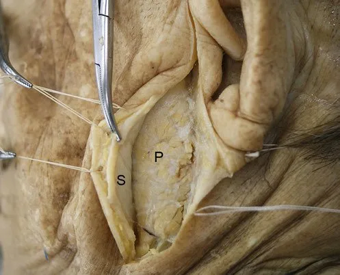 Fig 1. Cadaveric dissection of the lateral cheek: a whitish, translucent fibrous lamina (S) has been incised and separated with traction sutures, and below it the surface of the parotid plane (P) is exposed. The image documents that the SMAS is a dissectable, independent sheet, not a virtual plane — (Watanabe, 2016, p. 117).
Fig 1 is the physical proof of the previous argument: if the SMAS can be caught with sutures and lifted as a complete sheet, then it separates two fat compartments that do not communicate, and a product deposited above it will never migrate to the deep plane on its own. Layer 3 is not a descriptive convention.
What is inside each layer, and why it changes the decision
Layer 1. Thickness, pigmentation and adherence vary by region [3]. In the infraorbital region the skin is thin, transparent and firmly attached to the orbicularis; in the buccal and parotideomasseteric it rests on a variable thickness of fat with loose connections; in the perioral it is directly attached to muscle [3,26].
Layer 2. The subcutaneous fat is partitioned into compartments by fibrous septa that do two things at once: they are the protected route by which cutaneous nerves and vessels ascend from the depth, and they are the anchoring points of the skin to muscle, to other fasciae or to the skeleton [3,27]. This fat is continuous with general body fat and increases with obesity [3]. The superficial compartments descend with age [6]; the deep ones do not change their position relative to bone [21,28,6].
Layer 3. It contains the platysma in the neck, the SMAS in the face, the superficial temporal fascia in the temple, the galea in the cranium and the orbicularis oculi in the periorbit [4,3]. Described in 1976 [18] and revised histologically in 2003 [19], it is not a flat lamina but a three-dimensional composite of fat and collagen tissue [6]. In 2021 the presence of striated muscle fibers within the midfacial SMAS itself was demonstrated [29], which gives an anatomical basis to treatment with intradermal microtoxin. The zygomaticus major and minor muscles originate on the lateral cheek below this layer and perforate it on entering the medial cheek, from there being included in it [3].
Layer 4. It contains the deep fat, whose adipocytes are smaller and of different morphology from the superficial ones [30]. The limits of these compartments are the transit routes of the branches of the facial nerve and of the branches of the facial artery and vein [3]. In the temple, this layer is almost absent in the superior temporal compartment and varies with age in the inferior one, where it protects the temporal branches of the facial nerve [3,17].
Layer 5. Its name changes with the region and that is where many people get lost: it is periosteum in the scalp, deep temporal fascia in the temple, parotideomasseteric fascia in the lateral cheek, and in the neck it continues with the investing layer of the deep cervical fascia [4,3]. Medial to the facial vein it becomes periosteum again and connects with the epineurium of the infraorbital nerve [3].
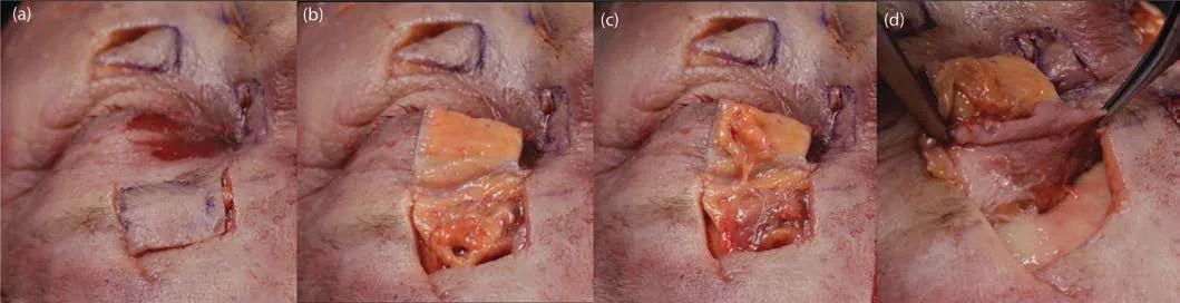 Fig 2. Four-panel dissection sequence over the forehead and supraorbital rim of a specimen: (a) skin window opened with violet marking and a bleeding dermal bed; (b) and (c) the subcutaneous fat lamina, yellow and lobulated, is lifted en bloc as an independent flap; (d) the forceps hold the already-detached fat pad and reveal the underlying pearly-white deep plane — (Pirayesh, 2020, p. 107).
Fig 2 documents panel by panel what in the chair is only sensed through needle resistance: between the dermis and the deep plane there is a fat lamina that behaves as a unit and detaches whole. It is the anatomical argument for why a cannula advances effortlessly in the correct plane and meets resistance the moment it leaves it.
Operative translation layer by layer
| If the goal is… | Target layer | Expected effect | What happens if the plane is missed |
|---|---|---|---|
| Restore structural support | 4 (supraperiosteal) | Projection and foundation for the overlying mobile tissue [6] | If it moves up to layer 2: less projection per unit volume and an unnatural look with mimicry [6] |
| Reposition lateral tissue | 2-3 of the temple and lateral cheek | Upward pulling effect through fascial continuity [31,32,33] | If done medial to the line of ligaments: it projects instead of pulling [4,31] |
| Treat the fine static line | 1 (intradermal) | Direct filling of the cutaneous groove | In the glabella and forehead, the intermediate subcutaneous plane is exactly the one that contains the artery in its upper segment [2] |
| Relax mimicry | 3 (muscle) | Selective chemical denervation | The SMAS with striated fibers explains the effect of intradermal toxin [29] |
Classic trap: applying the vocabulary of the cheek to the lower eyelid. In the tear trough "subcutaneous" names no existing plane [4,3,25]; the product ends up inside the orbicularis, and the clinical signature is a bluish bulge from the Tyndall effect that persists, accentuates on smiling and does not respond to massage, because it is not in a compartment but inside a muscle that contracts over it.
A1.3 · Fat compartments, one by one
Schematic — the facial fat in two laminae and what each compartment does
| Superficial fat (layer 2) | Deep fat (layer 4) | |
|---|---|---|
| Separated by | SMAS (layer 3) [4,3] | |
| Adipocyte size | Larger [30] | Smaller, different morphology [30] |
| Behavior with age | Descends [6] | Does not change position relative to bone [21,6] |
| Mobility with mimicry (in vivo ultrasound) | Significantly greater [28] | Significantly less [28] |
| Continuity with body fat | Yes; increases with obesity [3] | Does not behave the same [16] |
| Filler effect | Smooths the surface, defines contour | Projects and creates a foundation for what lies above [6] |
| Risk of dynamic overcorrection | Low | High if the transverse facial septum is ignored [34] |
Superficial compartments, with their septum and their neighbor
| Superficial compartment | Upper limit | Lower limit | Medial / anterior limit | Lateral / posterior limit | Roof / floor | What happens when filling it |
|---|---|---|---|---|---|---|
| Central forehead | Superior frontal septum | Cutaneous insertion of the orbicularis and procerus | — | Lateral forehead compartments | Skin / frontalis muscle | Corrects central frontal hollowing; in a thin forehead, a visible nodule [1,14] |
| Lateral forehead (×2) | Superior frontal septum | Orbicularis complex | Central compartment | Superior temporal septum | Skin / frontalis muscle | Restores the forehead–temple transition; superficial to the STA in its lateral segment [4,2] |
| Superficial superior temporal | Superior temporal septum | Inferior temporal septum | Lateral forehead compartment | — | Skin / superficial temporal fascia | Plane of the superficial temporal technique: above the artery [4] |
| Superficial inferior temporal | Inferior temporal septum | Zygomatic arch adhesions | — | — | Skin / superficial temporal fascia | Same; lateral pulling effect through fascial continuity [31,33] |
| Superficial nasolabial | Tear trough | Cutaneous insertion of the orbicularis oris | Nasal side and ala | Medial cheek compartment | Skin / SMAS | Worsens the nasolabial fold: not filled a priori [6] |
| Medial cheek (malar fat) | Tear trough | Cutaneous adhesions to the platysma | Nasolabial fold | Middle cheek compartment | Skin / SMAS and orbicularis | Rounds the anterior cheekbone; in excess, a flat, wide cheek |
| Middle cheek | Zygomatic adhesions | Cutaneous adhesions to the platysma | Medial compartment | Lateral compartment | Skin / SMAS | Anterolateral contour |
| Lateral cheek | Zygomatic adhesions | Platysma | Middle compartment | Auricle | Skin / SMAS | Lateral repositioning vector [31,32] |
| Jowl | Labiomandibular fold and platysma adhesions | Mandibular border | Nasolabial compartment | Middle compartment | Skin / modiolar platysma | Not filled: worsens the labiomandibular fold and the jowl itself [6] |
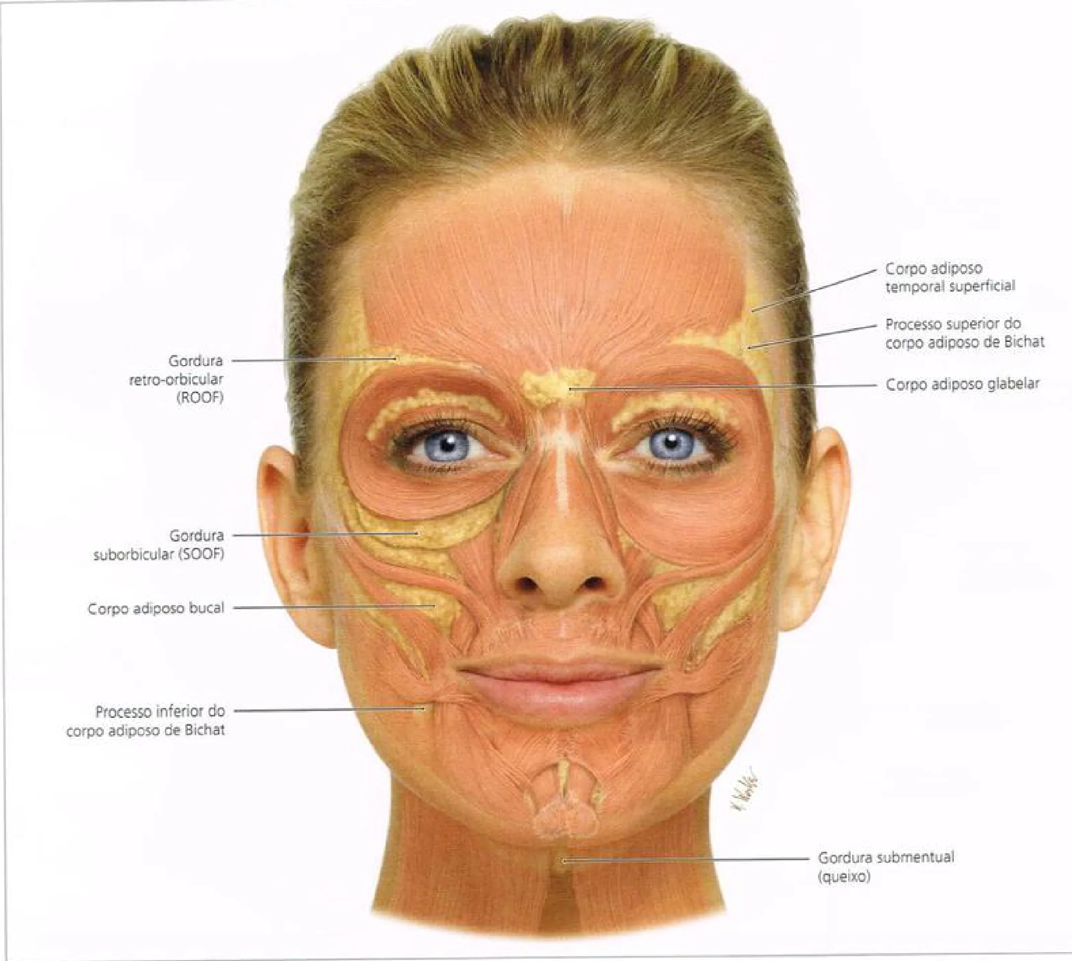 Fig 3. Female face with the skin and superficial fat removed: over the muscular plane (pink) the deep fat bodies stand out in yellow, labeled one by one — retro-orbicularis fat (ROOF) above the brow, glabellar fat body in the midline, suborbicularis fat (SOOF) below the inferior orbital rim, superficial temporal fat body and the superior process of the buccal fat body of Bichat in the temple, buccal fat body and its inferior process in the cheek, and submental fat in the chin — (Radlansky, clinical facial anatomy atlas, p. 48).
Fig 3 makes visible the asymmetry this block lives on: the deep fat is not a continuous layer but a set of discrete bodies, separated and with identifiable neighbors. That is what allows a bolus placed in one of them to project a specific zone, and also what explains why a bolus placed in the one next door gives a result the patient describes as "odd" without being able to point to where.
Deep compartments, anterior view
| Deep compartment | Upper limit | Lower limit | Medial limit | Lateral limit | Critical content / neighbor |
|---|---|---|---|---|---|
| Deep central and lateral forehead | Superior frontal septum | Middle frontal septum | — | — | Between the frontal complex and the periosteum; the supraorbital pedicle defines its walls [4,1] |
| ROOF (retro-orbicularis) | Inferior frontal septum | Orbicularis retaining ligament | Emergence of the supraorbital bundle | Lateral orbital thickening | Communicates with the inferior temporal compartment through the superior interval [4] |
| Medial SOOF | Orbicularis retaining ligament | Zygomatic-cutaneous ligament | Angular vein | Lateral SOOF | Communicates with the inferior temporal compartment through the temporal tunnel [4] |
| Lateral SOOF | Orbicularis retaining ligament | Zygomatic-cutaneous ligament | Medial SOOF | Midfacial extension of the superficial lamina of the deep temporal fascia | Rests on that lamina, not on the periosteum [4,35] |
| Premaxillary space (deep nasolabial fat pad) | Fascial fusion of the midfacial SMAS with the common elevator | Bony insertion of the common elevator | Lateral nasal wall | — | Roof: orbicularis and cheek SMAS; floor: common elevator of the lip and nasal ala [4,22] |
| Deep pyriform space | — | — | Medial to the infraorbital foramen | Deep medial fat pad | Contains the angular artery in its variable segment; roof, the common elevator [4,36] |
| Deep medial cheek fat pad | Zygomatic-cutaneous ligament | Levator anguli oris | Infraorbital foramen | Angular vein | Anterior malar projection; the infraorbital bundle is its superior neighbor [4,37] |
| Deep lateral cheek fat pad | Zygomatic-cutaneous ligament and/or zygomaticus minor | Transverse facial septum | Angular vein | Zygomaticus major | The transverse septum is the cause of the "apple cheeks" on smiling [4,34] |
| Deep mental fat | — | — | Midline | — | Gliding space over the periosteum [4,10] |
| Deep labiomandibular fat | — | — | — | — | Surrounds the mental foramen and protects the bundle as it emerges [4,10] |
Deep compartments, lateral view
| Compartment | Limits | Critical content |
|---|---|---|
| Superior temporal (deep) | Superior temporal septum / inferior temporal septum; roof superficial temporal fascia, floor superficial lamina of the deep temporal fascia | No relevant neurovascular structures [4] |
| Inferior temporal (deep) | Inferior temporal septum / zygomatic arch adhesions | Frontal branches of the facial nerve, zygomaticotemporal branches, sentinel vein [4,17] |
| Superficial temporal fat pad | Between the two laminae of the deep temporal fascia | Medial zygomaticotemporal vein [4,24] |
| Deep temporal fat pad | Deep to the deep lamina of the deep temporal fascia | Temporal extension of the buccal fat pad of Bichat [4] |
| Masticator space | Separated from the premasseteric spaces by the masseter and from the buccal spaces by the facial vein canal | Buccal fat pad of Bichat with its four extensions [4,38] |
| Superior premasseteric | Over the masseter, below the SMAS | Transverse facial artery, which gives an axial branch to the SMAS within the zygomatic ligament (McGregor) [4] |
| Middle premasseteric | — | Fascial connections of the buccal branch of the facial nerve [4] |
| Inferior premasseteric | — | Fascial connections of the marginal mandibular branch [4] |
| Buccal space | Deep: buccinator; superficial: platysma; anterior: modiolus; posterior: facial vein canal; inferior: mandibular ligament; superior: transverse facial septum | Facial artery; the facial vein is NOT content, it runs in its own canal [4,38] |
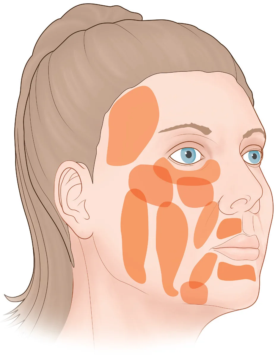 Fig 4. Outline schematic of a face in three-quarter view with the fat compartments shown as discrete orange patches: a single temporal plate, three staggered vertical bands on the lateral and middle cheek, an infraorbital oval, a nasolabial band, two perilabial bands and a patch in the jowl region. The territories are shown unlabeled, separated by corridors of uncolored tissue that correspond to the septa — (Carruthers, 2018, p. 179).
Fig 4 is useful for what it leaves blank: the uncolored corridors between patches are septa, and a product injected there sits in no compartment but on a wall. It is the graphic representation of why the same milliliter performs differently with two millimeters of difference at the entry point.
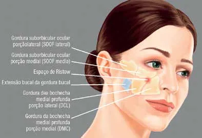 Fig 5. Three-quarter-view illustration with the deep compartments of the mid third labeled in Portuguese: suborbicularis oculi fat lateral portion (lateral SOOF) and medial portion (medial SOOF) below the orbital rim, Ristow's space in blue next to the pyriform aperture, the buccal extension of the buccal fat pad of Bichat, and the deep cheek fat in its two portions, lateral (DCL) and medial (DMC) — (Lobo, 2022, p. 97).
Fig 5 locates Ristow's space, which in the anatomical nomenclature of this chapter corresponds to the deep pyriform space, medial to the infraorbital foramen [4,36]: it is the only compartment whose filling acts directly on the base of the nasolabial fold without touching the superficial fat that worsens it.
Compartment rules that change the gesture
Consensus: the target is chosen by compartment, not by "zone"; applying the product in the wrong compartment gives an aesthetically undesirable result even when the volume and the product are correct [4].
Rule 1 — deep first. The deep compartments barely shift with mimicry or with age because their limits are muscular, ligamentous or fascial connections anchored to bone [21,28,6]. Filling them first rebuilds the foundation and lets the superficial tissue reposition and keep moving naturally [6].
Rule 2 — two compartments that are not filled. The superficial nasolabial, because it aggravates the fold itself; and the jowl one, because it aggravates the labiomandibular fold and the deformity [6]. Both are superficial and both are exactly where the patient points with the finger.
Rule 3 — the transverse facial septum rules the smile. It is a broad fascial membrane that joins the zygomaticus major to the underlying maxilla and forms the lower limit of the deep lateral fat pad [4]. On smiling it deforms and displaces cranially the compartments it supports: that is the physiological "apple cheek" and, overloaded, the facial overfilled syndrome, with normal volume at rest and exaggerated on the smile [34,6].
Rule 4 — the jowl does not originate where it is seen. Between the mandibular ligament and the masseter, the facial artery and vein cross the mandible wrapped in deep fat that communicates neither with the buccal space nor with the buccal fat pad of Bichat [4]. With age, the platysma glides over the mandible and drags the superficial jowl fat compartment downward [4,39,40]. The origin is the gliding, not the local excess of fat.
Rule 5 — ROOF and SOOF are connected to the temple. The ROOF communicates with the inferior temporal compartment through the superior interval, and the SOOF with the same compartment through the temporal tunnel, because both reside in layer 4 over the superficial lamina of the deep temporal fascia [4]. An excessive volume in the lower temple can present clinically in the periorbit.
Rule 6 — the surface reference of the medial end of the SOOF is the tear trough. The zygomatic-cutaneous ligament fuses with the orbicularis retaining ligament just as it crosses the mid-pupillary vertical line, and from there the whole is called the tear-trough ligament [4,3,41].
Volumetric changes with age, by compartment
| Finding | Direction | Source |
|---|---|---|
| Total facial fat | Decreases, independent of BMI | [6] |
| Superficial compartments | Descend; their position relative to bone changes | [27,6] |
| Deep compartments | Stable position relative to bone | [21,6] |
| Deep midfacial compartments on CT | Reduction with age | [42] |
| Superficial nasolabial | Apparent increase in prominence; debated whether it is real hypertrophy or an edge effect | [3,42,27] |
| Deep medial fat pad | Its loss unmasks the inferior orbital rim and creates the palpebromalar groove | [37,6] |
| Superficial vs deep ultrasound mobility | Statistically significant difference in favor of the superficial | [28] |
The debate about whether the superficial nasolabial compartment hypertrophies or simply accumulates against a barrier deserves precision, because it changes treatment: the fat cannot migrate below the fold because the mimetic muscles and the end of the SMAS anchor it to the skin, so it is forced upward and bulges above the fold [3]. If the mechanism is a barrier and not hypertrophy, the target is above and behind —deep compartments and lateral repositioning— and not the fold itself.
Classic trap: treating the dark circle with volume inside the SOOF when what is missing is the deep medial fat pad. The SOOF rests on a fascial lamina and has its medial limit at the tear trough [4]; loading it displaces the problem laterally and produces the "malar bag" bulge that worsens in the morning and with salt, because that compartment sits in the drainage territory that congests when supine.
A1.4 · Retaining ligaments
Schematic — the line of ligaments and the groove each one draws when it fails
| Ligament | Origin (deep) | Insertion (superficial) | Type | What it restrains | Groove or deformity that appears when it fails | Relative stiffness |
|---|---|---|---|---|---|---|
| Temporal ligamentous adhesion (TLA) | Temporal crest, periosteum | Superficial temporal fascia and dermis | True (osteocutaneous) | Descent of the brow tail and of the temporal flap | Brow-tail ptosis, excess skin in the temple | — |
| Superior temporal septum | Superior temporal line | Galea–superficial temporal fascia continuity | Fascial adhesion | Migration of product from temple to scalp | — | — |
| Inferior temporal septum | — | Superficial temporal fascia | Fascial adhesion | Separates the superior temporal compartment (avascular) from the inferior (with the frontal branch of the facial nerve) [4,17] | — | — |
| Lateral orbital thickening (LOT) | Lateral orbital rim, periosteum | Lateral dermis | True | Descent of the lateral orbicularis angle | Structural "crow's feet", drooping lateral brow | — |
| Orbicularis retaining ligament (ORL) | Orbital rim, bilaminar | Orbicularis oculi and dermis | True | Descent of the orbicularis; roof of the SOOF and floor of the ROOF [4,43] | Palpebromalar groove; visible eyelid–cheek junction | 2nd stiffest [3,44] |
| Tear-trough ligament | Maxilla, medial to the mid-pupillary line | Dermis of the tear trough | True | Skin–bone adhesion in the groove | Tear trough [41] | — |
| Zygomatic ligament (McGregor) | Zygomatic body and arch, next to the origin of the zygomaticus major | Malar dermis | True | Descent of the malar fat; forms the SOOF hammock [3] | Malar descent, "V-frame" | The stiffest [3,44] |
| Zygomatic-cutaneous ligament | Lower border of the zygomatic body | Dermis | True | Upper limit of the deep medial and lateral fat pads [4] | Mid-cheek groove | — |
| Masseteric ligament | Parotideomasseteric fascia, anterior border of the masseter | Dermis | False (fasciocutaneous) | Anteroposterior gliding of the SMAS over the masseter | Loss of the posterior jawline | — |
| Mandibular ligament | Mandibular parasymphysis | Dermis | True | Fixes skin and adnexa to bone; everything posterior is loose [4] | Jowl and labiomandibular fold [4,23] | 3rd [3,44] |
| Mandibular septum | Mandibular border | Prejowl subcutaneous fat | Adhesion | Contains the inferior migration of the jowl fat [39] | Prejowl notch | — |
| Zygomatic arch adhesions | Zygomatic arch | SMAS and superficial temporal fascia | Adhesion | Lower limit of the inferior temporal compartment [4] | — | — |
Consensus: the major ligaments are not scattered over the face but aligned along a single line immediately lateral to the lateral orbital rim, from the temporal crest to the mandible; from superior to inferior: temporal ligamentous adhesions, LOT, zygomatic ligament and mandibular ligament [4,6]. That line is at once a surface reference and a functional boundary between the medial and lateral face [33,6].
Discrepancy — do the ligaments loosen with age? · A (Cotofana 2016): the ligament that acts as a hammock for a compartment shows fatigue, bows along its course and favors the sagging of the corresponding compartment; it is the model that explains the jowl [3]. · B (Freytag 2022): there is no solid scientific evidence that facial ligaments increase their laxity with age; what changes is the bony surface where they insert, and the effect is wrongly attributed to the ligament [6]. As indirect support, in the rabbit medial collateral ligament the content of collagen, GAG and water did not change with age, while the expression of lubricin/PRG4, which affects interfascicular lubrication, did [45]. · Decide: if the patient has documented bone resorption (short mandible, decreased maxillary angle, widened pyriform aperture), model B dictates treating the bone first with supraperiosteal product and not chasing the groove. If the skeleton is preserved and the descent is purely lateral and postural, model A justifies the lateral pulling vector. Do not average: they are two distinct targets.
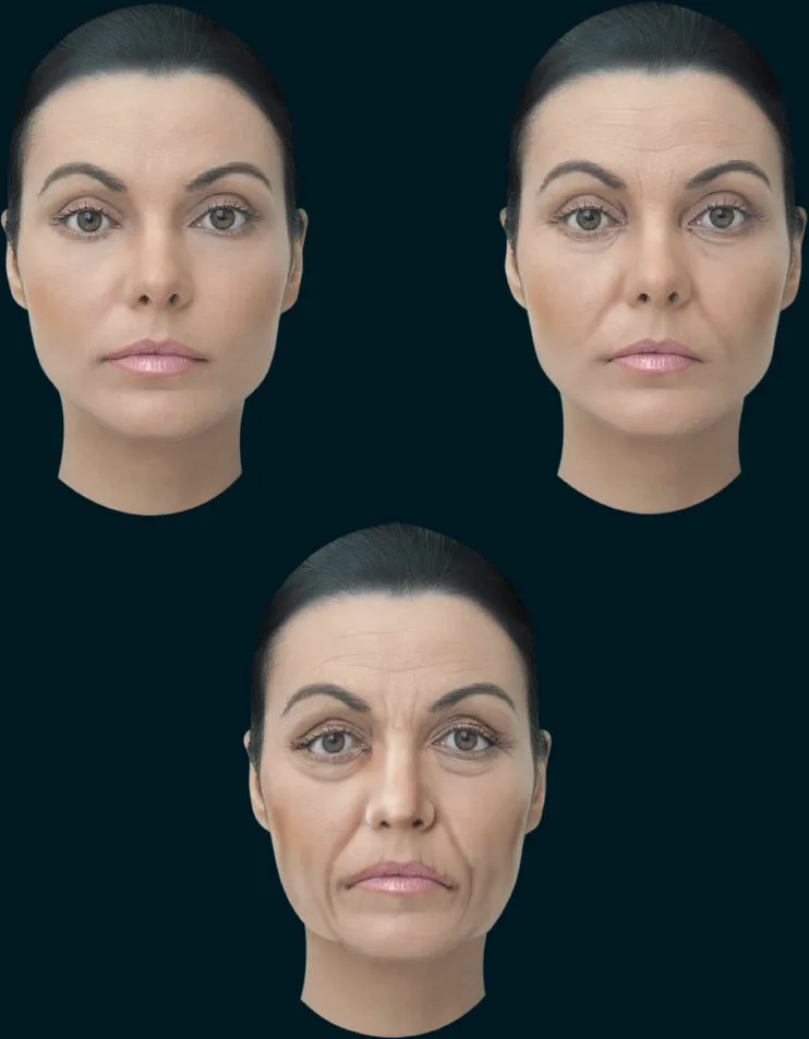 Fig 6. Three frontal renders of the same woman on a dark background in increasing stages of aging: in the first the oval is continuous and the eyelid–cheek junction is not marked; in the second, tear trough, nasolabial fold and loss of jaw definition appear; in the third the lower third widens, the marionette lines are drawn, the upper lip lengthens and the brow descends — (Vieira Braz, 2017, p. 122).
Fig 6 chains in three images what the tables describe separately: the first visible change is a groove (a ligament marking its trace over a receding bone), the second is a superficial compartment that descends, and the third is the loss of mandibular perimeter. That order is what justifies the craniocaudal treatment sequence of the next block.
Measured biomechanics and what it implies
In a biomechanical test to failure, the zygomatic ligament resists the greatest force; it is followed by the orbicularis retaining and the mandibular [3,44]. Translation: the point where a surgical traction or a product vector meets most resistance is the lateral malar, not the submandibular. The ligaments are composed of collagens, proteoglycans, glycosaminoglycans and water, and their mechanical competence depends on the whole plus intrafascicular lubrication [3].
The face medial to the line moves laterally and the lateral face moves medially, converging on the line; three-dimensional vector analysis of the skin surface confirmed it, so the line is not only a drawing but a boundary of movement [33]. Moreover, passive mobility —the one that depends on gravity and age— is greater in the lateral face, where there are no mimetic muscles or major ligaments anchoring to bone, and where all the layers are continuous from the scalp to the neck, allowing the lower tissue to pull the upper [6].
The three principles that come out of here
| Principle | Statement | Anatomical basis | Clinical verification |
|---|---|---|---|
| 1 · Upper third first | Treat above the region of aesthetic interest before the region itself | The final result of aging is descent; restoring above repositions what is below [6] | Viscoelastic product in the subdermal plane of the upper temple reduces midfacial volume and accentuates the jawline [6] |
| 2 · Lateral face first | Treat lateral to the line of ligaments before medial | The lateral layers continue with the SMAS, the orbicularis and the platysma; the medial ones do not influence their neighbors because of the interposition of the eye and the mouth [6] | Controlled hemiface study: preconditioning laterally achieved symmetry with less product [31] |
| 3 · Deep first | Supraperiosteal before subcutaneous | The deep compartments do not shift; the superficial ones do [28,6] | Also, the supraperiosteal plane is generally the least vascularized, with regional exceptions [2,6,46] |
Vector: lifting versus volumizing
Consensus: medial to the line of ligaments filler projects; lateral to it it lifts structures located lower down [4,31,32]. The mechanism is the fascial arrangement: oblique in the medial face, because the muscles run from bone to skin; parallel in the lateral, because there are no mimetic muscles [6].
Operative consequence for the order of work: the indication to fill the nasolabial fold directly frequently disappears after volumizing the upper mid third, that is the prezygomatic space of the SOOF [6].
The same anatomical grid governs thread-lifting techniques: the review of their anatomical considerations lists exactly the four elements of this chapter —navigation among vascular structures, protection of the facial nerve branches, manipulation of fat compartments and engagement of the retaining ligaments— and places ligamentous engagement as the condition for a sustainable lift without tissue distortion [47]. The instrument changes; the target does not.
Classic trap: chasing the tear trough with product in the groove itself. The groove exists because a ligament anchors the skin to the maxilla along that trace [41]; adding volume in front of the anchor makes it more visible by contrast, and edema in a zone with no real subcutaneous layer or deep fascia [6] translates into a morning bulge the patient attributes to sleep for months.
A1.5 · Muscles
Schematic — origin, insertion, vector, antagonist and layer
| Muscle | Origin | Insertion | Contraction vector | Direct antagonist | Layer |
|---|---|---|---|---|---|
| Frontalis | No bony origin; continuation of the galea aponeurotica | Brow dermis, interwoven with orbicularis, procerus and corrugator | Brow upward; transverse wrinkle | Procerus, corrugator, depressor supercilii, orbital orbicularis | 3-4; with a thin superficial fascia and a thick deep fascia [4,1] |
| Procerus | Fascia of the nasal bone and upper lateral cartilage | Glabellar dermis | Glabella downward; horizontal wrinkle | Frontalis | 3 |
| Corrugator supercilii | Medial end of the superciliary arch (frontal bone) | Dermis of the middle third of the brow | Brow medially and inferiorly; vertical wrinkles | Frontalis | Deep to the orbicularis and the frontalis; perforated by the supratrochlear artery [2] |
| Depressor supercilii | Frontal bone next to the medial canthus | Dermis of the head of the brow | Head of the brow downward | Frontalis | 3 |
| Orbicularis oculi (orbital, preseptal, pretarsal) | Medial canthal ligament, lacrimal crest, orbital border | Lateral canthal raphe, dermis | Eyelid closure; the orbital part lowers the brow | Frontalis (for the orbital portion) | 3 — it is periorbital SMAS [4,3] |
| Nasalis (transverse and alar portions) | Maxilla over the canine fossa | Dorsal aponeurosis; ala border | Compresses the dorsum / dilates the nostril | — | 3 |
| Depressor septi nasi | Incisive fossa of the maxilla | Mobile nasal septum | Nasal tip downward on smiling | Common elevator | Deep, next to the pyriform space [4] |
| Levator labii superioris alaeque nasi (common elevator) | Frontal process of the maxilla | Nasal ala and upper lip | Elevates the ala and lip; opens the nostril | Depressor septi | 5 — roof of the premaxillary space and floor of the deep pyriform [4] |
| Levator labii superioris | Inferior orbital rim over the infraorbital foramen | Upper lip | Elevates the lip | Depressor labii inferioris | Deep, over the deep medial fat pad [4] |
| Levator anguli oris | Canine fossa of the maxilla | Modiolus | Elevates the commissure | Depressor anguli oris | Deep, lower limit of the deep medial fat pad [4] |
| Zygomaticus major | Zygomatic body, next to the zygomatic ligament | Modiolus; interweaves with buccinator and orbicularis oris [26] | Commissure superolaterally | Depressor anguli oris, platysma | Originates deep to the SMAS on the lateral face and perforates it on entering the medial [3] |
| Zygomaticus minor | Zygomatic body, more medial | Upper lip | Elevates the lip superolaterally | — | Variable presence; follows the course of the zygomatic-cutaneous ligament [4] |
| Risorius | Parotideomasseteric fascia (variable origin) | Modiolus | Commissure purely laterally | Orbicularis oris | 3; anatomical origin variable relative to the facial planes [48] |
| Buccinator | Buccinator crest of the mandible, maxilla and pterygomandibular raphe | Modiolus and orbicularis oris | Compresses the cheek against the teeth | — | 6; the only facial muscle with its own epimysium [3] |
| Orbicularis oris | Muscular complex with no single bony origin; receives fibers from all the converging muscles | Perioral dermis and mucosa | Lip closure and protrusion | All the elevators and depressors | 3 |
| Depressor anguli oris | Oblique line of the mandible | Modiolus | Commissure inferolaterally | Levator anguli oris, zygomaticus major | 3 |
| Depressor labii inferioris | Oblique line of the mandible, medial to the previous | Lower lip | Lower lip downward and lateral | Mentalis, levator labii | 3 |
| Mentalis | Mandibular incisive fossa | Chin dermis | Elevates and protrudes the lower lip; wrinkles the chin | Depressor labii inferioris | Deep, next to the deep mental fat [4,10] |
| Platysma | Deltopectoral and pectoral fascia | Mandibular border, modiolus (horizontal modiolar portion), dermis | Lowers the commissure and jaw; tenses the neck | Modiolus elevators | 3 — it is layer 3 of the neck and the lower face [4,40] |
| Masseter | Zygomatic arch | Mandibular angle and ramus | Jaw closure | Digastric, lateral pterygoid | Deep to the parotideomasseteric fascia (layer 5) |
| Temporalis | Temporal fossa | Coronoid process | Jaw closure and retrusion | Lateral pterygoid | 9 in the temporal count [4] |
Consensus: almost all mimetic muscles have a direct bony connection and end in the dermis or in another muscular complex, which gives them an oblique deep-to-superficial course in the medial face; in the lateral face there are no mimetic muscles and the layers are parallel [6]. The facial muscles lack an epimysium, with the exception of the buccinator [3].
Discrepancy — "the deep fat is the gliding plane of the muscle". · A: several authors attribute to the deep fat a gliding function for the mimetic muscles, because it surrounds them [3]. · B (Cotofana 2016): that function is questionable, because most facial muscles change plane from layer 5 to layer 2 and end up surrounded by superficial fat too; besides, their function is to transmit contraction to the skin or the SMAS, which a protective wrapping would hinder [3]. · Decide: if B is accepted, the goal of deep filling is not to lubricate the muscle but to recreate the lost bony foundation [6], and then the amount is calculated by the skeletal deficit, not by the muscle volume.
The two balances that govern the face
Brow balance. A single elevator, the frontalis; depressors, the corrugator, the depressor supercilii, the procerus and the orbital portion of the orbicularis [6]. Any intervention on one of the depressors changes the resting position of the brow: that is what makes the glabella a functional unit and not a sum of muscles.
Modiolus balance. Depressors: depressor anguli oris, platysma and depressor labii inferioris. Elevators: levator anguli oris, zygomaticus major and minor, levator labii superioris [6]. The position of the commissure —and with it the reading of sadness or fatigue— is the result of that balance, not of lip volume.
Muscle aging: what is measured and what is not
| Parameter | Finding | Source |
|---|---|---|
| Muscle length and tone | The facial muscles lengthen, increase tone and reduce range of movement; resting tone approaches that of maximal contraction | [3] |
| Clinical consequence | Conversion of dynamic lines into static ones and accentuation of folds by fat displacement | [3] |
| Signal-to-noise ratio on surface electromyography (young vs old) | No statistically significant difference overall | [6,49] |
| Subanalysis: corrugator and procerus | Increased signal in older subjects; consistent with more marked glabellar wrinkles | [6,49] |
| Subanalysis: zygomaticus major | Decreased signal in older subjects; lower motor unit potential | [6,49] |
| Ultrasound correlate of the zygomaticus major | Lower ability to compress the angular vein against the maxilla in older subjects | [6,50] |
| Voluntary facial exercise | Limited efficacy | [3] |
| Neuromuscular electrostimulation | Promising results | [3] |
The combined reading matters because it contradicts intuition: facial muscle aging is not a global atrophy, but a redistribution of activity, with more active glabellar depressors and a less active midfacial elevator [6,49]. A plan that relaxes the glabellar depressors and does not compensate for the loss of midfacial support accentuates exactly the measured imbalance.
Anatomical variability: why a fixed schematic fails
The variability of the midfacial muscles is documented in dissection series: 50 hemifaces in one series [51] and 52 fresh-cadaver hemifaces of a Persian population in another, the latter correlating muscle variation with the morphology of the nasolabial fold [52]. The insertion of the zygomaticus major at the commissure has different arrangements between individuals [53], the origin of the risorius varies relative to the facial planes [48] and the zygomaticus minor may be absent [4]. The variations also differ between ethnic groups [3,52]. This is why neuromodulator treatment moved from rigid point schemes to a plan individualized according to each person's anatomy [4].
The histological substrate of the nasolabial fold reinforces the same point: the zygomaticus major and the buccinator connect to each other and have strong adhesions toward the skin forming the fold, and then both fuse with the orbicularis oris [3,26]. A nasolabial fold is not an excess of fat above it: it is the insertion line of a muscular system.
The mapping of motor innervation refines that idea: with Sihler staining on dissections, the motor plates of the platysma cluster in the upper two thirds of the muscle, while the lower third is mostly sensory [54]. The anatomical consequence is that the muscle is not homogeneous as a target, and the same effect is obtained by concentrating the action where the motor plate is.
Classic trap: treating the platysmal band as a neck-only problem. The platysma has a horizontal modiolar portion that participates in the dynamics and contour of the lower face [40], and glides over the mandible dragging the jowl fat compartment [4]. Relaxing only the vertical cervical bands leaves intact the vector that produces the jowl, and the patient returns describing that "the neck improved and the jaw did not".
A1.6 · Arteries
Schematic — where the artery is and where the product goes, region by region
| Region | Most likely location of the artery | Recommended plane for the product | Anatomical note |
|---|---|---|---|
| Upper forehead | Superficial (layer 2, superficial fat) | Supraperiosteal (layer 4) and intradermal (layer 1) | Above the middle frontal septum the vessel has already perforated the frontalis [2,1] |
| Lower forehead | Deep (layer 4, supraperiosteal) | Supraperiosteal and intradermal | Below the middle frontal septum the vessel is in bone contact [2] |
| Glabella | Superficial (layer 2) | Supraperiosteal and intradermal | No major arteries were identified deep to the procerus [2] |
| Superficial temple | Superficial (layer 3, superficial temporal fascia) | Subcutaneous (layer 2) | The STA runs within layer 3, below the injection plane [4,2] |
| Deep temple | Deep (layer 9, supraperiosteal) | Supraperiosteal, "one up and one down" | Places the product in front of the deep temporal arteries [2] |
| Medial mid third | Deep (layer 4, supraperiosteal) | Supraperiosteal, between the infraorbital foramen and the inferior orbital rim | In the tear trough there are only 3 layers [2] |
| Lateral mid third | Deep (layer 4) | Subcutaneous (layer 2); bone contact allowed at the zygomatic arch | [2] |
| Nasal dorsum (midline) | Superficial (layer 2) | Supraperiosteal / supraperichondral | Most nasal arteries are subcutaneous: the deep plane is relatively avascular [2,55] |
| Nasolabial fold | Superficial (layer 2) | Supraperiosteal (canine fossa) or intradermal | On CT arteries are seen less than 4 mm from the skin [2] |
| Jawline | Deep (layer 4) | Subcutaneous; bone contact allowed at the mandibular angle | [2] |
| Lips | Deep (submucosal) | Subcutaneous and intradermal | Labial arteries submucosal in 78.1 %, subcutaneous in 2.1 % [2,56] |
| Chin | Superficial (layer 2) | Supraperiosteal | [2] |
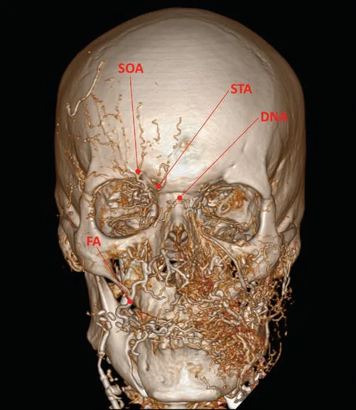 Fig 7. Three-dimensional reconstruction of a contrast cranial CT, frontal view: the arterial tree appears in orange over the gray bone after injecting the right internal carotid and the left facial artery. Labeled are the supraorbital (SOA) and supratrochlear (STA) emerging at the superior orbital rim, the dorsal nasal (DNA) at the nasal root and the facial (FA) crossing the mandibular border. The contralateral filling of the right facial tree from a single left injection is visible — (Cotofana, 2019, p. 417).
Fig 7 is the image that justifies the whole block: the filling of the opposite side from a single injection shows that the territories are not sealed compartments and that an embolus introduced into the face can reach the ophthalmic circulation. The labeled arrows also mark the exact point where the internal carotid system surfaces on the face.
Relevant extracranial-intracranial anastomoses
| Extracranial branch | Intracranial / ophthalmic branch it anastomoses with |
|---|---|
| Middle meningeal artery (internal maxillary) | Orbital and anterior branch of the ophthalmic |
| Anterior deep temporal (internal maxillary) | Lacrimal artery |
| Infraorbital (internal maxillary) | Medial and lateral muscular branches of the ophthalmic; dorsal nasal artery |
| Sphenopalatine | Anterior and posterior ethmoidals |
| Anterior branch of the superficial temporal | Supraorbital and supratrochlear |
| Facial artery | Dorsal nasal artery |
| Transverse facial artery | Dorsal nasal artery |
| Superior pharyngeal | Lateral clival artery |
| Occipital | Stylomastoid; vertebral artery (segments 1-2) |
Table adapted from the corpus anatomical analysis [2] and consistent with the neurointerventional description of the dangerous anastomoses [57]. The functional importance is documented: in patients with symptomatic internal carotid occlusion, focal brain regions may depend on the supply of the ipsilateral external carotid through a patent route from the ophthalmic artery [2].
Ophthalmic artery and central retinal artery
| Data | Value | Source |
|---|---|---|
| First intracranial branch of the internal carotid | Yes | [2,58] |
| Number of branches | 13 | [2] |
| Retinal supply | Central retinal + medial and lateral posterior ciliaries | [2] |
| Portions | Intracranial · intracanalicular · intraorbital | [2] |
| Intraorbital segments | 1st lateral to the optic nerve · 2nd crosses the nerve (83 % above, 17 % below) · 3rd parallel to the medial wall, ends connecting with the contralateral dorsal nasal | [2] |
| Central retinal artery diameter | 0.1-0.6 mm | [2] |
| Length | 7-20 mm | [2] |
| Narrowest point | Where it perforates the dura mater and arachnoid of the optic nerve, on its medial or inferior face | [2] |
| Terminal branches in the retina | 4 (nasal and temporal, superior and inferior) | [2] |
| Retinal perfusion | 0.52 cc/min per mg of tissue (brain: 0.48) | [2] |
| Experimental retinal survival (primate) | No detectable damage up to 97 min; afterward, progressive irreversible damage | [2,59] |
| Complete occlusion on fluorescein angiography | Rare | [2] |
The anatomical consequence for the conduct facing filler-related visual loss: the point where it matters to deposit hyaluronidase is the vicinity of the narrowest segment of the central retinal artery, that is the point of penetration into the optic nerve, which lies outside the nerve and surrounded by intraorbital tissue [2]. The anatomical description of the retrobulbar approach published in the corpus palpates the inferolateral orbital rim at a maximum of 1 cm medial to the lateral canthus, rests the injector on the bony margin and advances hugging the orbital floor, slightly medially and superiorly because of the conical shape of the orbit, crossing skin, subcutaneous fat, orbicularis and orbital septum, to 30-45 mm of depth depending on the physiognomy [2]. (P) The anatomical detail is gathered here because it defines the route; the indication, the dilution and the consent belong to the complications chapter and are not resolved with anatomy.
Course and depth, artery by artery
Facial, mandibular segment. It arises from the external carotid, alone or as a linguofacial trunk. It passes next to the submandibular gland, deep to the investing layer of the deep cervical fascia. At the mandibular angle it crosses the bone in 100 % of cases in front of the vein, deep to the platysma and deep to the marginal mandibular branch of the facial nerve, protected by a deep fat that communicates neither with the buccal fat pad of Bichat nor with the buccal space [2,38].
Facial, buccal segment. It enters the buccal space, bounded by the buccinator (deep), platysma (superficial), modiolus (anterior), facial vein canal (posterior), mandibular ligament and platysma adhered to the mandible (inferior) and transverse facial septum (superior) [2,38]. There it gives the inferior labial and/or the horizontal labiomental [60]. Its depth depends on the amount of fat in the subcutaneous compartments and within the buccal space [2].
Facial at the modiolus. Identifiable in 100 % of cases next to the corner of the mouth, held by a muscular band from the buccinator that fixes it 1.5 cm behind the commissure, between the buccinator (deep) and the modiolar portion of the platysma plus the converging muscles: levator anguli oris, zygomaticus major, levator labii superioris and depressor anguli oris (superficial) [2].
Labial arteries. Three possible positions: submucosal between mucosa and orbicularis (78.1 %), intramuscular between the two laminae of the orbicularis, and subcutaneous between skin and orbicularis (2.1 %) [2,56]. The proposed explanation is embryological: the muscular precursors form after the arteries have taken their definitive position [2].
Preinjection ultrasound adds a nuance of course that dissection averages out: located by sonography, the superior and inferior labial arteries arise on the deep, lateral slope of the lip and progressively approach the midline becoming more superficial, with superficial branches identifiable in the submucosal plane near the midline [61]. The point of greatest risk of the lip is therefore not the same in the lateral third as in the central.
Angular at the nasolabial fold. It runs deep in the fold but close to the dermis; its two-dimensional position varies between individuals and between sides of the same individual, so that it cannot be predicted [2,62]. On a contrast CT slice perpendicular to the fold, arteries are identified less than 4 mm below the skin [2].
Angular between the nasal ala and medial canthus. Very variable course in two and three dimensions. A series of 12 fresh hemifaces identified it within the deep pyriform space, bounded above and in front by the common elevator [2,36]. As it approaches the medial canthus it gives branches toward the nasal side and connects with the infraorbital as it emerges from the foramen [2].
Dorsal nasal. Bilateral terminal branch of the ophthalmic. It leaves the orbit and perforates the orbital septum above the medial canthal ligament, runs deep to the orbicularis and connects with the contralateral at the origin of the procerus. It has a direct connection with the angular, with the superior and inferior medial palpebrals and with the supratrochlear; it has no direct connection with the supraorbital, only indirect through the supratrochlear [2].
Supratrochlear. Terminal branch of the ophthalmic. It emerges through the supratrochlear notch or foramen at the superomedial angle of the orbit, in bone contact, perforates the corrugator and the inferior frontal septum and enters the subfrontal fat. On reaching the middle frontal septum —at 1.5 ± 0.17 cm (1.3-1.7) in the midline and 3.0 ± 0.24 cm (2.7-3.3) laterally from the supraorbital rim— it perforates the frontalis and its fascia and passes to the subcutaneous plane [2,1,8].
Supraorbital. It emerges from the supraorbital foramen or notch 1-3 mm medial to the mid-pupillary vertical line, in bone contact and lateral to the supratrochlear bundle. Its course is variable: it may perforate the corrugator (medial course) or not contact it (lateral course) [2,8]. Accompanied by the nerve and vein of the same name, it becomes more superficial on perforating the inferior frontal septum, where it gives branches to the frontal periosteum. After perforating the frontalis at the middle frontal septum it runs between the lateral and central superficial forehead compartments, and there gives a branch that connects with the anterior branch of the superficial temporal [2,1].
Superficial temporal. Most cranial branch of the external carotid. It emerges from deep planes toward the superficial temporal fascia in 100 % of observed cases, 1 cm in front of and 1 cm above the apex of the tragus [2]. Throughout the temporal region it runs within layer 3. After crossing the temporal crest it changes plane and lies in the subcutaneous of the forehead, where it connects with branches of the supraorbital [2].
Deep temporals. Anterior and posterior, deep in the temporal fossa and in close relation with the periosteum, with a longitudinal course at 1.5-2 cm (anterior) and 2.5-3.0 cm (posterior) from the lateral orbital rim [2].
Transverse facial. It runs in the superior premasseteric space and gives a branch that runs within the zygomatic ligament (McGregor) to give axial supply to the overlying SMAS [4].
Nose. Three arterial patterns according to the dominant source: facial, dorsal nasal or infraorbital [2,55]. It also receives from the superior labial, the columellar and contralateral branches. Most of the arteries are in the subcutaneous plane, which leaves the supraperiosteal and supraperichondral plane relatively avascular [2].
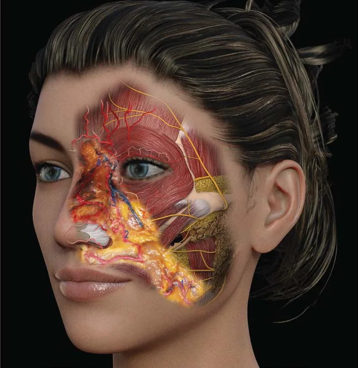 Fig 8. Three-dimensional model of a female face with the right hemiface dissected: over the forehead and orbit the frontalis and orbicularis are seen in red, crossed by fine yellow nerve branches; in the mid third, the deep fat appears yellow and over it the red arterial axis ascends from the nasolabial fold toward the medial canthus, with the lateral nasal branches heading to the ala; in the preauricular region the masseter and parotid are identified — (Pirayesh, 2020, p. 172).
Fig 8 shows the point the table summarizes in one cell: the facial-angular axis climbs the nasolabial fold superficial to the deep fat, so that a supraperiosteal deposit in the canine fossa passes below it, while a deposit "at mid depth" crosses it.
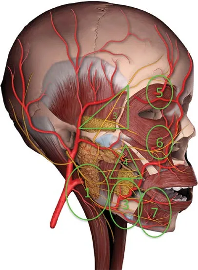 Fig 9. Lateral view of a schematic head dissection with seven numbered green circles over the risk regions: 1 in the retromandibular and cervical area, 2 and 3 in the parotid and mandibular region, 4 in the middle cheek, 5 in the high temple, 6 in the lateral periorbital region and 7 in the perioral region. The red tree of the superficial temporal and occipital, the yellow mesh of the nerve branches and the temporalis muscle with the parotid gland labeled below are visible — (Pirayesh, 2020, p. 107).
Fig 9 is read alongside the plane table: each numbered circle corresponds to a region where artery and nerve share a corridor, and that is why the plane criterion cannot be set by "aesthetic zone" but by the vascular segment that crosses that zone.
Why anatomy alone does not explain the incidence
In the glabella there are multiple connections between the dorsal nasal, supratrochlear and supraorbital of both sides, with high two-dimensional variability, which makes it the area of maximal risk of visual compromise [2,12]. But anatomy is not enough: most injections there are done with a sharp needle, and two independent studies showed that the needle distributes product into every plane it crosses, with material identifiable in the subdermal plane even when the tip is in bone contact [2,63,64]. Added to the double-perforation phenomenon of the arterial wall, a negative aspiration does not exclude intra-arterial deposit [2].
The rest of the risk picture is not only anatomical either: the variability is such —between individuals and between sides of the same individual— that it is not possible to guarantee absolute safety in any injectable procedure, and that sentence belongs to the consent, not to the technique chapter [2,12,65]. The safety guidelines moved from being authority-based to being evidence-based precisely because of the anatomical work of the last decade [2,46].
Classic trap: believing the nasolabial fold is a "deep" zone. It is exactly the opposite: it is one of the regions where the artery is more superficial than the operator expects, less than 4 mm from the skin [2]. The clinical signature of the error is an immediate blanching in the nasal ala and lateral dorsum —territory of the lateral nasal— that appears during the injection and not after, and which an operator expecting a "deep" problem is slow to interpret.
A1.7 · Veins and lymphatics
Schematic — the system that explains edema and the one that explains venous blindness
| Structure | Course | Depth | Why it matters in aesthetics |
|---|---|---|---|
| Angular vein | From the medial canthus toward inferolateral; oblique from inferolateral to superomedial | 4.2 ± 0.7 mm below the inferior orbital rim, deep to the orbital portion of the orbicularis [4,66] | Lateral limit of the deep medial fat pad and medial limit of the deep lateral fat pad: it divides the two compartments where the cheekbone is projected [4] |
| Angular vein at the medial canthus | Communicates with the superior ophthalmic vein through the nasofrontal vein | Deep | Direct anatomical route toward the cavernous sinus; the facial system lacks competent valves [67,68] |
| Facial vein | Continuation of the angular; descends over the cheek in its own facial vein canal | Its canal is the posterior limit of the buccal space, and the vein is not content of that space [4,38] | Reference that separates the buccal space from the masticator space; medial to it layer 5 becomes periosteum again [3] |
| Facial vein at the mandible | Crosses the mandibular border behind the artery | Deep to the platysma, next to the marginal mandibular branch | The three elements share a critical point at the mandibular border [2] |
| Sentinel vein / medial zygomaticotemporal | Ascends from the superficial temporal fat pad (layer 6), changes plane in the inferior temporal compartment and passes deep to the orbicularis (layer 4) toward the forehead | Deeper than the superficial temporal arteries [2,24,69] | Surgical reference point in the temple and cause of hematoma in deep temporal injection |
| Supraorbital vein | Accompanies the artery and nerve of the same name | Becomes superficial on perforating the inferior frontal septum [2] | Shares a corridor with artery and nerve: a single plane error affects all three |
| Superficial temporal vein | Drains the scalp and forehead; joins the maxillary to form the retromandibular | Layer 3 | The retromandibular descends through the parotid, sends a branch to the facial and joins the posterior auricular to form the external jugular [67] |
| Superior and inferior ophthalmic veins | Drain the orbit toward the cavernous sinus | Intraorbital | Explain why an orbital problem has an intracranial expression |
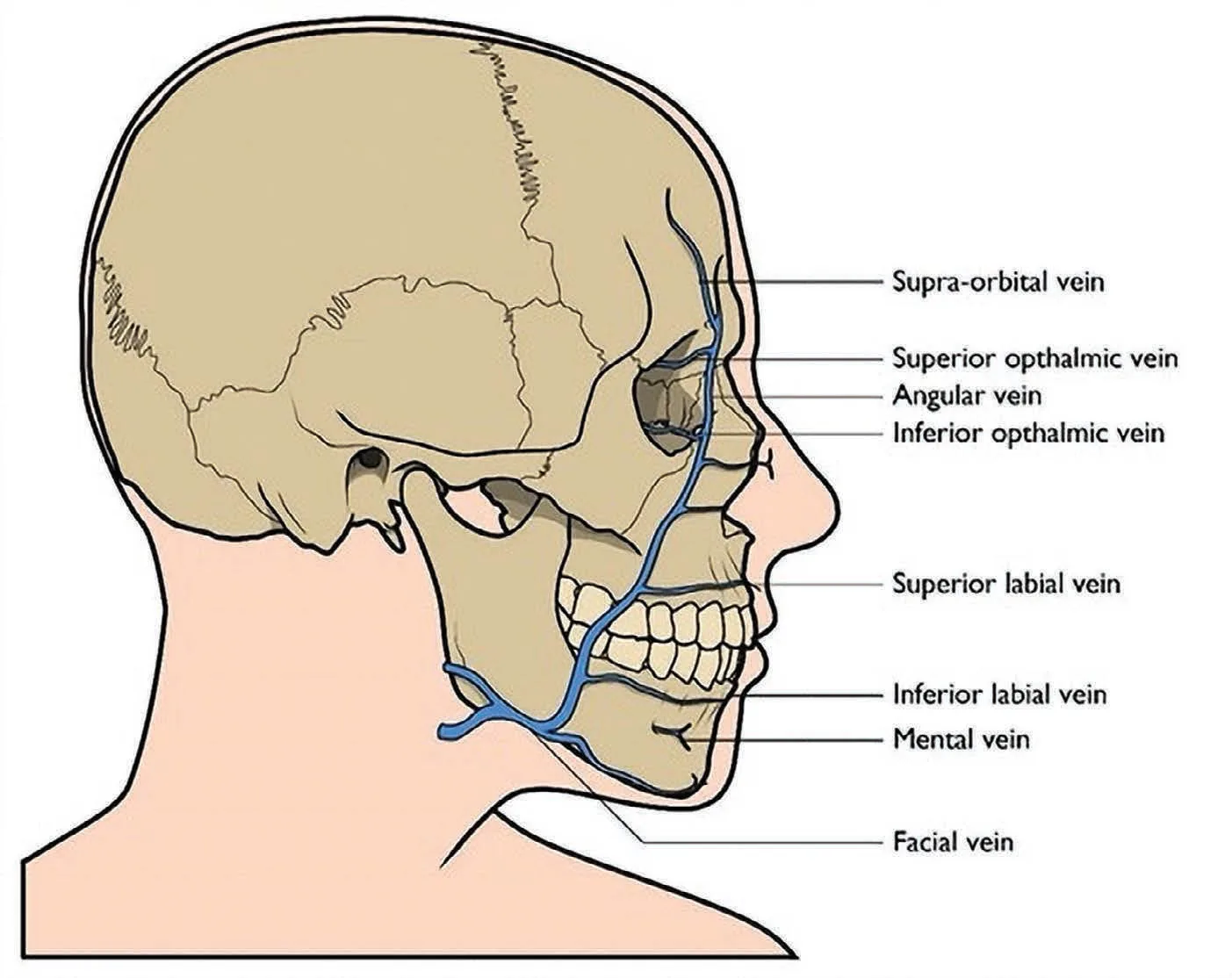 Fig 10. Lateral-view skull schematic with the facial venous tree labeled in blue: the supraorbital vein descends from the forehead to the medial angle, where it receives the superior ophthalmic vein and continues as the angular vein; the inferior ophthalmic vein is labeled on the orbital floor; the trunk descends obliquely over the cheek receiving the superior and inferior labial veins and the mental vein, and crosses the mandibular border as the facial vein toward the neck — (Parker, 2022, p. 29).
Fig 10 shows the detail that decides conduct in the periorbit: the angular vein does not end at the canthus, but continues into the orbit with the ophthalmic veins. That continuous blue line is the route by which a midface problem reaches the orbit and the cavernous sinus.
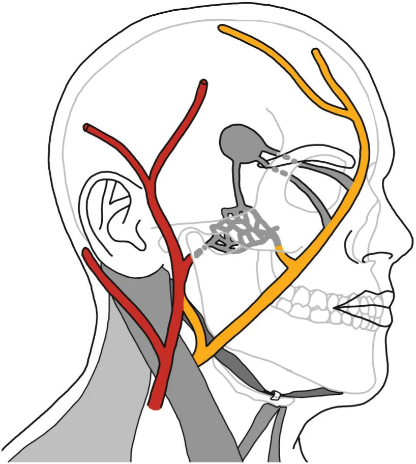 Fig 11. Outline schematic of the head and neck: the facial venous axis is shown in orange descending from the frontotemporal region in front of the eye and cheek to cross the mandibular border and converge in the neck, while the posterior system appears in red behind the auricle and over the sternocleidomastoid. In gray the facial skeleton and the muscular planes of the neck are drawn — (van Gijn, 2022, p. 360).
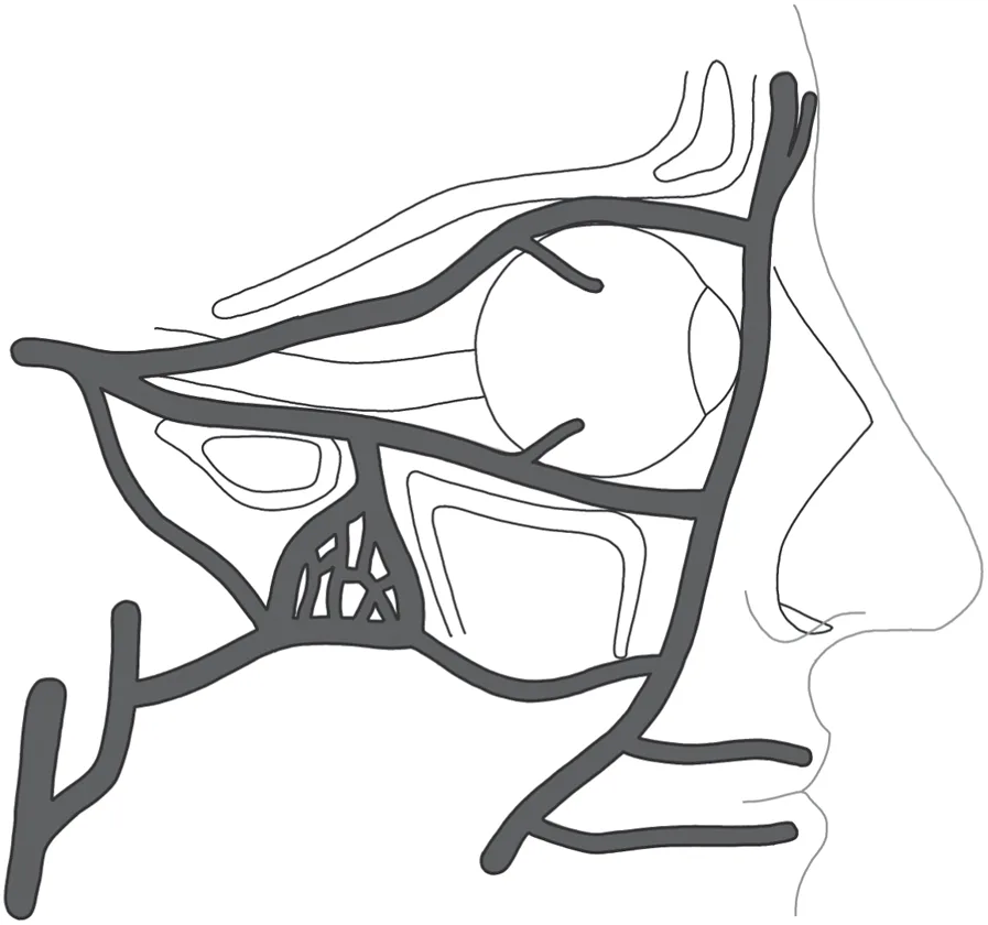 Fig 12. Lateral-view schematic of the orbit: the dark-gray venous trunks surround the globe forming a perimeter ring, with a superior trunk running along the orbital roof, an inferior trunk over the floor, and an anterior confluence at the medial angle that continues toward the face; posteriorly, the network converges toward the orbital fissure. The reticular plexus in the region of the posterior orbital floor is visible — (van Gijn, 2022, p. 414).
Fig 11 and Fig 12 together explain periorbital edema more than any table does: the drainage of the orbit converges forward and toward the medial angle, that is toward the same point where the tear trough is injected, and from there it follows the facial venous axis of Fig 11. A volume that compresses that corridor produces an edema that is not inflammatory and that is why it does not respond to anti-inflammatories.
Lymphatics: four rules and one declared limitation
| Rule | Statement | Source |
|---|---|---|
| 1 | The superficial lymphatics accompany the veins; the deep ones accompany the arteries | [67] |
| 2 | Lateral face → parotid nodes; upper lip and lateral half of the lower lip → submandibular; chin and central part of the lower lip → submental; everything ends in the deep cervical nodes | [67] |
| 3 | The eyelids have dual drainage: upper eyelid, lateral canthus and lateral half of the lower eyelid → preauricular/parotid nodes; nasal slope of both eyelids and medial canthus → along the facial vein toward the submandibular nodes | [70] |
| 4 | The palpebral lymphatic system is delicate and not prepared for trauma or for volume procedures; edema after large volumes in the tear trough, after periocular sculpting or after surgery in that area is an expected finding, just like the complaint of "swelling" after toxin | [71] |
Declared limitation, with the query that proves it. In the run A1-20260809 (evaluation/runs/A1.jsonl, 200 records, 3 027 contexts retrieved), 86 contexts contain a lymphatic term versus 395 that contain facial or angular vein. The best-scored facet of the chapter is anatomy_planes_danger (mean global_top_score 0.745) and no lymphatic-specific facet exists in the program. Honest translation: facial lymphatic anatomy is in the corpus as an atlas topographic description [67,70,68] and as a clinical observation [71], not as an anatomical series measured to the level of detail this chapter offers for arteries. Any quantitative claim about facial lymphatic flow would have to come from outside the corpus.
Angular vein: the detail that changes periorbital filling
The angular vein is identified along the medial wall of the zygomatic-cutaneous ligament after emerging deep to the zygomaticus major, and its relation with the zygomaticus minor is variable, superficial or deep [4]. Its position defines two compartments that are filled with different goals: the deep medial fat pad lies between the infraorbital foramen and the vein; the deep lateral, between the vein and the zygomaticus major [4]. An operator who does not have it in mind is not choosing a compartment, is choosing a depth.
There is also a measured functional datum: the ability to compress the angular vein against the underlying maxilla on smiling decreases with age, in parallel with the drop in the electromyographic signal of the zygomaticus major [6,49,50]. That is, the muscular mechanism that aids midfacial venous return weakens precisely in the population that consults for morning eyelid edema.
Why malar edema is not a product problem
(P) With the four rules above the picture orders itself, without invoking properties of the filler: the lateral malar region drains through superficial lymphatics that accompany veins [67], rests on the SOOF, which in turn rests on a fascial lamina and not on periosteum [4,35], and has above it a ligament that compartmentalizes it [41]. A volume placed there occupies a drainage space, not a support space. The clinical signature —worse in the morning, worse with salt, better through the day, without erythema or pain— corresponds to a positional and drainage problem, and the anatomical conduct is not to keep adding volume in the same plane.
Convergence points to memorize
| Point | What converges | Consequence |
|---|---|---|
| Medial canthus | Angular vein + superior ophthalmic vein (nasofrontal route) + dorsal nasal artery + angular artery | Zone of direct orbit–face communication [2,68] |
| Mandibular border, in front of the masseter | Facial artery (anterior) + facial vein (posterior) + marginal mandibular branch of the facial nerve | Three structures within one centimeter, all deep to the platysma [2] |
| Inferior temporal compartment | Frontal branch of the facial nerve + zygomaticotemporal branch + sentinel vein | The superior temporal compartment is the one that lacks relevant structures [4] |
| Supraorbital rim | Supraorbital artery, vein and nerve in a single bundle | A wrong plane compromises all three [2,8] |
Classic trap: interpreting as "inflammation" the periorbital edema that appears days after a tear-trough filling and treating it with corticosteroid. The described substrate is a palpebral lymphatic system that tolerates neither volume nor trauma [71] over a region with no real subcutaneous layer [4,3]; the corticosteroid does not reopen an occupied drainage corridor, and the signature that gives it away is the hourly and postural fluctuation, absent in a true inflammatory picture.
The dangerous triangle, the valves and the compressing hematoma
The facial venous system communicates with the cavernous sinus through the superior and inferior ophthalmic veins via the angular and the nasofrontal vein [67,68]. That communication is the anatomical route of two serious pictures distinct from arterial blindness: cavernous sinus thrombosis and intracranial spread of an infection from the so-called dangerous triangle —nasal root to commissures—, which a furuncle or a cellulitis from infected filler can follow retrograde [67,68].
| Statement | Nuance that is taught badly | Consequence |
|---|---|---|
| "Facial veins have no valves" | It is a simplification: valves exist, but scarce or incompetent, and the flow can reverse in the face of an increase in distal pressure [67] | The risk of retrograde ascent is real; the popular explanation is imprecise, not false. (P) correct it when you explain it |
| The angular vein is superficial and palpable at the medial canthus | It is the usual cause of the dramatic hematoma in dark-circle filling [4,66] | Hematoma ≠ occlusion, but a tense hematoma can compress and produce secondary ischemia through a mechanism distinct from the arterial embolus |
| Periorbital drainage converges toward the medial canthus | There the tear trough is injected, over a lymphatic system that does not tolerate volume [71] | Extensive periorbital ecchymosis after tear-trough filling mandates re-examination at 24-48 h to separate a benign hematoma from compromise [12] |
(P) The combined reading: the venous route does not blind by embolus, but turns a local infection or a tense hematoma into a regional or intracranial problem through the same corridor without competent valves. Management lives in J1-J2; here the substrate remains.
A1.8 · Nerves
Schematic — motor and sensory, what is blocked and what is injured
| Nerve | Origin | Emergence point / reference | Territory | Blocked for | Injured when… | Signature of the injury |
|---|---|---|---|---|---|---|
| VII — temporal (frontal) branch | Parotid plexus | Crosses the zygomatic arch and travels in the inferior temporal compartment (layer 4) [4] | Frontalis, upper orbicularis, corrugator | — | Dissecting or injecting in the inferior temporal compartment; temporal technique at mid depth [4,11] | Unilateral drooping brow, asymmetric smooth forehead |
| VII — zygomatic branch | Parotid plexus | Between parotideomasseteric fascia and SMAS, in fibrous septa [4] | Orbicularis oculi | — | Lateral sub-SMAS dissection | Incomplete eyelid closure |
| VII — buccal branch | Parotid plexus | In the fibrous septum that connects the parotideomasseteric fascia and SMAS; roof of the middle premasseteric space [4] | Buccinator, lip elevators, orbicularis oris | — | Dissection of the middle premasseteric space | Elevator weakness, air escape |
| VII — marginal mandibular branch | Parotid plexus | Crosses the mandibular border superficial to the facial artery and vein [2] | Depressor anguli oris, depressor labii inferioris, mentalis | — | Injecting or dissecting at the mandibular border anterior to the masseter | Lower-lip asymmetry when speaking and smiling |
| VII — cervical branch | Parotid plexus | Deep to the platysma | Platysma | — | Cervical dissection | Loss of platysmal tightening |
| V1 — supraorbital | Ophthalmic | Supraorbital foramen or notch, 1-3 mm medial to the mid-pupillary vertical [2] | Forehead, anterior scalp, upper eyelid | Forehead and anterior scalp | Compressing the bundle in the inferior frontal septum [2,8] | Frontal dysesthesia, projected headache |
| V1 — supratrochlear | Ophthalmic | Supratrochlear notch, superomedial angle of the orbit; perforates the corrugator [2] | Glabella and medial forehead | Glabella, medial forehead | Deep glabellar injection with volume | Transient glabellar anesthesia |
| V1 — infratrochlear and external nasal | Ophthalmic | Medial canthus; the external nasal emerges between the nasal bone and the lateral cartilage | Nasal dorsum and tip | Non-surgical rhinoplasty | Compression by supraperichondral product | Hypoesthesia of the nasal tip |
| V2 — infraorbital | Maxillary | Infraorbital foramen, on the mid-pupillary vertical; it is the medial limit of the deep medial fat pad [4] | Lower eyelid, nasal ala, upper lip, medial cheek | Upper lip, ala, lower eyelid | Filling the deep medial compartment with excessive volume | Paresthesia and pain of the upper lip |
| V2 — zygomaticofacial and zygomaticotemporal | Maxillary | Foramina of the zygomatic body; the zygomaticotemporal travels in the inferior temporal compartment [4] | Cheekbone and anterior temple | Temporal block | Temporal injection in the inferior compartment | Anterior temporal dysesthesia |
| V3 — mental | Mandibular | Mental foramen, surrounded by the deep labiomandibular fat that protects it [4,10] | Lower lip, chin | Lower lip, chin | Filling the chin without respecting the periosteal gliding space | Hypoesthesia of the lower lip |
| V3 — buccal | Mandibular | Crosses the buccinator | Buccal mucosa | Mucosal procedures | — | — |
| V3 — auriculotemporal | Mandibular | Behind the condyle, ascends with the superficial temporal | Posterior temple, ear | Posterior temporal block | — | Frey syndrome if injured with aberrant reinnervation |
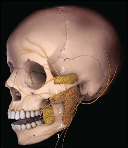 Fig 13. Three-dimensional model of a skull in lateral view with the parotid gland shown in tan over the mandibular ramus and the five branches of the facial nerve drawn in yellow and numbered from inferior to superior: 1 cervical, toward the neck below the mandibular angle; 2 marginal mandibular, following the lower border of the mandible; 3 buccal, toward the commissure; 4 zygomatic, toward the orbit; and 5 temporal, crossing the zygomatic arch toward the temple and the brow — (Pirayesh, 2020, p. 158).
Fig 13 locates the fact that orders motor safety: the five branches leave the same plexus and diverge like a fan, so that their distance from each other is minimal in the preauricular region and maximal in the periphery. The zone of risk of multiple injury is the parotid; that of isolated injury, the periphery.
The principle of the septa: where the motor branches travel
The marginal mandibular, buccal and zygomatic branches of the facial nerve do not run free in the fat: they travel within laminae of fibrous connective tissue that connect the parotideomasseteric fascia with the SMAS, and those laminae are precisely the walls that bound the superior, middle and inferior premasseteric spaces used in lift surgery [4]. Direct consequence for the injector: the interior of a premasseteric space is empty of nerve; its wall is not. The safe dissection or injection in the lateral cheek is the one that respects the compartment, not the one that respects a depth in millimeters.
In the temple the logic is identical: the superior temporal compartment contains no relevant neurovascular structures, while the inferior houses the frontal branch of the facial nerve, the sensory zygomaticotemporal branches and the sentinel vein [4]. The deep temporal technique works because it passes below all that content, in bone contact [2].
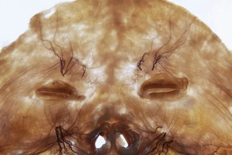 Fig 14. Anatomical specimen of the forehead and glabella seen backlit against a light background: over the translucent amber tissue the nerve filaments stand out in dark brown, emerging in two symmetric tufts above the superior orbital rims and fanning out upward and laterally; in the lower midline the nasal openings and another network of filaments ascending from the nasal root are visible — (Kim, 2016, p. 37).
Fig 14 explains why the supraorbital block works with so little volume and why its failure is always lateral: the filaments leave concentrated in a tuft and open out afterward; an infiltration below the point of divergence covers the whole fan, and an infiltration above covers only the sector reached.
The three foramina and their alignment
The three sensory emergence points of the anterior hemicranium are aligned approximately in the same vertical plane: the supraorbital foramen 1-3 mm medial to the mid-pupillary vertical line [2], the infraorbital foramen on that same vertical —it is the medial limit of the deep medial cheek fat pad [4]— and the mental foramen, surrounded by the deep labiomandibular fat that acts as a gliding space and protects it as it emerges [4,10]. That alignment is the surface reference on which the three trunk blocks of the face are built [72,9].
A connection datum that changes the gesture in the mid third: medial to the facial vein, layer 5 continues with the periosteum of the facial skeleton and connects with the epineurium of the infraorbital nerve [3]. The deep medial fat pad is not simply near the nerve: it shares a fascial continuity with it, and that is why an excessive volume there produces dysesthesia and not only mechanical compression.
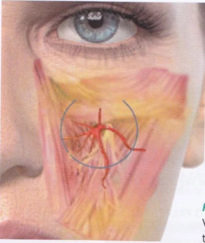 Fig 15. Detail of the left infraorbital region with the skin removed: below the lower eyelid the muscular plane is seen in pink and the fat in yellow, and a blue circle marks the emergence point of the infraorbital bundle, from which a red arterial tuft departs opening in several directions —toward the nasal ala, laterally and downward, toward the upper lip— accompanied by descending yellow nerve fibers — (Libro Zonas Peligrosas en Medicina Estética Facial, p. 20).
Fig 15 corrects a frequent scale error: the infraorbital bundle is not a point but a fan that occupies much of the upper medial cheek. A deposit "lateral to the foramen" may still be inside the fan.
What is blocked and with what anatomical purpose
| Block | Target | Anesthetized territory | Anatomical note |
|---|---|---|---|
| Supraorbital + supratrochlear | Notches of the supraorbital rim | Forehead, glabella, anterior scalp | Both bundles emerge in bone contact and ascend in the subfrontal fat [2] |
| Infraorbital | Infraorbital foramen, cutaneous or intraoral route | Lower eyelid, nasal ala, upper lip, medial cheek | Also a target for upper-lip and nasolabial-fold procedures |
| Mental | Mental foramen, intraoral route | Lower lip and chin | The foramen is wrapped in deep fat; the vestibular approach respects that plane [4,10] |
| Zygomaticotemporal | Inferior temporal compartment | Anterior temple | Shares the compartment with the frontal branch of the facial nerve [4] |
What gets injured, in order of practical frequency
(P) Ordered by what the anatomical corridor predicts, not by a series: frontal branch of the facial nerve when working on the temple at intermediate depth, because the inferior temporal compartment is exactly that plane [4,11]; marginal mandibular branch when injecting the mandibular border anterior to the masseter, where it runs superficial to the facial vessels [2]; and infraorbital nerve, through fascial continuity with the deep medial fat pad [4,3]. The three share the same mechanism: it is not a failure of lateral aim, it is a failure of plane.
The corridor can also be objectified in real time. In a systematic review of 16 studies on ultrasound-guided chemical denervation in muscles innervated by the facial nerve, the cadaver trials reached up to 88 % deposit accuracy versus 50 % for the technique based only on surface references, and the anatomical studies included identified variation in muscle depth and vascular risk zones [73]. The datum measures exactly what this block describes: the variability of the corridor, not the operator's aim.
Classic trap: looking for the "mid plane" in the temple. The two described planes —subcutaneous above the superficial temporal fascia and supraperiosteal in bone contact— exist precisely because between them is the compartment that contains the frontal branch of the facial nerve, the sensory zygomaticotemporal branches and the sentinel vein [4,2]. The signature of the error is a brow asymmetry that appears hours or days later, not immediately, and which the patient attributes to the toxin that was not given.
A1.9 · Bone
Schematic — what moves, in which direction and what sign it produces
| Bony structure | Change with age | Consequence on the soft tissue | Clinical sign | Source |
|---|---|---|---|---|
| Calvaria | Decreases in volume in both sexes | Less overall support; the soft tissue follows gravity | Panfacial descent | [6,74] |
| Frontal bone | Glabellar protrusion and expansion of the supraorbital arches; differences by age and sex on CT | Changes the brow–rim relation | Apparently lower brow with the same muscular position | [3,75,76] |
| Orbital aperture | Increases in area; the superomedial and inferolateral quadrants are the ones that expand | The orbital septum loses support | Appearance of the palpebromalar groove and scleral show | [3,77,78] |
| Orbits | Lateral translation | Increases the apparent intercanthal distance | A more "separated" gaze | [3,75] |
| Maxillary angle | Decreases | Expansion of the inferior orbital rim and anterior positioning of the orbital septum | Pseudoprolapse of the retroseptal fat pads → eyelid bags | [3,79] |
| Maxilla | Retrusion; loss of anterior projection | Loss of mid-third support | Midfacial descent, nasolabial fold | [6,80,79] |
| Pyriform aperture | Decreased angle and recession | Loss of base for the nasal ala and upper lip | Descent of the nasal tip, lengthening of the lip | [3,79] |
| Midfacial height (nasion to nasal spine) | Decreases in both sexes | Less support for the midfacial soft tissue | "Accordion" effect: the tissue folds over itself | [6,81,82,83] |
| Occlusal region and chin | Increase in vertical height with chin prominence in the classic series; loss of alveolar height and dentition in the current description | Loss of support of the lower lip and the jowl | Prejowl notch, loss of jawline | [3,6,75] |
| Cranial cortical thickness | Changes with age and sex | Relevant for deep temporal injection | — | [84,85] |
| Globe–orbital rim position | Changes with age | Modifies the eyelid–cheek relation | — | [86] |
Consensus: the facial skeleton is not static; it undergoes continuous remodeling throughout life and those changes modify appearance and expression [3,6,81]. The bone is therefore the foundation of treatment and not a backdrop [6].
Discrepancy — magnitude of the angular change by population. · A (Caucasian population): the glabellar, orbital, maxillary and pyriform angles decrease with age, and the maxilla, the pyriform aperture and the infraorbital rim recede [3,79]. · B (Asian population): similar results in direction, but the orbital and maxillary angles change less, and the pyriform angle changes more than in Caucasian series [3,87]. · Decide: in an Asian-phenotype patient, the expected dominant deficit is at the pyriform base and not in the malar projection; inverting the priority reproduces an aged Caucasian face instead of rejuvenating the one at hand. The angles are not averaged across populations.
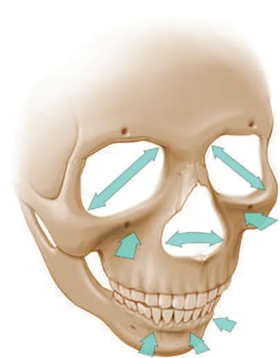 Fig 16. Frontal-view skull with superimposed blue arrows indicating the directions of remodeling: in each orbit, two opposing diagonal arrows mark the expansion of the superomedial and inferolateral quadrants; a bidirectional horizontal arrow marks the widening of the pyriform aperture; converging arrows point toward the maxilla below the orbital rim and toward the lateral region; and in the mandible, arrows directed toward the alveolar border and toward the chin region — (Azizzadeh, 2018, p. 255).
Fig 16 translates the table into an examination gesture: the pattern is not a homogeneous loss of bone but an expansion in some quadrants and a recession in others. Palpating the orbital rim in its four quadrants and the pyriform base indicates where support is missing before deciding a single milliliter.
Fig 17. Two facing profile illustrations, with the skull drawn beneath the soft-tissue contour. On the left, a young subject: full dentition, tall mandibular ramus and body, projected chin and a taut skin contour over the mandibular border. On the right, an older subject: gray hair, reduced mandibular height, recession of the chin and maxilla, and a soft-tissue contour that protrudes below the mandibular border instead of resting on it, with the nose relatively more prominent — (Standring, Gray's Anatomy, 2016, p. 959).
Fig 17 is the argument against the purely gravitational explanation: the soft tissue of the right panel has not increased, and yet it overflows the mandibular border because the bone that supported it has receded. That is the mechanism that turns a resorption into a visible jowl [4,39].
The complete causal chain, in order
- Bone remodeling → decrease of the maxillary angle and expansion of the inferior orbital rim [3,79].
- → Anterior positioning of the orbital septum, with reduction of its retention capacity [3].
- → Pseudoprolapse of the retroseptal fat pads: the eyelid bag is not new fat, it is poorly contained fat [3].
- → The orbicularis retaining ligament loses its horizontal position and tilts inferiorly [3].
- → Loss of stability of the orbicularis, which forms the anterior wall of the ROOF (above the aperture) and the SOOF (below) [3].
- → Malar mounds, palpebromalar groove and visible eyelid–cheek junction [3,41].
This chaining is the reason block A1.4 keeps open the discrepancy about ligamentous laxity: if step 1 explains steps 4 to 6, the ligament has not changed, its anchoring point has changed [6].
Overarching theories
Clockwise rotation of the viscerocranium (Lambros). The sum of the changes described by the classic series —lateral translation of the orbits, glabellar protrusion, expansion of the supraorbital arches, increased depth and lateral expansion of the cheeks, increased length, width and vertical dimension of the nose, and increase in vertical height in the occlusal region— was summarized as a clockwise rotation of the viscerocranium seen from the right [3,75]. The relation between bone remodeling and that rotation was later quantified on CT [81]. Pessa verified Lambros's theory by three-dimensional stereolithography in 12 skulls, measured the significant decrease of the maxillary and pyriform angles with age (p = 0.004 and 0.005) and formalized the result as an algorithm of facial aging that still orders the diagnosis [88].
Accordion effect (concertina). A small craniofacial skeleton —from advanced age or congenital skeletal insufficiency— reduces the space available for the soft tissue of the mid third, which folds over itself with a bellows-like appearance; a young face represents the point at which the skeletal proportions are adequate for the volume of soft tissue that covers them [83]. That formulation has a practical consequence: a young patient with skeletal insufficiency presents signs of premature aging that are not aging.
Rhythm and chronology: what the corpus does NOT support
Declared gap, with the query that proves it. The run A1-20260809 (evaluation/runs/A1.jsonl) was searched for the terms maxillary angle, glabellar angle, pyriform aperture/angle, orbital aperture, decade, década, reabsor, resorption over the 3 027 retrieved contexts. The returned contexts describe the direction of the change and the regions affected, but none provides a decade-by-decade resorption table with magnitudes: the highest-scoring hits were aesthetic-practice manuals and technique chapters, not anthropometric series stratified by age. The dose_parameters facet —the one that would capture quantitative magnitudes— is moreover the lowest-scored in the chapter (mean global_top_score 0.536, minimum 0.357).
(P) Therefore this chapter does not publish a decade-by-decade chronology. What the retrieved evidence does support is an order, not a calendar:
| Order | What changes first | Basis |
|---|---|---|
| 1st | Orbital aperture and infraorbital rim: they produce the first visible sign at the eyelid–cheek junction | [3,77,78] |
| 2nd | Maxillary angle and pyriform aperture: they trigger midfacial descent and the deepening of the nasolabial fold | [3,79,87] |
| 3rd | Midfacial height and calvarial volume: they reduce overall support | [6,81,74] |
| 4th | Alveolar height and dentition: loss of lower-third support | [6] |
Anyone needing magnitudes by decade must go to the original CT series [80,77,79,87,82] and check how they stratified age; the own corpus does not contain them at that detail.
Bone and safety: thickness matters
The cortical thickness of the skull changes with age and sex [84,85], and the overall morphology of the adult skull also [85]. This is not an academic detail in the temple: it has been explicitly investigated whether intracranial penetration is possible during temporal filler injection [89]. The safety reference remains bone contact with a short injector and depth control, not confidence in an assumed thickness.
A second bone–technique intersection point: repeated toxin injection into the masseter has been associated in recent literature with a risk of bone resorption, among other complications described for that indication [90]. It is the same principle in the reverse direction: muscular loading shapes the bone, and removing it shapes it too.
Classic trap: compensating with soft-tissue volume for a skeletal projection deficit. The result is a face that at rest looks correct and in motion behaves like a heavy tissue over an insufficient support [6,83]; the signature is the exaggerated increase of midfacial volume on smiling from cranial displacement of the compartments over the transverse facial septum [34], and the correction is not more product but product in another plane or support surgery.
A1.10 · Proportions and variants
> This is the block that D1-D13 consume. The thirteen regions link here for thirds, fifths, landmarks and variants; none of them repeats these.
Schematic — vertical and transverse proportions
| Proportion | Exact definition | Reference value | Source |
|---|---|---|---|
| Upper third | Hairline (trichion) → glabella | 1/3 | [91,83,92] |
| Mid third | Glabella → subnasale | 1/3 | [91,83,92] |
| Lower third | Subnasale → soft-tissue chin | 1/3 | [91,83,92] |
| Subdivision of the lower third | Subnasale → stomion of the upper lip | 1/3 of the lower third | [91,83,92] |
| Subdivision of the lower third | Stomion of the lower lip → soft-tissue chin | 2/3 of the lower third | [91,83,92] |
| Trichion–glabella distance | Anterior hairline to glabella | 7.5-9.5 cm usually | [93] |
| Facial fifths | Facial width divided into five vertical columns | Each fifth = width of one eye | [91,83,92] |
| Transverse equalities | Intercanthal distance = alar base width = palpebral fissure = intercanine width | Equal to each other | [91] |
| Mouth | Mouth width = interpupillary distance | Equal | [91] |
| Nasal dorsum | Dorsum width | 1/2 of the intercanthal distance | [91] |
| Nasal lobule | Lobule width | 2/3 of the intercanthal distance | [91] |
| Nasal length | Radix → tip | Equal to stomion → chin | [91] |
| Midlines | Facial, nasal, chin, labial and dental | Should coincide | [91] |
| Bizygomatic width / craniofacial height (vertex–chin) | Zy-Zy over V-Me | ≈ 60 % | [7] |
| Bizygomatic width / physiognomic facial height (trichion–chin) | Zy-Zy over Tr-Me | ≈ 70-75 % | [7] |
| Bitemporal / bizygomatic width | Most lateral point of each side of the forehead | 80-85 % | [7] |
| Bigonial / bizygomatic width | Soft-tissue gonion of each side | 70-75 % | [7] |
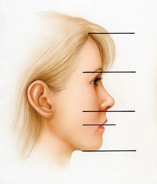 Fig 18. Female profile illustration with five black horizontal lines drawn from left to right over the face: the first at the level of the hairline, the second at the level of the glabella and brow, the third at the subnasale, the fourth at the labial stomion and the fifth at the soft-tissue chin. The lines delimit the three vertical thirds and the subdivision of the lower third — (Master Techniques in Facial Rejuvenation, 2018, p. 35).
Fig 18 sets the examination gesture that replaces the general impression: five lines on a profile photo tell whether the patient has a short lower third, a lengthened mid third or a long upper lip, and each of those three findings leads to a distinct target.
Landmarks (nomenclature for D1-D13)
| Point | Definition |
|---|---|
| Trichion (Tr) | Anterior hairline in the midline [93] |
| Glabella (G) | Flat area of bone between the brows [91] |
| Soft-tissue nasion (N′) | Point overlying the midpoint of the nasofrontal suture [7] |
| Radix | Nasal root |
| Tip (T) | Most anterior projection of the nose [93] |
| Columellar point (Cm) | Most anterior soft-tissue point of the columella [93] |
| Subnasale (Sn) | Junction of the columella with the upper cutaneous lip [93] |
| Labrale superius (LS) | Mucocutaneous junction of the upper lip in the midsagittal plane [93] |
| Superior stomion (STMS) | Lower border of the upper lip in the midsagittal plane [93] |
| Inferior stomion (STM) | Upper border of the lower lip in the midsagittal plane [93] |
| Soft-tissue chin (Me′) | Most inferior point of the midline of the chin border [7] |
| Zygion (Zy′) | Most lateral soft-tissue point over each zygomatic arch [7] |
| Soft-tissue gonion | Most lateral soft-tissue point over each mandibular angle [7] |
| Vertex (V) | Highest point of the head in natural position [7] |
Golden ratio: what it is and what it is not for
Formal definition: the section of a line such that the lesser part is to the greater as the greater is to the whole [92]. It is a descriptive criterion, not a therapeutic target.
Consensus: knowledge of the theoretically ideal facial proportions is useful in planning [94]. And the same text sets the limit: those ideals must not be used alone, but together with the consultation in which the patient's subjective concerns and ideas are gathered, and moreover the differences between male and female faces, between ethnicities and between age groups must be considered [94]. (P) A fifths template applied without that correction produces faces that are correct on paper and alien in the mirror.
Variation by sex
| Trait | Pattern | Source |
|---|---|---|
| Frontal bone | Measured differences by age and sex on CT | [76] |
| Lip anatomy | Layer thickness and filler behavior vary significantly with sex | [95] |
| Cranial cortical thickness | Changes with age and sex | [84] |
| Overall cranial morphology | Changes with age and sex | [85] |
| Midfacial height | Decreases in both sexes | [6,81] |
| Calvarial volume | Decreases in both sexes | [6,74] |
(P) The combined reading is that the sexual difference is in the starting shape (forehead, jaw, lip), not in the direction of aging, which is common. A feminization or masculinization plan works on the former; a rejuvenation plan, on the latter. Confusing them is what produces an aged result but of another sex.
Variation by ethnicity
| Finding | Content | Source |
|---|---|---|
| Asian skeleton | The orbital and maxillary angles change less with age; the pyriform angle changes more than in Caucasian series | [3,87] |
| Midfacial musculature | Series of 52 fresh-cadaver hemifaces in a Persian population: anatomical variation of the midfacial muscles correlated with the morphology of the nasolabial fold | [52] |
| General muscle variation | The mimetic muscles vary in position and course, and also between individuals of different ethnic groups | [3] |
| Lip anatomy | Varies significantly with race and ethnicity, in addition to sex, BMI and age, in ultrasound measurement of 126 participants | [95] |
| Methodological caveat | The proportion ideals must be corrected for ethnicity, sex and age before use | [94] |
Discrepancy — which target to expect by phenotype. · A (Caucasian series): the dominant deficit with age is maxillary and infraorbital rim retrusion, with midfacial descent [3,79]. · B (Asian series): more marked change of the pyriform angle and less of the maxillary and orbital [3,87]. · Decide: the patient's phenotype and palpation of the pyriform base versus the infraorbital rim. The angles are not averaged across populations, and the aesthetic goal of one population is not imported into another.
Variation by age: which proportions move
| Proportion | Direction with age | Mechanism |
|---|---|---|
| Lower third | Loss of effective height from alveolar resorption and tooth loss | [6] |
| Lip–chin subdivision | The upper lip lengthens and the vermilion shortens | [6,96] |
| Bigonial width | Loss of mandibular perimeter and appearance of the prejowl notch | [3,39,96] |
| Bitemporal width | Temporal hollowing from bone, muscle and fat-pad loss | [4,6] |
| Apparent intercanthal distance | Increases from lateral translation of the orbits | [3,75] |
| Mid-third height | Decreases (nasion to nasal spine) | [6,81] |
| Apparent nasal length | Increases; the nose gains relative prominence as the maxilla recedes | [3,75] |
Layer thickness: the datum that changes lip technique
In a cross-sectional series of 126 participants evaluated with high-frequency ultrasound in two cities, the subcutaneous layer of the lip measured less than 1 mm on average in both the upper and lower lip, so that filler ended up being intramuscular in the majority of treated people [95]. Treated patients showed greater thickness of the connective-tissue layer and of the pars peripheralis, and vertical injection techniques were associated with deeper deposit, hypervascularity and signs of migration [95].
(P) That measurement connects with block A1.2: the lip is not the only region where the nominal subcutaneous plane does not exist at the thickness the operator assumes; it is the same lesson as the tear trough, now quantified with in vivo ultrasound. The consequence for D5 (perioral) is that "subcutaneous" in the lip describes an intention, not a compartment.
How a region uses this block
(P) Reading protocol for D1-D13, so that no region repeats general anatomy:
1. Take from here the thirds and fifths and the landmarks with their exact definition.
2. Take from A1.2 the target layer and its regional exceptions.
3. Take from A1.3 the specific compartment and its neighbor.
4. Take from A1.6 the arterial depth by segment and the recommended plane.
5. The region only adds: product, instrument, movement, volume, school, non-injectable alternative and the discrepancy between schools.
Classic trap: measuring thirds on a photograph without natural head position. The vertical references are defined relative to the vertex with the subject in natural position [7]; a cephalic tilt of a few degrees lengthens or shortens a third in the image and produces a diagnosis of "short lower third" that disappears when the photo is repeated. The signature of the error is a chin-projection plan that the patient rejects on seeing themselves in the mirror, where the head returns to its natural position.
Classic canons, Ogee curve and angles
| Element | Statement | Scientific status | Source |
|---|---|---|---|
| Vitruvian canon | "The well-proportioned man has a face divided into three symmetric parts: one from the start of the hair to the brows, another from the brows to the tip of the nose, and the third from the tip of the nose to the chin. The distance between the eyes must correspond exactly to their width, and the width of the eyes correspond to the width of the nose" | It is the classic canon followed by aesthetic physicians and surgeons, and the UPO teaching itself declares it disparaged by science as untenable | [97] [D] |
| Golden ratio (phi) | Section of a line in which the lesser part is to the greater as the greater is to the whole | Descriptive principle of harmony and growth; not a therapeutic target | [92,97] |
| Tetrakis / facial triangle of beauty | Grid of equilateral triangles from an equidistant point; aesthetic medicine equates it with the facial triangle | Geometric-philosophical construct, without anthropometric validation | [97] [D] |
| Symmetry | Averages turn out attractive, but very attractive faces are not a middle ground | Experimental observation cited in the teaching; the average of the four most beautiful faces scored above the average of the 32 | [97] [D] |
| Ogee curve | S-shaped curve of the malar contour in three-quarter view, from cheekbone to cheek | Contour reference used in planning; UPO teaches it as a template | [97] [D] |
| Mimetic muscles | The teaching cites 43 mimetic muscles and more than 10 000 facial expressions | A teaching figure; the muscle count varies according to whether complexes or individual bellies are counted | [97] [D] |
| Facial angles | Profile angle templates (nasofrontal, nasolabial, mentolabial, cervicomental) | UPO teaches them with diagrams; this chapter does not reproduce their numerical values — see the declared limitation below | [97] [D] |
Declared limitation on the reference angles. The corpus pass A1-20260809 was queried for nasolabial angle, ángulo nasolabial, nasofrontal, nasofacial, mentolabial, cervicomental, regla de los tercios, proporción áurea and E-line over the 3 027 retrieved contexts. It returned definitions and proportions —thirds, fifths, transverse equalities, bizygomatic percentages— but no normative angular ranges with their source series; the master's angular diagrams are slides with no figure in the converted text [97]. (P) An angular range supported only by a slide is never_sufficient_alone, and that is why this chapter publishes the definitions of the angles and refers to the corpus cephalometric monograph [7] for the values, instead of printing figures it cannot trace.
Classic trap of the canons block: using the Vitruvian canon as a treatment goal. The very teaching that presents it calls it scientifically untenable [97], and the clinical anatomy monograph that provides the templates warns that the ideals must not be used alone, but together with the patient's subjective concerns and corrected for sex, ethnicity and age [94]. The signature of the error is a patient who at the review describes the result as "correct but I don't recognize myself".
Regional anatomy: neck, décolletage and dorsum of the hand
> The thirteen facial regions live in D1-D13; neck, décolletage and hands have no chapter of their own and their applied anatomy is closed here. The technique and the laser/peeling parameters live in the dermatological lane (F3), not in anatomy.
Schematic — three extrafacial regions and what rules in each
| Region | Critical structures | Working plane and consequence | Source |
|---|---|---|---|
| Neck | Platysma (layer 3 continuous with the face), external jugular vein (superficial, crosses the sternocleidomastoid), great auricular nerve (Erb's point, over the SCM), marginal mandibular branch | Superficial intraplatysmal toxin; excessive depth reaches the infrahyoid muscles and the larynx and produces dysphagia and dysphonia [40,67] | [40,67] |
| Décolletage | Thin skin, few adnexa (scarce follicles and glands), poor repair reservoir; thin reticular dermis over a loose subcutaneous plane | Heals worse than the face: the laser and peeling parameters are set below the facial ones, and the reason is anatomical, not of indication (parameters in F3) [94,98] | [94,98] |
| Dorsum of the hand | Three superimposed fat laminae; dorsal veins and nerves in the intermediate lamina; extensor tendons in the deep lamina; total thickness (epidermis+dermis+subcutaneous) of 1-2 mm | The only safe plane: subdermal in the superficial lamina, which contains no structures; never between the tendons or over them [98,99] | [98,99] |
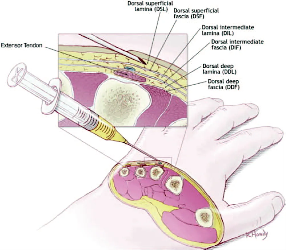 Fig 19. Schematic illustration of the dorsum of the hand with a needle advancing in the most superficial plane; the inset enlarges the cross-section with the three fat laminae labeled —dorsal superficial lamina (DSL, no structures), dorsal intermediate lamina (DIL, with a blue vein inside it) and dorsal deep lamina (DDL, with the extensor tendon)— separated by the dorsal superficial (DSF), intermediate (DIF) and deep (DDF) fasciae; below, a section through the five metacarpals locates the extensor tendon and the intertendinous fat reservoir — (UPO, hand rejuvenation, p. 13).
Fig 19 sets the plane the table summarizes in one cell: the needle goes in the superficial lamina, above the dorsal vein of the intermediate lamina and well above the tendon of the deep one. It is the same principle as the temple —two safe planes and a dangerous intermediate one— transferred to the hand.
The dorsum of the hand, lamina by lamina
The cadaveric investigation of the dorsum of the hand (Bidic) identified three distinct fat laminae beneath the dermis, divided by thin fasciae and joined by fibrous walls and 8-10 perforating vessels that cross them perpendicularly [99]. From superficial to deep: the dorsal superficial lamina (DSL) contains no structures and is the recommended deposit site; the dorsal intermediate lamina (DIL) houses the dorsal veins and nerves; the dorsal deep lamina (DDL) contains the extensor tendons [98,99]. The injection site is bounded by the 2nd to 5th metacarpals and the dorsal wrist crease [99].
Consensus: the deposit goes subdermal, in the superficial lamina, adjacent and superficial to the dorsal veins; the skin pinch ("tenting") separates the laminae and facilitates cannula advance [98,99]. Discrepancy — to pinch or not to pinch? · A: the pinch magnifies the subdermal areolar plane and moves the tip away from veins and tendons [98]. · B (Rosen, dissection of 19 fresh hands + duplex ultrasound): the pinch lifts the dorsal veins and the fascial plane itself too along with the skin, and that is why his subdermal scraping technique avoids it so as not to increase the vascular risk [98]. · Decide: with a cannula and faint veins, the pinch helps; with prominent veins and a needle, the pinch can bring the vein up to the plane of the tip. Do not average: it is a decision per hand.
Classic trap: treating the décolletage and the dorsum of the hand with facial parameters. Both regions have fewer adnexa and a worse repair reservoir, and the result ages worse if the facial energies are copied [94,98]. It is a parameter error, not an indication error.
Cadaveric dissection as a curricular standard
The reference academies (AAFE, ECAMS, Archidemia and equivalent centers such as the ICLO in Verona) place dissection as a curricular requirement, not as a complement [100]. (P) The rationale is that of the whole chapter: the anatomical variability of A1.6 —arteries that change plane by segment, displaced foramina, absent muscles— is not internalized in an atlas, which draws the mode; it is internalized by seeing it several times on real tissue.
What a course must deliver for dissection to teach real anatomy
| Requirement | Why | Source |
|---|---|---|
| Fresh-frozen, not formalin-fixed | Formalin hardens and discolors the tissue and requires more pressure to cut than in vivo, so it lies about the planes and the elasticity; the fresh preserves the mechanics the injector will find in the chair [101] | [101] |
| Layered dissection from the skin, not regional windows | It is the only way to run through the stratification of A1.2 and see where each layer changes name (temporal fascia, parotideomasseteric, platysma) [1,3] | [1,3] |
| Dye injection and subsequent dissection | Injecting a dye (methylene blue or homogenized with filler) and dissecting afterward is the only way to see where the bolus really went, not where it was intended [15,100] | [15,100] |
| In vivo ultrasound correlation before or after | Closes the loop between the live patient's image (A1.9) and the real anatomy beneath | [102] |
| Ratio ≤ 4 students per specimen | Below that ratio each student dissects; above it, they watch someone dissect (P) [D] — a teaching-quality criterion, never_sufficient_alone, never a clinical parameter |
— |
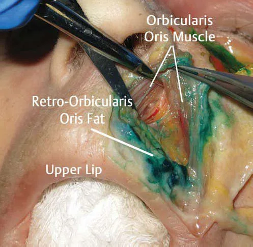 Fig 21. Cadaveric dissection of the perioral region and upper lip after percutaneously injecting hyaluronic acid homogenized with green dye into the retro-orbicularis oris fat compartment (ROOrF); the staining delimits exactly the territory the bolus occupied and forceps separate the overlying orbicularis oris — (Lamb, 2018, p. 66).
Fig 21 is the argument for the method: the green dye marks the compartment the bolus really occupied. Without staining and going to look, the operator believes they deposited in a plane and has no way of knowing whether they hit it. The Paternostro study formalizes it: 9 non-embalmed donors, methylene blue in the supraorbital region, subsequent dissection to measure the real diffusion of the volume [100].
Classic trap: validating a technique on a formalin-fixed cadaver. The fixed tissue behaves like a rigid, homogeneous plane, different from the living; a maneuver that "works" there may reproduce neither the real plane nor the real diffusion [101].
Anatomical orientation by ultrasound
High-frequency cutaneous ultrasound turns anatomy from statistics into a patient datum: where A1.6 gives the most likely location of an artery, the probe gives that of this patient, this side and today. Its full development lives in G6; here its place in the anatomical map remains.
Schematic — settings and what is reliably identified
| Parameter | Usual setting | Source |
|---|---|---|
| Probe | Linear, 15-22 MHz (18-22 MHz for dermis and eyelid) | [102] |
| Depth | 10-20 mm for the face; 5-10 mm for the eyelid | [102] |
| Doppler | Color + power Doppler, essential before filling the temple, glabella or nose | [102] |
| Gel | Thick layer or stand-off in irregular zones (nose, orbital rim) | [102] |
What is reliably identified: layers 1-5 of A1.2, the artery (pulsatile, Doppler+), the vein (compressible) and the filler material [102,103]. Ultrasound is the diagnostic tool that recognizes a previous filler the patient does not recall or denies, and the guide for targeted hyaluronidase (J3, J7).
Ultrasound appearance of filler, material by material
| Material | Sonographic pattern | Source |
|---|---|---|
| Hyaluronic acid | Anechoic collection (pseudocyst) with posterior wall enhancement; if cross-linked, internal permeation echoes | [102,103,104,105] |
| Calcium hydroxyapatite (CaHA) | Hyperechoic band with variable degrees of posterior acoustic shadow from the calcium component | [102,103,104] |
| PMMA | Heterogeneous granular hyperechoic deposits with a comet-tail mini-artifact | [103,104] |
| Silicone / permanent biopolymers | "Snowstorm": diffuse, granular hyperechoic pattern, with a dirty shadow that erases the deep planes and may migrate to nodes | [103,104,105] |
| PLLA | Initially hyperechoic from the microspheres; then iso- or hypoechoic, often without expression except a palpable nodule | [102,103] |
| Autologous fat | Well-defined isoechoic nodule with anechoic permeation areas | [104] |
 Fig 20. B-mode ultrasound of a superficial deposit with a "snowstorm" pattern: a dome of diffuse granular hyperechoic echoes in the subcutaneous plane with a posterior "dirty" acoustic shadow that prevents differentiating the deep planes, the characteristic appearance of silicone and permanent biopolymers — (UPO, filler adverse effects, p. 216).
Fig 20 shows why ultrasound changes conduct facing an unknown filler: the snowstorm pattern identifies a permanent biopolymer without the patient needing to recall what was placed, and that single image reorders the diagnosis and the management (J7). Hyaluronic acid, in contrast, is seen as an anechoic collection that hyaluronidase can reach in a targeted way.
Classic trap: assuming that "nothing is palpable" means "there is no filler". Integrated PLLA and hydrated hyaluronic acid may not be palpable and still be seen —or their absence confirmed— on the probe; a pre-treatment ultrasound assessment in an operated zone avoids injecting over an unexpected material [103].
Danger zones by region (operative synthesis)
Ordered by consequence, not by frequency. Beleznay maintains the reference series of filler blindness: in the review of 98 cases the glabella (38.8 %) and the nasal region (25.5 %) led, followed by the nasolabial fold (13.3 %) and the forehead (12.2 %) [12]; the update of 48 new cases inverted the order toward the nose (56.3 %) and the glabella (27.1 %), with the forehead (18.8 %) and the nasolabial fold (14.6 %) behind, and hyaluronic acid as the causal material in 81.3 % [106]. The two sites that lead are constant across reviews: glabella and nose.
| Zone | Vessel at stake | Operative rule | Source |
|---|---|---|---|
| Glabella | Supratrochlear (ophthalmic branch) | Minimal bolus, very superficial dermis, needle in motion; aspiration is not enough. Many avoid filler here | [2,12] |
| Nose (dorsum, tip, columella) | Dorsal nasal, lateral nasal | Supraperiosteal/supraperichondral in the midline, low volume, cannula; operated nose = multiplied risk | [2,55,106] |
| Tear trough | Angular, palpebrals | Deep supraperiosteal, cannula, slow retro-injection; remember there is no layer 2 | [4,66] |
| Temple | Superficial temporal (superficial) / deep temporal (deep) | Two safe planes —very superficial subdermal or bone contact—; the intermediate houses the frontal branch of the facial nerve | [4,2] |
| Nasolabial fold | Facial / angular | Cannula, subdermal plane; the artery is less than 4 mm away, never a blind deep bolus | [2,62] |
| Lip | Superior/inferior labials | Submucosal; the vessel is deeper than believed, and the subcutaneous plane is the least vascularized | [2,56] |
| Lateral cheek / arch | Transverse facial | Supraperiosteal over bone, bone contact allowed | [2,4] |
The two rules that summarize all the above: (1) if one is in the mid subcutaneous of a named territory, one is in the worst possible plane —Lee measured that the facial artery itself changes plane along its course and that there is no universally safe plane for it [107]—; (2) a negative aspiration does not rule out intravascular position: it is weak information, not a permission, because the needle seeds product into every plane it crosses and the double-perforation phenomenon of the wall exists [2,63,64]. The safe conduct is not to aspirate but to choose a plane, move the needle and retro-inject slowly.
Schools
The reader wants to know what is done; the schools answer differently to a single question —"what is aging made of?"— and that is why they are not averaged. The comparative development lives in B5; here their anatomical stance remains.
| Current | Anatomical stance | Logic | Source |
|---|---|---|---|
| Structural / supraperiosteal (Swift, MD Codes) | Deep deposits over bone, at fixed points | Rebuilds the lost skeletal support; the deep plane is generally the least vascularized [6,46] | [6,46] |
| Layers / layering (Sattler, German school) | Deep volume first + subdermal refinement | More natural result; assumes ultrasound mastery of the plane [6] | [6] |
| Anti-volume / skin-first (growing since ~2020) | Minimal volume, priority to skin quality and biostimulation | Reaction to the facial overfilled syndrome; aging is not only volume loss [6,34] | [6,34] |
| Ultrasound-guided (expanding Iberian and Latin American school) | No plane is assumed: it is seen before injecting | The anatomical variability of A1.6 makes "blind" injection in a risk zone unacceptable [102,107] | [102,107] |
Consensus among all: treat lateral to the line of ligaments before medial, and deep before superficial, because the lateral layers continue with the SMAS and lift, and the deep compartments do not shift [31,32,6]. The discrepancy is in how much weight to give volume versus skin quality, and that is decided per patient, not per school.
Errors and how to avoid them
| Error | Why it happens | How to avoid it | Source |
|---|---|---|---|
| Treating anatomy as fixed | It is studied in atlases, which draw the norm | Assume a distribution; Doppler in a risk zone [2,102] | [2,102] |
| Marking supine | Operator comfort | Mark always seated at 90°, verify standing [6] | [6] |
| Trusting aspiration | Taught as a safety gesture | Aspirate, but treat it as weak information; the plane and the volume rule [63,64] | [63,64] |
| Injecting into the mid subcutaneous of the temple | It is where the tissue "gives" | Only two planes: very superficial subdermal or bone contact [4,2] | [4,2] |
| Believing the glabella is superficial and therefore safe | Superficial ≠ safe; the supratrochlear surfaces | Minimal bolus, needle in motion, or do not treat [2,12] | [2,12] |
| Filling the groove instead of the compartment | The patient points to the groove | Treat the donor compartment; the groove is a ligament edge [6,41] | [6,41] |
| Compensating bone loss with subcutaneous volume | Gives immediate improvement in the mirror | Supraperiosteal support; the subcutaneous swells, does not project [6,83] | [6,83] |
| Using facial parameters on the décolletage and hands | The same skin is assumed | Reduce energy and dose; they heal worse [94,98] | [94,98] |
| Injecting inside the foramen when blocking | The "click" is sought | Deposit next to the foramen, aspirating, without seeking paresthesia [72,9] | [72,9] |
| Taking an operated nose for granted as safe | Surgery alters the vascularization and creates scar | Increased risk; many consider it a relative contraindication [2,106] | [2,106] |
Step-by-step protocol — anatomical mapping before any infiltration
(P) It gathers the landmarks of A1.1, the layers of A1.2 and the arterial depth of A1.6 into a repeatable sequence. No region (D1-D13) repeats it; all of them assume it.
- Seated at 90°, overhead light, no makeup, gaze to the horizon (Frankfort plane).
- Standardized photography: frontal, bilateral ¾, bilateral profile, at rest and in dynamics.
- Palpate and mark bone: orbital rim in its four quadrants, zygomatic arch, temporal fusion line, mandibular border, pyriform aperture. With the bone marked, half the decisions are already made.
- Mark the three foramina on the mid-pupillary vertical (supraorbital 1-3 mm medial, infraorbital 5-10 mm below the rim, mental).
- Mark Pitanguy's line if the temple, brow tail or upper third are to be worked.
- Find the superficial temporal with the finger in front of the tragus and follow it upward; mark it.
- Doppler / ultrasound if the plan includes glabella, nose, temple or tear trough, or if there is previous surgery, previous filler of unknown origin or unexplained asymmetry.
- Declare the plane of each point in writing before loading: supraperiosteal / deep subcutaneous / subdermal / intradermal. A point with no declared plane is not injected.
- Check the escape route: where the hyaluronidase is, how many IU and at what distance. If the answer is not immediate, do not begin (J2).
- Reassess standing when finished: gravity is part of the anatomy.
Classic trap: starting to load without having declared the plane of each point. Step 8 is the one that separates a map from an intention, and its absence is the silent signature of almost everything the previous sections describe as a plane error.
Coverage vs UPO
| Topic UPO teaches | Status in this chapter | What the atlas adds |
|---|---|---|
| Treatment by facial thirds as an anatomical approach scheme [97] | Covered — A1.1 and A1.10 | Adds the exact definition of each third with its points and the 1/3-2/3 subdivision of the lower third, with an external source [91,83,92] |
| Perception of beauty, symmetry and harmony as a framework prior to technique [97] | Covered in its operative consequence — A1.10 | Adds the explicit limit: the ideals are not used alone and are corrected for sex, ethnicity and age [94] |
| Vitruvian canon [97] | Covered — A1.10, classic canons | Adds that the teaching itself declares it untenable and contrasts it with measured proportions [91,7] |
| Golden ratio / phi / tetrakis [97] | Covered — A1.10 | Adds the formal definition from the anatomy and filling atlas [92] |
| Ogee curve [97] | Covered as a contour reference — A1.10 | Adds the anatomical substrate of the contour: deep lateral fat pad, transverse facial septum and its dynamic behavior [4,34] |
| Angle diagrams and templates [97] | Covered in definition; values NOT published — A1.10 | Declares the limitation with the query that proves it and refers to the corpus cephalometric source [7] |
| 43 mimetic muscles and more than 10 000 expressions [97] | Covered — A1.5 | Adds origin, insertion, vector, antagonist and layer muscle by muscle, plus the variability measured in dissection [51,52,53,48] |
| Upper-third anatomy applied to filling (T8.1, Arenas) [108] | Covered — A1.1, A1.2, A1.6 | Adds the temple layer count, the position of the middle frontal septum with its measurement and the plane-by-arterial-segment table [4,2,1] |
| Mid-third anatomy applied to filling (T8.1, Arenas; midface anatomy article) [108] | Covered — A1.3, A1.6, A1.7 | Adds the complete map of deep compartments with limits, the angular vein as a boundary and the compartment rules [4,21,22,66] |
| Jawline and chin: supraperiosteal plane at the chin, subdermal with a microcannula at the jawline [108] | Covered as anatomy — A1.1, A1.6, A1.9 | Adds why: arteries deep to the platysma in the lower third and bone contact allowed at the mandibular angle [2] |
| Facial biomechanics and treatment principles (assigned reading Freytag 2022) [6] | Covered — A1.4 | Adds the discrepancy about ligamentous laxity and the hemiface study that quantifies the product saving [31,45] |
| Filler complications (T10) | Out of scope | This chapter provides only the anatomical substrate (A1.6, A1.7); management lives in the complications chapter |
| Vascular pathology of the limbs (M5) | Not applicable | Different territory; no facial anatomical overlap |
What UPO does NOT cover and this chapter does:
| Absent in UPO | Where it is here |
|---|---|
| Layer model by region with its three exceptions (temple, tear trough, perioral) | A1.2 |
| Inventory of superficial and deep fat compartments with limits one by one | A1.3 |
| Line of ligaments as a functional boundary and its effect on the vector | A1.4 |
| Arterial depth by segment and recommended-plane table region by region | A1.6 |
| Retinal time window and anatomy of the ophthalmic route | A1.6 |
| Facial venous and lymphatic anatomy applied to edema | A1.7 |
| Neurovascular corridors by compartment (where each branch of VII travels) | A1.8 |
| Bone remodeling with its causal chain and the variation by population | A1.9 |
| Ultrasound measurement of layer thickness and its effect on the real deposit plane | A1.10 |
Currency caveat on the UPO lane. The master's material is slide teaching and theoretical chapters; no figure supported only by that source is published here as a clinical parameter (never_sufficient_alone). Its value in this chapter is as a syllabus: it defines what had to be covered, not how much each number is worth.
Self-assessment
Ten active-recall questions. All are answered only with data published in this chapter.
1. Which four structures form the line of ligaments, from superior to inferior, and what changes in the result depending on whether one injects medial or lateral to it?
Answer
Temporal ligamentous adhesions, lateral orbital thickening (LOT), zygomatic ligament and mandibular ligament. **Medial** to the line filler **projects**; **lateral** to it it **lifts** structures located lower down, because of the oblique arrangement of the layers in the medial face versus the parallel one in the lateral.2. At what distance from the supraorbital rim is the middle frontal septum and why does that figure change the injection plane in the forehead?
Answer
**1.5 ± 0.17 cm** (range 1.3-1.7) in the midline and **3.0 ± 0.24 cm** (range 2.7-3.3) laterally. Below that septum the supratrochlear and supraorbital arteries are supraperiosteal; above, they have already perforated the frontalis and are subcutaneous. The forehead does not admit a single plane.3. How many layers does the tear trough have and which are they?
Answer
Three: skin, orbital portion of the orbicularis oculi muscle and periosteum. There is no subcutaneous layer or deep fascia; what is injected "subcutaneously" ends up intramuscular.4. Where is the facial artery fixed at the modiolus, with what frequency and by which structure?
Answer
At **1.5 cm behind the oral commissure**, identifiable in **100 %** of cases, fixed by a muscular band from the buccinator, between the buccinator (deep) and the modiolar portion of the platysma plus the converging muscles (superficial).5. In what percentage are the labial arteries submucosal and in what percentage subcutaneous, and what plane remains the least vascularized in the lip?
Answer
Submucosal in **78.1 %**, subcutaneous in **2.1 %**; the rest are intramuscular between the two laminae of the orbicularis. The subcutaneous plane is the least vascularized. Measured by ultrasound, the subcutaneous layer of the lip measures **less than 1 mm**, so that the deposit ends up intramuscular in the majority.6. Which temporal compartment contains the frontal branch of the facial nerve and which is free of relevant neurovascular structures?
Answer
The **inferior** temporal compartment contains the frontal branch of the facial nerve, the sensory zygomaticotemporal branches and the temporal portion of the sentinel vein. The **superior** temporal compartment contains no relevant neurovascular structures.7. What is the retinal survival time window measured experimentally and why is complete occlusion rare on angiography?
Answer
No detectable damage until **97 minutes** in the primate; beyond that the damage is irreversible and progressive, with 90 minutes as the operating reference point. Complete occlusion is rare because the retina receives collateral supply from the posterior ciliary arteries and the variable cilioretinal ones. Retinal perfusion is **0.52 cc/min per mg**, versus 0.48 in the brain.8. Where is the angular vein located relative to the inferior orbital rim and which two compartments does it separate?
Answer
**4.2 ± 0.7 mm** below the inferior orbital rim, with an oblique course from inferolateral to superomedial and deep to the orbital portion of the orbicularis. It separates the **deep medial fat pad** of the cheek (between the infraorbital foramen and the vein) from the **deep lateral fat pad** (between the vein and the zygomaticus major).9. Which two superficial fat compartments are not filled and why?
Answer
The **superficial nasolabial**, because it aggravates the nasolabial fold itself; and the **jowl** one, because it aggravates the labiomandibular fold and the jowl deformity. Both are superficial and both are where the patient points with the finger.10. What difference is there between Caucasian and Asian populations in the angular changes of the facial skeleton with age?
Answer
In the Asian population the **orbital and maxillary angles change less** and the **pyriform angle changes more** than in Caucasian series, where the glabellar, orbital, maxillary and pyriform angles decrease, and the maxilla, the pyriform aperture and the infraorbital rim recede. The angles are not averaged across populations.What's new and trends (2022-2026)
| Year | What changed | Consequence for this chapter | Source |
|---|---|---|---|
| 2022 | The three treatment principles derived from facial biomechanics are formulated explicitly for the first time: upper third first, lateral face first, deep first | A1.4 moves from describing ligaments to ordering the treatment sequence | [6] |
| 2022 | The absence of solid evidence of ligamentous laxity with age is declared, and the descent is reattributed to bone remodeling | Introduces the discrepancy maintained in A1.4 and reinforces A1.9 | [6] |
| 2024 | Mapping of the platysma's motor innervation by Sihler staining: motor plates concentrated in the upper two thirds, lower third mostly sensory | A1.5 stops treating the platysma as a homogeneous target | [54] |
| 2024 | Preinjection sonography of the labial arteries: deep, lateral origin with progressive approach to the midline in a superficial plane | A1.6 gains a risk gradient within the lip itself | [61] |
| 2024 | Review of the anatomical considerations of the thread lift: vessels, facial nerve branches, compartments and ligamentous engagement as the condition of a sustainable lift | A1.4 is applied to a different instrument without changing the target | [47] |
| 2024 | The risk of bone resorption associated with repeated masseter denervation is noted | A1.9 incorporates the reverse direction: muscular loading shapes the bone | [90] |
| 2025 | Non-surgical modification of chin and prejowl systematized by bone, muscle and compartment anatomy | A1.9 and A1.10 gain the lower-third correlate | [96] |
| 2026 | High-frequency ultrasound in 126 participants: subcutaneous lip layer < 1 mm, intramuscular deposit in the majority, and significant variation by sex, race and ethnicity, BMI and age | A1.10 replaces the descriptive variant with a measured one; A1.2 gains a second region where the nominal plane does not exist | [95] |
| 2026 | Systematic review of 16 studies: ultrasound guidance reaches up to 88 % deposit accuracy versus 50 % for the surface-reference technique | A1.8 gains a measure of the variability of the neurovascular corridor | [73] |
What did NOT change, and why the old references remain the state of the art. The description of the superficial fat compartments [14] and their septa, the layer model [4,3], the inventory of retaining ligaments [109,13] and their biomechanical hierarchy [44], the course and depth of the facial arteries by segment [2] and the anatomy of the ophthalmic route [2,58] have not been replaced: they have been confirmed with another modality, first by imaging [15,21] and now by in vivo ultrasound [28,95]. A dissection article from 2007 or from 1989 remains the primary source of an anatomical limit because the limit has not changed; what has changed is the ability to see it without opening. (P) The correct reading of this chapter's reference list is that the median year is low because the macroscopic anatomy is stable, and that what ages fast is not the description of the compartment but the recommendation of what to do with it.
What remains open. The decade-by-decade chronology of bone resorption (declared as a gap in A1.9), the quantification of facial lymphatic flow (declared in A1.7) and the real existence of measured ligamentous laxity (open discrepancy in A1.4).
Unexplored directions (AI speculation)
> Notice. What follows is not evidence. These are hypotheses generated by the model from the data already cited in this chapter, marked with [IA-ESPEC] so they cannot be confused with the [A]-[D] tags or with a recommendation. No proposal includes a dose, a product or an applicable protocol. Each one declares its anchor (a fact already cited here), the proposal and what would settle it.
1 · Ultrasound of the middle frontal septum as a substitute for the metric reference. [IA-ESPEC]
· Anchor: the middle frontal septum is at 1.5 ± 0.17 cm in the midline and 3.0 ± 0.24 cm laterally [2,1], and above it the artery becomes subcutaneous.
· Proposal: that the individual position of the septum be locatable by high-frequency ultrasound as a recognizable interface, instead of being estimated from a population mean.
· What would settle it: an ultrasound–dissection concordance study in the same specimen, measuring the deviation between the real position and the one predicted by the mean, with the dispersion (±0.17 and ±0.24 cm) as the utility threshold.
2 · The line of ligaments as a drainage boundary, not only of movement. [IA-ESPEC]
· Anchor: the line is a functional boundary of movement demonstrated by surface vector analysis [33], and the superficial lymphatics accompany the veins [67].
· Proposal: that the same line separates superficial lymphatic drainage territories, which would explain why edema after lateral filling behaves differently from edema after medial filling.
· What would settle it: indocyanine fluorescence lymphography before and after a standardized volume load on each side of the line, measuring transit time.
3 · Reattribution of the descent: ligament versus bone, in the same subject. [IA-ESPEC]
· Anchor: the open discrepancy between ligamentous fatigue [3] and remodeling of the bony anchor [6], with no series that separates them.
· Proposal: that the relative contribution be measurable by comparing, in matched cohorts, subjects with documented and preserved bone resorption, with the same degree of soft-tissue descent.
· What would settle it: paired volumetric CT [82] with simultaneous measurement of the superficial compartment descent by ultrasound [28]; if the descent correlates with the maxillary angle and not with age, the ligament is exonerated.
4 · Submillimeter subcutaneous thickness as a plane-exclusion criterion. [IA-ESPEC]
· Anchor: the subcutaneous layer of the lip measures less than 1 mm and the deposit ends up intramuscular in the majority [95]; the tear trough outright lacks layer 2 [4,3].
· Proposal: that there be a thickness threshold below which the subcutaneous plane ceases to be considered a technical option and is declared nonexistent on the regional map, just as is already done in the tear trough.
· What would settle it: ultrasound mapping of layer-2 thickness by region and by demographic group, with the rate of unintended deposit as the contrast variable.
5 · Motor plates as a target in more muscles than the platysma. [IA-ESPEC]
· Anchor: Sihler staining showed clustering of motor plates in the upper two thirds of the platysma [54], and surface electromyography showed that corrugator and procerus increase signal with age while the zygomaticus major reduces it [49].
· Proposal: that the motor-plate map, and not the muscle volume, explains the variability of response between patients in the muscles with measured signal change.
· What would settle it: Sihler mapping of corrugator, procerus and zygomaticus major in age-stratified specimens, correlated with the electromyographic signal of the same muscle.
6 · The transverse facial septum as a dynamic examination variable. [IA-ESPEC]
· Anchor: the transverse facial septum displaces the compartments cranially on smiling and is the substrate of the facial overfilled syndrome [34,6].
· Proposal: that its individual stiffness, measurable by elastography, predicts which patients will develop exaggerated volume on the smile with a normal volume load.
· What would settle it: shear-wave elastography of the septum at rest and on smiling, comparing patients with and without dynamic overfilling at equal received volume.
7 · Aging of the zygomaticus major and venous return. [IA-ESPEC]
· Anchor: the ability to compress the angular vein against the maxilla decreases with age, in parallel with the drop in signal of the zygomaticus major [6,49,50].
· Proposal: that this loss of muscular pump contributes measurably to morning periorbital edema, independent of the injected volume.
· What would settle it: Doppler of the angular vein at rest and during a held smile, in separate age cohorts and with no history of filler, correlated with edema measured by morning volumetry.
8 · A phenotype-specific layer map, not a correction over the Caucasian map. [IA-ESPEC]
· Anchor: the angular changes differ by population [3,87], the midfacial muscles vary between ethnic groups [3,52] and lip anatomy varies by race and ethnicity in a statistically significant way [95].
· Proposal: that the layer model by region has systematic differences by phenotype, and not only proportional differences, which would make proportional correction over a single template inadequate.
· What would settle it: ultrasound measurement of thickness by layer and region in cohorts of different phenotypes with the same protocol, checking whether the difference is of scale or of structure.
Safety
What this chapter is. An anatomy chapter. It contains no indications, doses, volumes or injection protocols, and none of its recommended-plane tables replaces individual assessment: the plane recommendations gathered from the anatomical literature are based on anatomy, not on the clinical presentation or the needs of the specific patient, which may require a different approach [2].
The five anatomical red lines
- No technique is absolutely safe. The facial arterial vasculature varies greatly between individuals and between sides of the same individual; because of that lack of predictability it is impossible to guarantee absolute safety in an injectable procedure. The risk must be communicated to the patient, and the operator must be trained and equipped to treat the complication [2].
- A negative aspiration does not exclude intra-arterial passage. The needle distributes product into every plane it crosses, with material identifiable in the subdermis even when the tip is on bone, and the double-perforation phenomenon of the arterial wall exists [2,63,64].
- Glabella and periorbital region are internal carotid territory. The anastomoses between dorsal nasal, supratrochlear, supraorbital and infraorbital connect directly with the ophthalmic circulation [2,57]. The deep glabellar plane is the least vascularized described, not a safe plane [2].
- The retinal window is short. No detectable damage up to 97 minutes in the experimental model; afterward, progressive irreversible damage [2,59]. Prior preparation —not an improvised reaction— is what fits within that window.
- The temple has two planes and no intermediate one. Subcutaneous above the superficial temporal fascia, or bone contact. Between them is the compartment that houses the frontal branch of the facial nerve, the sensory branches and the sentinel vein [4,2]; and the possibility of intracranial penetration in that region has been explicitly investigated [89].
On this chapter's figures. Every measurement published here comes from a source identified in the reference list. The magnitudes the corpus pass did not return with a traceable source —decade-by-decade bone-resorption chronology (A1.9), quantification of facial lymphatic flow (A1.7) and normative profile angle ranges (A1.10)— are declared as a gap with the query that proves it, instead of being filled in. No figure supported solely by the master's teaching material is published as a clinical parameter: never_sufficient_alone.
On the (P) material. The sentences marked (P) are the model's reasoning ordering data already cited. They contain no dose, no product and no new plane, and must not be cited as a source.
Sources of the figures
The 21 figures were resolved with figure_pick.py and with targeted search over the corpus, and each one was opened with Read before writing its caption; the captions describe what the image shows, not the corpus caption, which in several of them is a dump of the surrounding markdown and in two cases (a clinical photograph attributed to a ligament schematic and a dissection panel attributed to a proportions text) did not correspond to the image; those two were discarded. Fig 19-21 were added in the salvage for the new sections (dorsum of the hand, cadaveric dissection and ultrasound) and were also opened with Read before being captioned.
> Sources: Fig 1 — Watanabe [110] · Fig 2, 8, 9, 13 — Pirayesh [111] · Fig 3 — Radlansky [94] · Fig 4 — Carruthers [112] · Fig 5 — Lobo [113] · Fig 6 — Vieira Braz [92] · Fig 7 — Cotofana [2] · Fig 10 — Parker [114] · Fig 11, 12 — van Gijn [91] · Fig 14 — Kim [115] · Fig 15 — Libro Zonas Peligrosas en Medicina Estética Facial [116] · Fig 16, 18 — Master Techniques in Facial Rejuvenation [83] · Fig 17 — Standring [117] · Fig 19 — UPO, hand rejuvenation [99] · Fig 20 — UPO, filler adverse effects [105] · Fig 21 — Lamb [118].
Corpus readings assigned to this chapter
Corpus documents assigned to A1 in the brief that are not cited in the body because their content is regional and belongs to D1-D13; they are listed so that the regions do not search for them again:
| Document | What it provides and to which region |
|---|---|
| Sykes, upper third with fillers [119] | Regional anatomy of the upper third applied to injection → D1 |
| Cotofana, mid third with fillers [120] | Regional anatomy of the mid third → D3 |
| Braz, lower third with fillers [121] | Regional anatomy of the lower third → D6 |
| de Maio, assessment and injection guide [122] | Origin of the "one up and one down" reference in the temple → D1 |
| Malherbe, ultrasound protocol in facial aesthetics [102] | Ultrasound correlate of the layers described in A1.2 → cross-cutting |
| Fehrenbach, illustrated head and neck anatomy [72] | General head and neck reference → cross-cutting |
| Moore, clinically oriented anatomy [9] | Distribution of VII and the trigeminal → A1.8 |
References
- Cotofana S, Mian A, Sykes JM, et al. An update on the anatomy of the forehead compartments. Plast Reconstr Surg. 2017;139(4):864e-872e.
[B] - Cotofana S, Lachman N. Arteries of the face and their relevance for minimally invasive facial procedures: an anatomical review. Plast Reconstr Surg. 2019;143(2):416-426.
[B]· corpus MEDLIB (Cotofana). - Cotofana S, Fratila AAM, Schenck TL, Redka-Swoboda W, Zilinsky I, Pavicic T. The anatomy of the aging face: a review. Facial Plast Surg. 2016;32(3):253-260.
[B]· corpus MEDLIB (Cotofana). - Cotofana S, Lachman N. Anatomy of the facial fat compartments and their relevance in aesthetic surgery. J Dtsch Dermatol Ges. 2019;17:399-413.
[B]· corpus MEDLIB (Cotofana). - Mendelson B, Wong CH. Changes in the facial skeleton with aging: implications and clinical applications in facial rejuvenation. Aesthetic Plast Surg. 2012;36(4):753-760.
[B] - Freytag L, Alfertshofer MG, Frank K, et al. Understanding facial aging through facial biomechanics: a clinically applicable guide for improved outcomes. Facial Plast Surg Clin North Am. 2022;30(2):125-133. PMID 35501049 · DOI 10.1016/j.fsc.2022.01.001.
[B]· corpus MEDLIB (Freytag). - Naini FB. Facial Aesthetics: Concepts and Clinical Diagnosis. Wiley-Blackwell; 2011.
[C]· corpus MEDLIB. - Berchtold V, Stofferin H, Moriggl B, Brenner E, Pauzenberger R, Konschake M. The supraorbital region revisited: an anatomic exploration of the neuro-vascular bundle with regard to frontal migraine headache. J Plast Reconstr Aesthet Surg. 2017;70(9):1171-1180.
[B] - Moore KL, Dalley AF, Agur AMR. Clinically Oriented Anatomy. 8th ed. Wolters Kluwer; 2018.
[C]· corpus MEDLIB. - Pilsl U, Anderhuber F. The chin and adjacent fat compartments. Dermatol Surg. 2010;36(2):214-218.
[B] - Sabini P, Wayne I, Quatela VC. Anatomical guides to precisely localize the frontal branch of the facial nerve. Arch Facial Plast Surg. 2003;5(2):150-152.
[B] - Beleznay K, Carruthers JDA, Humphrey S, Jones D. Avoiding and treating blindness from fillers: a review of the world literature. Dermatol Surg. 2015;41(10):1097-1117. PMID 26356847 · DOI 10.1097/DSS.0000000000000486.
[B] - Alghoul M, Codner MA. Retaining ligaments of the face: review of anatomy and clinical applications. Aesthet Surg J. 2013;33(6):769-782.
[B] - Rohrich RJ, Pessa JE. The fat compartments of the face: anatomy and clinical implications for cosmetic surgery. Plast Reconstr Surg. 2007;119(7):2219-2227. PMID 17519724 · DOI 10.1097/01.prs.0000265403.66886.54.
[B] - Schenck TL, Koban KC, Schlattau A, et al. The functional anatomy of the superficial fat compartments of the face: a detailed imaging study. Plast Reconstr Surg. 2018;141(6):1351-1359.
[B]· corpus MEDLIB (Schenck). - Kruglikov I, Trujillo O, Kristen Q, et al. The facial adipose tissue: a revision. Facial Plast Surg. 2016;32(6):671-682.
[B]· corpus MEDLIB (Cotofana). - O'Brien JX, Ashton MW, Rozen WM, Ross R, Mendelson BC. New perspectives on the surgical anatomy and nomenclature of the temporal region: literature review and dissection study. Plast Reconstr Surg. 2013;131(3):510-522.
[B] - Mitz V, Peyronie M. The superficial musculo-aponeurotic system (SMAS) in the parotid and cheek area. Plast Reconstr Surg. 1976;58(1):80-88.
[B] - Ghassemi A, Prescher A, Riediger D, Axer H. Anatomy of the SMAS revisited. Aesthetic Plast Surg. 2003;27(4):258-264.
[B] - Moss CJ, Mendelson BC, Taylor GI. Surgical anatomy of the ligamentous attachments in the temple and periorbital regions. Plast Reconstr Surg. 2000;105(4):1475-1490.
[B] - Cotofana S, Gotkin RH, Frank K, et al. The functional anatomy of the deep facial fat compartments: a detailed imaging-based investigation. Plast Reconstr Surg. 2019;143(1):53-63.
[B] - Wong CH, Mendelson B. Facial soft-tissue spaces and retaining ligaments of the midcheek: defining the premaxillary space. Plast Reconstr Surg. 2013;132(1):49-56.
[B] - Mendelson BC, Freeman ME, Wu W, Huggins RJ. Surgical anatomy of the lower face: the premasseter space, the jowl, and the labiomandibular fold. Aesthetic Plast Surg. 2008;32(2):185-195.
[B] - Yang HM, Jung W, Won SY, Youn KH, Hu KS, Kim HJ. Anatomical study of medial zygomaticotemporal vein and its clinical implication regarding the injectable treatments. Surg Radiol Anat. 2015;37(2):175-180.
[B] - Mojallal A, Cotofana S. Anatomy of lower eyelid and eyelid-cheek junction. Ann Chir Plast Esthet. 2017;62(5):365-374.
[B] - Beer GM, Manestar M, Mihic-Probst D. The causes of the nasolabial crease: a histomorphological study. Clin Anat. 2013;26(2):196-203.
[B] - Gierloff M, Stöhring C, Buder T, Wiltfang J. The subcutaneous fat compartments in relation to aesthetically important facial folds and rhytides. J Plast Reconstr Aesthet Surg. 2012;65(10):1292-1297.
[B] - Schelke L, Velthuis PJ, Lowry N, et al. The mobility of the superficial and deep midfacial fat compartments: an ultrasound-based investigation. J Cosmet Dermatol. 2021;20(12):3849-3856.
[B] - Sandulescu T, Weniger J, Philippou S, et al. Immunohistochemical evidence of striated muscle cells within midfacial superficial musculoaponeurotic system. Ann Anat. 2021;234:151647.
[B] - Wan D, Amirlak B, Giessler P, et al. The differing adipocyte morphologies of deep versus superficial midfacial fat compartments: a cadaveric study. Plast Reconstr Surg. 2014;133(5):615e-622e.
[B] - Casabona G, Frank K, Koban KC, et al. Lifting vs volumizing — the difference in facial minimally invasive procedures when respecting the line of ligaments. J Cosmet Dermatol. 2019;18(5):1237-1243.
[B]· corpus MEDLIB (Casabona). - Casabona G, Bernardini FP, Skippen B, et al. How to best utilize the line of ligaments and the surface volume coefficient in facial soft tissue filler injections. J Cosmet Dermatol. 2020;19(2):303-311.
[B] - Freytag DL, Alfertshofer MG, Frank K, et al. The difference in facial movement between the medial and the lateral midface: a 3-dimensional skin surface vector analysis. Aesthet Surg J. 2021;42(1):1-9.
[B] - Cotofana S, Gotkin RH, Frank K, et al. Anatomy behind the facial overfilled syndrome: the transverse facial septum. Dermatol Surg. 2020;46(8):e16-e22.
[B] - Rohrich RJ, Arbique GM, Wong C, Brown S, Pessa JE. The anatomy of suborbicularis fat: implications for periorbital rejuvenation. Plast Reconstr Surg. 2009;124(3):946-951.
[B] - Surek CK, Vargo J, Lamb J. Deep pyriform space. Plast Reconstr Surg. 2016;138(1):59-64.
[B] - Rohrich RJ, Pessa JE, Ristow B. The youthful cheek and the deep medial fat compartment. Plast Reconstr Surg. 2008;121(6):2107-2112.
[B] - Schenck TL, Koban KC, Schlattau A, et al. Updated anatomy of the buccal space and its implications for plastic, reconstructive and aesthetic procedures. J Plast Reconstr Aesthet Surg. 2018;71(2):162-170.
[B] - Reece EM, Pessa JE, Rohrich RJ. The mandibular septum: anatomical observations of the jowls in aging — implications for facial rejuvenation. Plast Reconstr Surg. 2008;121(4):1414-1420.
[B] - de Almeida ART, Romiti A, Carruthers JDA. The facial platysma and its underappreciated role in lower face dynamics and contour. Dermatol Surg. 2017;43(8):1042-1049.
[B] - Wong CH, Hsieh MKH, Mendelson B. The tear trough ligament: anatomical basis for the tear trough deformity. Plast Reconstr Surg. 2012;129(6):1392-1402.
[B] - Gierloff M, Stöhring C, Buder T, Gassling V, Açil Y, Wiltfang J. Aging changes of the midfacial fat compartments: a computed tomographic study. Plast Reconstr Surg. 2012;129(1):263-273.
[B] - Muzaffar AR, Mendelson BC, Adams WP Jr. Surgical anatomy of the ligamentous attachments of the lower lid and lateral canthus. Plast Reconstr Surg. 2002;110(3):873-884.
[B] - Brandt MG, Hassa A, Roth K, Wehrli B, Moore CC. Biomechanical properties of the facial retaining ligaments. Arch Facial Plast Surg. 2012;14(4):289-294.
[B] - Thornton GM, Lemmex DB, Ono Y, et al. Aging affects mechanical properties and lubricin/PRG4 gene expression in normal ligaments. J Biomech. 2015;48(12):3306-3311.
[B] - Freytag DL, Frank K, Haidar R, et al. Facial safe zones for soft tissue filler injections: a practical guide. J Drugs Dermatol. 2019;18(9):896-902.
[B] - Hong GW, Kim SB, Park Y, et al. Anatomical considerations for thread lifting procedure. J Cosmet Dermatol. 2024;24(1):e16618. PMID 39376117 · DOI 10.1111/jocd.16618.
[A] - Bae JH, Lee JH, Youn KH, et al. Surgical consideration of the anatomic origin of the risorius in relation to facial planes. Aesthet Surg J. 2014;34(7):NP43-NP49.
[B] - Cotofana S, Assemi-Kabir S, Mardini S, et al. Understanding facial muscle aging: a surface electromyography study. Aesthet Surg J. 2021;41(9):NP1208-NP1217.
[B] - Cotofana S, Lowry N, Devineni A, et al. Can smiling influence the blood flow in the facial vein? An experimental study. J Cosmet Dermatol. 2020;19(2):321-327.
[B] - Pessa JE, Zadoo VP, Adrian EK Jr, et al. Variability of the midfacial muscles: analysis of 50 hemifacial cadaver dissections. Plast Reconstr Surg. 1998;102(6):1888-1893.
[B] - Farahvash MR, Abianeh SH, Farahvash B, et al. Anatomic variations of midfacial muscles and nasolabial crease: a survey on 52 hemifacial dissections in fresh Persian cadavers. Aesthet Surg J. 2010;30(1):17-21.
[B] - Shim KS, Hu KS, Kwak HH, et al. An anatomical study of the insertion of the zygomaticus major muscle in humans focused on the muscle arrangement at the corner of the mouth. Plast Reconstr Surg. 2008;121(2):466-473.
[B] - Yi KH, Wong IKJ, Wan J. Optimizing botulinum toxin injections in the platysma muscle based on motor nerve distribution. J Cosmet Dermatol. 2025;24(7):e70301. PMID 40571678 · DOI 10.1111/jocd.70301.
[A] - Pilsl U, Anderhuber F. The external nose. Plast Reconstr Surg. 2016;138(5):830e-835e.
[B] - Cotofana S, Pretterklieber B, Lucius R, et al. Distribution pattern of the superior and inferior labial arteries: impact for safe upper and lower lip augmentation procedures. Plast Reconstr Surg. 2017;139(5):1075-1082.
[B] - Geibprasert S, Pongpech S, Armstrong D, Krings T. Dangerous extracranial-intracranial anastomoses and supply to the cranial nerves: vessels the neurointerventionalist needs to know. AJNR Am J Neuroradiol. 2009;30(8):1459-1468.
[B] - Michalinos A, Zogana S, Kotsiomitis E, Mazarakis A, Troupis T. Anatomy of the ophthalmic artery: a review concerning its modern surgical and clinical applications. Anat Res Int. 2015;2015:591961.
[B] - Hayreh SS, Zimmerman MB, Kimura A, Sanon A. Central retinal artery occlusion: retinal survival time. Exp Eye Res. 2004;78(3):723-736.
[B] - Lee SH, Lee HJ, Kim YS, Kim HJ, Hu KS. What is the difference between the inferior labial artery and the horizontal labiomental artery? Surg Radiol Anat. 2015;37(8):947-953.
[B] - Kim JS. 9-point injection technique for lip augmentation and lip corner lifting using sonographic imaging of the labial artery pathway. Aesthet Surg J. 2024;44(10):1080-1090. PMID 38649792 · DOI 10.1093/asj/sjae086.
[A] - Pilsl U, Anderhuber F, Neugebauer S. The facial artery: the main blood vessel for the anterior face? Dermatol Surg. 2016;42(2):203-208.
[B] - van Loghem JAJ, Humzah D, Kerscher M. Cannula versus sharp needle for placement of soft tissue fillers: an observational cadaver study. Aesthet Surg J. 2018;38(1):73-88.
[B] - Pavicic T, Frank K, Erlbacher K, et al. Precision in dermal filling: a comparison between needle and cannula when using soft tissue fillers. J Drugs Dermatol. 2017;16(9):866-872.
[B] - Scheuer JF 3rd, Sieber DA, Pezeshk RA, Gassman AA, Campbell CF, Rohrich RJ. Facial danger zones: techniques to maximize safety during soft-tissue filler injections. Plast Reconstr Surg. 2017;139(5):1103-1108.
[B] - Cotofana S, Steinke H, Schlattau A, et al. The anatomy of the facial vein: implications for plastic, reconstructive, and aesthetic procedures. Plast Reconstr Surg. 2017;139(6):1346-1353.
[B] - Netter FH. Atlas of Human Anatomy. 7th ed. Elsevier; 2022.
[C]· corpus MEDLIB. - Mannan's Regional Dissection and Surface Anatomy. 14th ed. 2017.
[C]· corpus MEDLIB. - Trinei FA, Januszkiewicz J, Nahai F. The sentinel vein: an important reference point for surgery in the temporal region. Plast Reconstr Surg. 1998;101(1):27-32.
[B] - de Castro CC. Midface Surgery. Elsevier; 2009.
[C]· corpus MEDLIB. - Dermal Fillers for Facial Harmony. 2019.
[C]· corpus MEDLIB. - Fehrenbach MJ, Herring SW. Illustrated Anatomy of the Head and Neck. 5th ed. Elsevier; 2017.
[C]· corpus MEDLIB. - Chowdhury R, Schiff B, Lee YH, Mohan S. Ultrasound guidance for botulinum toxin injection of muscles innervated by the facial nerve: a systematic review of anatomical precision, safety, and outcomes. Aesthet Surg J. 2026;46(2):195-201. PMID 40971765 · DOI 10.1093/asj/sjaf175.
[A] - Cotofana S, Gotkin RH, Ascher B, et al. Calvarial volume loss and facial aging: a computed tomographic (CT)-based study. Aesthet Surg J. 2018;38(10):1043-1051.
[B] - Enlow DH. A morphogenetic analysis of facial growth. Am J Orthod. 1966;52(4):283-299.
[B] - Frank K, Gotkin RH, Pavicic T, et al. Age and gender differences of the frontal bone: a computed tomographic (CT)-based study. Aesthet Surg J. 2019;39(7):699-710.
[B] - Kahn DM, Shaw RB Jr. Aging of the bony orbit: a three-dimensional computed tomographic study. Aesthet Surg J. 2008;28(3):258-264.
[B] - Pessa JE, Chen Y. Curve analysis of the aging orbital aperture. Plast Reconstr Surg. 2002;109(2):751-755.
[B] - Richard MJ, Morris C, Deen BF, Gray L, Woodward JA. Analysis of the anatomic changes of the aging facial skeleton using computer-assisted tomography. Ophthal Plast Reconstr Surg. 2009;25(5):382-386.
[B] - Shaw RB Jr, Kahn DM. Aging of the midface bony elements: a three-dimensional computed tomographic study. Plast Reconstr Surg. 2007;119(2):675-681. PMID 17230106 · DOI 10.1097/01.prs.0000246596.79795.a8.
[B] - Cotofana S, Gotkin RH, Morozov SP, et al. The relationship between bone remodeling and the clockwise rotation of the facial skeleton. Plast Reconstr Surg. 2018;142(6):1447-1454.
[B] - Karunanayake M, To F, Efanov JI, et al. Analysis of craniofacial remodeling in the aging midface using reconstructed three-dimensional models in paired individuals. Plast Reconstr Surg. 2017;140(3):448e-454e.
[B] - Azizzadeh B, Murphy MR, Johnson CM, Massry GG, Fitzgerald R. Master Techniques in Facial Rejuvenation. 2nd ed. Elsevier; 2018.
[C]· corpus MEDLIB. - Lillie EM, Urban JE, Lynch SK, Weaver AA, Stitzel JD. Evaluation of skull cortical thickness changes with age and sex from computed tomography scans. J Bone Miner Res. 2016;31(2):299-307.
[B] - Urban JE, Weaver AA, Lillie EM, et al. Evaluation of morphological changes in the adult skull with age and sex. J Anat. 2016;229(6):838-846.
[B] - Pessa JE, Desvigne LD, Lambros VS, et al. Changes in ocular globe-to-orbital rim position with age: implications for aesthetic blepharoplasty of the lower eyelids. Aesthetic Plast Surg. 1999;23(5):337-342.
[B] - Kim SJ, Kim SJ, Park JS, Byun SW, Bae JH. Analysis of age-related changes in Asian facial skeletons using 3D vector mathematics on picture archiving and communication system computed tomography. Yonsei Med J. 2015;56(5):1395-1400.
[B] - Pessa JE. An algorithm of facial aging: verification of Lambros's theory by three-dimensional stereolithography, with reference to the pathogenesis of midfacial aging, scleral show, and the lateral suborbital trough deformity. Plast Reconstr Surg. 2000;106(2):479-488. PMID 10946949 · DOI 10.1097/00006534-200008000-00040.
[B] - Philipp-Dormston WG, Bieler L, Hessenberger M, et al. Intracranial penetration during temporal soft tissue filler injection — is it possible? Dermatol Surg. 2018;44(1):84-91.
[B] - Popescu MN, Beiu C, Iliescu CA, et al. Ultrasound-guided botulinum toxin-A injections into the masseter muscle for both medical and aesthetic purposes. Toxins (Basel). 2024;16(10):413. PMID 39453189 · DOI 10.3390/toxins16100413.
[A] - van Gijn DR, Dover S. Oxford Handbook of Head and Neck Anatomy. Oxford University Press; 2022.
[C]· corpus MEDLIB. - Vieira Braz A, Sakuma TH. Atlas de Anatomia e Preenchimento Global da Face. Guanabara Koogan; 2017.
[C]· corpus MEDLIB. - Truswell WH. Surgical Facial Rejuvenation. Thieme; 2009.
[C]· corpus MEDLIB. - Radlansky RJ, Wesker KH. Atlas Ilustrado de Anatomia Clínica da Face.
[C]· corpus MEDLIB. - Harris S, Schelke L, Orlovska M, Wortsman X, Cotofana S, Velthuis P. Ultrasound evaluation of lip anatomy and filler placement: a cross-sectional study of injection accuracy, migration, and demographic variation. Plast Reconstr Surg. 2026;158(1):65e-75e. PMID 41494527 · DOI 10.1097/PRS.0000000000012772.
[A] - Melley LE, Altman A, Bloom JD. Nonsurgical chin and prejowl modification. Facial Plast Surg. 2025;41(5):583-594. PMID 40389235 · DOI 10.1055/a-2600-6759.
[A] - Anido J, Arenas Escribano D. Valoración inicial del paciente en Medicina Estética, Cosmética y Regenerativa. Módulo 2, Asignatura 4. Máster en Medicina Estética, Universidad Pablo de Olavide (UPO).
[D]· corpus MEDLIB, UPO teaching material —never_sufficient_alone. - Alam M, ed. Evidence-Based Procedural Dermatology. Springer; 2019.
[C]· corpus MEDLIB. - Universidad Pablo de Olavide (UPO). Técnicas de rejuvenecimiento de manos (M4/T16.5) — captures the cadaveric anatomy of the dorsum of the hand from Bidic et al. Máster en Medicina Estética.
[D]· corpus MEDLIB, UPO teaching material —never_sufficient_alone. - Paternostro F, Hong WJ, Zhu GS, Green JB, Milisavljevic M, Cotofana MV, Alfertshofer M, et al. Simulating upper eyelid ptosis during neuromodulator injections: an exploratory injection and dissection study. J Cosmet Dermatol. 2024;23(12):3936-3941. DOI 10.1111/jocd.16631.
[B]· corpus MEDLIB. - Yogesh. Textbook of General Anatomy with Early Clinical Exposure. [ISBN 9789354667930].
[C]· corpus MEDLIB. - Malherbe C. Ultrasound Protocol for Facial Aesthetics. Springer; 2024.
[C]· corpus MEDLIB. - van Loghem J, ed. Soft Tissue Filler Complications. CRC Press; 2023.
[C]· corpus MEDLIB. - Cavallieri FA, Balassiano LKA, de Bastos JT, da Fontoura GHM, de Almeida AT. Edema tardío intermitente y persistente (ETIP): reacción adversa tardía al ácido hialurónico. Surg Cosmet Dermatol. 2017;9(3). DOI 10.5935/scd1984-8773.201793931.
[B]· corpus MEDLIB. - Universidad Pablo de Olavide (UPO). Efectos adversos de los materiales de relleno (M2/T10, Tejero, 2024). Máster en Medicina Estética.
[D]· corpus MEDLIB, UPO teaching material —never_sufficient_alone. - Beleznay K, Carruthers JDA, Humphrey S, Carruthers A, Jones D. Update on avoiding and treating blindness from fillers: a recent review of the world literature. Aesthet Surg J. 2019;39(6):662-674. PMID 30805636 · DOI 10.1093/asj/sjz053.
[A] - Lee JG, Yang HM, Choi YJ, Favero V, Kim YS, Hu KS, Kim HJ. Facial arterial depth and relationship with the facial musculature layer. Plast Reconstr Surg. 2015;135(2):437-444. PMID 25626791 · DOI 10.1097/PRS.0000000000000991.
[B] - Arenas Escribano D. Revisión de materiales de relleno · Tercio superior · Tercio medio · Técnicas avanzadas de rellenos. Módulo 2, T8.1. Máster en Medicina Estética, Universidad Pablo de Olavide (UPO).
[D]· corpus MEDLIB, UPO teaching material —never_sufficient_alone. - Furnas DW. The retaining ligaments of the cheek. Plast Reconstr Surg. 1989;83(1):11-16.
[B] - Watanabe K, Shoja MM, Loukas M, Tubbs RS. Anatomy for Plastic Surgery of the Face, Head, and Neck. Thieme; 2016.
[C]· corpus MEDLIB. - Pirayesh A, Bertossi D, Heydenrych I. Aesthetic Facial Anatomy Essentials for Injections. CRC Press; 2020.
[C]· corpus MEDLIB. - Carruthers J, Carruthers A. Soft Tissue Augmentation. 4th ed. Elsevier; 2018.
[C]· corpus MEDLIB. - Lobo T. Harmonização Orofacial. 2022.
[C]· corpus MEDLIB. - Parker E. Fundamentals for Cosmetic Practice. Routledge; 2022.
[C]· corpus MEDLIB. - Kim HJ, Seo KK, Lee HK, Kim J. Clinical Anatomy of the Face for Filler and Botulinum Toxin Injection. Springer; 2016.
[C]· corpus MEDLIB. - Libro Zonas Peligrosas en Medicina Estética Facial.
[C]· corpus MEDLIB. - Standring S, ed. Gray's Anatomy: The Anatomical Basis of Clinical Practice. 41st ed. Elsevier; 2016.
[C]· corpus MEDLIB. - Lamb J, Surek C. Facial Volumization: An Anatomic Approach. Thieme; 2018.
[C]· corpus MEDLIB. - Sykes JM, Cotofana S, Trevidic P, et al. Upper face: clinical anatomy and regional approaches with injectable fillers. Plast Reconstr Surg. 2015;136(5 Suppl):204S-218S.
[B]· corpus MEDLIB (Cotofana). - Cotofana S, Schenck TL, Trevidic P, et al. Midface: clinical anatomy and regional approaches with injectable fillers. Plast Reconstr Surg. 2015;136(5 Suppl):219S-234S.
[B]· corpus MEDLIB (Cotofana). - Braz A, Humphrey S, Weinkle S, et al. Lower face: clinical anatomy and regional approaches with injectable fillers. Plast Reconstr Surg. 2015;136(5 Suppl):235S-257S.
[B] - de Maio M, Swift A, Signorini M, Fagien S; Aesthetic Leaders in Facial Aesthetics Consensus Committee. Facial assessment and injection guide for botulinum toxin and injectable hyaluronic acid fillers. Plast Reconstr Surg. 2017;140(2):265e-276e.
[B]
Verification: corpus pass A1-20260809 with medrag.retrieval_program.cli run over _CURRICULUM_MAP.md, subtopics A1.1-A1.10, --all-facets --k 8 --figure-k 6, overlay aesthetic-regenerative-dermatology-v1; on-disk artifact ~/Data/Aesthetic-Medicine-cheatsheets/evaluation/runs/A1.jsonl (200 records, 3 027 text contexts, 1 124 figure hits) plus the ten partials A1.1.jsonl-A1.10.jsonl; doctor medlib → VERDICT: usable (419 129 text chunks, 145 877 figures). Facets by mean global_top_score: anatomy_planes_danger 0.745 · definition_scope 0.744 · mechanism_foundations 0.721 · assessment 0.717 · cutaneous_ultrasound 0.702 … and at the thin end dose_parameters 0.536 and contraindications_interactions 0.553, which was declared as a gap in A1.9 (bone chronology) and A1.10 (angular ranges). Year profile of the retrieved corpus: median 2017, range 2000-2026, 19 % from 2022 on — that is why the external lane was opened expressly with PubMed (7 references 2024-2026 verified, with PMID and DOI linked). Queries in ES, EN and PT. Silences proved and declared: lymphatics 86 of 3 027 contexts versus 395 for facial/angular vein (A1.7); zero contexts with decade-by-decade resorption chronology (A1.9); zero traceable normative angular ranges (A1.10). Figures: figure_pick.py --top 486 --with-caption resolved 486 on-disk candidates; 18 were selected in the initial pass plus 3 in the salvage (21 in total), all opened with Read before writing their caption, and two were discarded whose corpus caption did not correspond to the image. Copied to _images/A1/ with space-free names and verified with Path.exists() and image_audit.py. UPO material treated as syllabus, never as a parameter (never_sufficient_alone).
Salvage (2026-08-11). Repair directed by the receipts docs/salvage/A1.rescate.json (numbers, PMID) and A1.concept.json (coverage by section). Recovered the 5 numbers the rewrite had lost —Pitanguy's line 0.5 cm, infraorbital foramen 5-10 mm, Ricketts' E-line 2 mm, supratrochlear 17-22 mm from the midline, McGregor's ligament 4.5 cm— each with its scenario in A1.1. Reverified against PubMed the 6 PMIDs lost before citing them (10946949 Pessa 2000, 17230106 Shaw-Kahn 2007, 17519724 Rohrich-Pessa 2007, 25626791 Lee 2015, 26356847 Beleznay 2015, 30805636 Beleznay 2019), three integrated into existing references and three as new references with DOI. Reconstructed the absent/thin sections: cadaveric dissection as a curricular standard, regional anatomy of neck/décolletage/dorsum of the hand, ultrasound orientation, danger zones by region, schools, errors and the prior-mapping protocol, with corpus retrieval (Malherbe, van Loghem, Cavallieri, Alam, Bidic via UPO, Yogesh, Paternostro). Added the Frankfort plane and the E-line to the landmarks and to the protocol. Salvage verified with salvage_diff.py against the previous version (--cross-lang, 0 lost) and against the archived stub (numbers and PMID at 0 lost).