C1 · Injection Planes, Tools & Technique Fundamentals
> Cross-reference chapter. Regions (D-series), MD Codes (C2), toxin maps and body chapters LINK here and do not repeat the generic technique. This is the how: the layer that separates the same product, in the same region, giving two different results and two different risk profiles.
> Tags: [A] guideline/consensus with year · [B] primary literature with PMID/DOI · [C] monograph · [D] slide or opinion, never sufficient alone · [MEDLIB] own corpus · [MODELO] structure only, never a figure · (P) model reasoning, never a dose · ⚠ disputed or stale figure.
> Currency and provenance — 41 references · median 2019, range 2011-2025, 17 % from 2022 on · provenance: verified external 56 % (23) · MEDLIB corpus 44 % (18, of which 1 from the UPO master's) · 1 flagged [D] never_sufficient_alone.
The three variables that govern injection safety, in order of weight:
- The plane. It sets the aesthetic result and it sets which structures are at risk. It is the single most important decision and the one most improvised. Deep on bone or superficial in dermis; the intermediate territory is where the arteries live [7][12].
- Pressure and speed. An arterial embolus needs injection pressure to exceed arterial pressure and volume to reach a critical territory. Low volume, low pressure, moving: the triad that limits harm even when the tip is misplaced [3][12].
- The tool. Needle or cannula, gauge and length. It matters, but less than the two above, and that is the order training tends to invert [1][5].
The claim this chapter defends against majority practice, stated plainly: no maneuver turns an unsafe injection into a safe one. Not aspiration, not the cannula, not bone contact. What lowers risk is anatomical knowledge, the correct plane, and low-volume, low-pressure technique. Everything else is a partial mitigation layer [1][3][5][6].
Subchapters
- [x] C1.1 — In 30 seconds
- [x] C1.2 — Instrumentation: needles and cannulas by gauge and length
- [x] C1.3 — Injection planes: identification by touch, by depth, and by ultrasound
- [x] C1.4 — Deposition movements, one by one
- [x] C1.5 — Ultrasound guidance: when it changes the decision, and when it is theater
- [x] C1.6 — Anaesthesia and comfort
- [x] C1.7 — Asepsis of the act and skin preparation
- [x] C1.8 — Aspiration, plunger pressure, and the safety maneuvers
- [x] C1.9 — Nomenclature: MD Codes and eponyms, an index
- [x] C1.10 — Technique errors and their clinical signature
C1.1 · In 30 seconds
Tool by task (choose by product and plane, not habit):
| Task | Default tool | Rationale |
|---|---|---|
| Structural bolus on bone (malar, chin, angle) | 27G sharp needle, or ≥25G cannula | Precise supraperiosteal depot; slow flow limits over-pressure |
| Intradermal / skinbooster / superficial lip | 30–32G sharp needle | Papule control, minimal tract |
| Long subcutaneous runs, high-risk zones (temple, tear trough, nose, glabella) | 25G (≥25G) blunt cannula | Fewer punctures, tends to displace vessels; NOT a safety guarantee [5][6] |
| Dense product / body / biostimulator volume | 22–18G cannula | Flow without raising plunger pressure |
The governing gesture, one line: inject on withdrawal, slowly, small volume, low pressure, and moving [12].
Hard red lines: - JVL bolus ceiling 0.025 mL per bolus or retrograde thread: the volume that blinds sits at 0.04–0.12 mL, so staying below 0.025 mL drives the chance of blinding "next to zero" [12]. - Aspiration is not a safety test. True positives 33% at ≤1 s, 128/340 false negatives after a full 10 s; a negative authorizes nothing [2][3]. - The cannula is not safe by itself. 25 of 28 severe HA embolism cases were injected with cannula (22–27G); avoid cannulas <25G [5]. A cannula can perforate an arterial wall [6]. - No HA injection without hyaluronidase on the trolley, in date and in quantity (stock ≥10 vials / >1500 U for a danger-zone practice) [24]. - Chlorhexidine-alcohol is the default skin antiseptic, but never periocular (keratitis risk); respect the dry time [19][20].
Ultrasound earns its place in four situations: unknown prior filler · nodule/asymmetry to characterize · high-risk zone · any hyaluronidase dissolution [12][24][29]. It reduces anatomical uncertainty; it does not remove vascular risk.
Plane rule: deep injections first (supraperiosteal scaffold), then superficial; the supraperiosteal plane is generally safer because the facial arteries mostly run more superficially [7].
Danger-zone one-liners (memorize these): - Nose / glabella: micro-droplets only, ≤0.025 mL, tip moving; "deep" is not "safe" here (ophthalmic anastomoses) [13][26]. - Temple: low force (>4 kg penetrates bone); sentinel vein; deep supraperiosteal or subdermal, not the plane between [14]. - Tear trough: low-hydrophilicity HA, low volume, on the rim; superficial placement swells and shows Tyndall [4][14]. - Nasolabial fold: the facial/angular artery ascends the fold; retrograde subdermal, deep support at the piriform [15]. - Any zone: bone contact does not protect; the cannula does not protect; a negative aspirate does not protect [1][3][5].
The chapter in one line each: - C1.2 Instrumentation: bore by product rheology; the cannula is not a safety device; the tool ranks third [1][5][13]. - C1.3 Planes: deep on bone or superficial in dermis; deep injections first; the intermediate territory holds the arteries [7][12]. - C1.4 Movements: retrograde by default, anterograde only at the vermilion; low-and-slow-and-moving beats any tool choice [12][15]. - C1.5 Ultrasound: map the danger zone, find prior product, guide hyaluronidase; it lowers uncertainty, not risk [24][27]. - C1.6 Anaesthesia: the goal is a still patient with the least drug that works; mark before you block [16][18]. - C1.7 Asepsis: chlorhexidine-alcohol, respect the dry time, no-touch; a late nodule is biofilm until proven otherwise [19][21]. - C1.8 Aspiration: not a safety test (33% at 1 s); a negative authorizes nothing; volume and pressure protect [2][3]. - C1.9 Nomenclature: a code resolves to a plane and a movement, never a dose [11]. - C1.10 Errors: read the sign backwards to the error; HA errors reverse, non-HA errors do not [12][14].
C1.2 · Instrumentation: needles and cannulas by gauge and length
Answer first: the selection grid.
| Gauge | Form | Typical use | Plane | Note |
|---|---|---|---|---|
| 30–33G | Sharp needle, 4–13 mm | Skinboosters, micropapules, superficial lip, fine rhytides | Intradermal / superficial subdermal | Slow flow; low deposited volume per second |
| 27G | Sharp needle, 13 mm | General filler workhorse; supraperiosteal depot on bone | Supraperiosteal / deep | Most versatile sharp needle |
| 25G | Sharp needle or cannula | Depot; short cannula runs | Deep / subcutaneous | The 25G watershed: at or above it a cannula behaves less like a needle [5] |
| 25–27G | Blunt cannula, 38–50 mm | Cheek, nasolabial, jaw, temple subdermal | Subcutaneous / subdermal | Face default cannula for long runs |
| 22–23G | Blunt cannula, 50–70 mm | Dense product, wide fields, jaw, body | Deep subcutaneous | Stiffer, holds plane; higher deposited volume |
| 18–21G | Blunt cannula, 70–100 mm | Body, biostimulator volume | Deep | Volume work; not facial danger zones |
Gauge is chosen by product rheology and target plane, not by what came in the box. The rule almost nobody keeps: a cannula too thin for the product bends, loses the plane, and forces the plunger pressure up, which erases the entire safety argument for using a cannula. If the product does not flow under gentle pressure, the gauge is wrong; the answer is a larger bore, not a harder push [13][14].
Needle vs cannula: geometry and behaviour
| Sharp needle | Blunt cannula | |
|---|---|---|
| Tip | Bevelled, cutting | Blunt, side port |
| Path | Perpendicular, short | Tangential, long from one entry point |
| Plane precision | High at the deposit point | Depends on tract control; flexible tip can stray [13] |
| Vessel trauma | Can puncture a vessel | Tends to displace the vessel; does not eliminate contact [5][6] |
| Bruising | More frequent | Less frequent (fewer entries) |
| Punctures | Multiple | One entry per region |
| Biofilm/infection | Higher with more punctures | Lower entry count, but no-touch handling still mandatory [12] |
| Intravascular risk | Real, well documented | Reduced, not abolished [5][6] |
| Typical use | Point depot, bone contact, intradermal, lip, midline | Long runs, high-risk zones, subcutaneous planes, body |
Fig 1 contrasts the two failure modes: the sharp needle (left) punctures the vessel wall in the subcutaneous plane (the spark), while the blunt cannula (right) bends and slides along the plane, tending to displace the vessel rather than transect it.
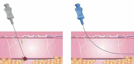 Fig 1. Needle transfixing a vessel versus cannula displacing it. (Lobo, 2022, p.163).
> Fuentes: Lobo, Harmonização Orofacial [2022], p.163
Fig 1. Needle transfixing a vessel versus cannula displacing it. (Lobo, 2022, p.163).
> Fuentes: Lobo, Harmonização Orofacial [2022], p.163 [C] [MEDLIB].
Why the 25G line matters. Below 25G a cannula is thin and flexible enough to behave like a needle: it can bend into an unintended layer, and its tip can no longer reliably discriminate a fibrous septum from a vessel wall. Hong documents the practical consequence in vitro: with a 27G/13 mm needle blood aspirated in 1 second, whereas with a 25G/40 mm cannula blood did not aspirate even after 10 seconds, so the same maneuver that "reassures" on a cannula is precisely where it fails [13].
Length. Length is chosen so the entry point sits outside the target and the deposit sits on it: a 38 mm cannula from a lateral cheek port reaches the medial cheek without a second puncture; a 50–70 mm cannula covers a jawline in one pass. A long thin cannula that whips at the tip has lost the plane before the product leaves the port. For a supraperiosteal bolus, a short 27G needle placed straight down to bone is more precise than a flexible cannula that may deposit off-periosteum [12][14].
Why the tool ranks third
Training tends to open with the instrument (needle or cannula), but the instrument is the third safety variable, after the plane and the pressure/volume, and treating it as the first is how the cannula acquired a reputation as a safety device it does not earn [1][5][6]. The instrument sets access geometry: a needle gives a short perpendicular path and a precise on-bone depot; a cannula gives a long tangential path from one port and fewer punctures. Neither changes the two facts that decide whether an injection blinds a patient: which plane the tip is in, and how much product at what pressure is delivered if that tip is intraluminal. A cannula in the wrong plane at high pressure is as dangerous as a needle; a needle in the right plane at low pressure and low volume is as safe as technique gets. The tool is chosen after the plane and the movement are decided, to serve them, and that ordering is the correction this chapter makes to the usual teaching sequence [1][12].
Cannula length and hub, chosen so the entry sits off-target
Length is selected so the entry point sits outside the treatment area and the deposit sits on it, minimizing punctures over a field:
| Length | Reach | Typical field |
|---|---|---|
| 38 mm (25–27G) | Lateral cheek to medial cheek in one pass | Midface, tear trough via a lateral port |
| 50 mm (22–25G) | One port covers a hemi-jawline or a nasolabial run | Jawline, nasolabial fold, marionette |
| 70 mm (22G) | Long single-port runs | Jaw, temple sweeps, neck |
| 70–100 mm (18–21G) | Body fields | Body contour, biostimulator volume |
A cannula that whips at the tip has lost the plane before the product leaves the port, so an over-long thin cannula for a dense product is the wrong tool twice: it bends and it forces the pressure up [13][14]. The luer-lock hub matters more with cannulas and dense products than injectors expect: under the pressure a high-G' product needs, a slip hub can separate and spray, so a locking hub is a safety item, not a preference [12]. For a supraperiosteal point-depot on bone, a short 27G needle placed straight down beats a flexible cannula that may deposit off-periosteum, which is the geometry the whole plane-precision row of the needle-vs-cannula table encodes [1][14].
Matching product rheology to bore and syringe
The bore is chosen by the product's mechanical class, not by habit. The variables that decide flow are G-prime (elastic modulus, stiffness), cohesivity and particle size; a high-G' volumizer needs a wider bore or it will not extrude at safe pressure.
| Product class | Example rheology | Typical bore | Plane / use |
|---|---|---|---|
| High-G' structural HA | 20 mg/mL, high cross-link, high lift (e.g. Voluma-class, Lyft-class) | 27G needle or 25G cannula | Supraperiosteal bolus, deep cheek/chin [14] |
| Mid-G' versatile HA | balanced lift and spread | 27–30G needle, 25G cannula | Subcutaneous, folds [14] |
| Low-G' soft HA | low lift, high spread (lip/fine-line class: Volbella, Refyne, Belotero) | 30–32G needle | Superficial lip, fine rhytides, dermis [15] |
| Calcium hydroxylapatite | biostimulator, thick | 25–27G cannula (often diluted/hyperdiluted) | Subdermal support, body |
| Poly-L-lactic acid | suspension, biostimulator | 25–26G needle/cannula, multi-plane | Cross-hatch/fanning, diffuse [15] |
Syringe mechanics. Pressure at the tip is force divided by plunger area, so the narrow plunger of a 1 mL syringe multiplies thumb force far above a 3 mL barrel; the same thumb pressure that is gentle on a 3 mL syringe is an embolic hazard on a 1 mL syringe with a fine needle and a stiff product [12]. A luer-lock hub prevents the needle blowing off under the pressure a viscous product needs; a slip hub can disconnect and spray. The practical rule: if a product will not flow under gentle pressure, the answer is a wider bore or a larger barrel, never a harder push, because the extra push is invisible pressure that only matters when the tip is intraluminal [12][13]. When a danger zone forces a small syringe for control, treat the reduced pressure margin as a reason to slow down and shrink the aliquot, not to push [12].
The cannula-versus-needle safety conflict, printed whole, not averaged ⚠
This is a live controversy. The two schools are kept with their scenarios because they measure different things and the question is not closed.
Consensus (what both sides do): minimize the number of dermal punctures, inject small aliquots, keep pressure low, and never treat the tool as a substitute for anatomy [5][12].
Discrepancy: the cannula-safer school [12]: - Fewer skin penetrations lower biofilm and infection risk, which rises (exponentially) with puncture count [12]. - A blunt, flexible tip tends to push vessels aside rather than transect them. - Preference: cannula ≥25G in high-risk facial zones. - Cadaver evidence (van Loghem, Humzah, Kerscher 2018): with a cannula the product stayed confined to the deep target plane, whereas with a sharp needle the tinted material appeared across multiple anatomical layers because it travelled retrograde up the needle tract from the periosteum toward superficial layers [1].
Discrepancy: the false-safety school [5][6]: - Zhou 2021: of 28 severe HA embolism cases (9 blindness, 1 blindness with stroke, 18 large-area necrosis), 25 of 28 were injected with a cannula (22–27G). The CMAC board consensus that followed: avoid cannulas <25G [5]. - Tansatit 2017 ("a dark side of the cannula"): a cannula can penetrate an arterial wall; a perpendicular artery-to-cannula angle is a key setup for injury, and during a blind insertion the injector cannot discriminate the resistance of a fibrous septum from the resistance of an artery at the cannula tip. Corollary: keep the cannula trajectory away from the region's main artery [6]. - Case evidence: fatal cerebral infarction and ophthalmic-artery occlusion after nasal HA delivered through a 25G blunt cannula (2 mL, unlicensed operator) [26].
Adjudication (P): the conflict does not resolve, it locates. The cadaver study measures tract and layering across multiple zones with tinted product; Zhou measures the tool used in a case series of catastrophes; Tansatit measures the mechanics of wall perforation. All three are valid in their own frame. What follows for the chair: - On long deep multi-zone runs, the cannula holds the plane and the needle seeds product along its withdrawal tract [1]. - In danger zones, fewer punctures and vessel displacement are real advantages, if the cannula is ≥25G and its trajectory avoids the named artery [5][6]. - The most important cadaver finding is the one that dismantles the reassuring gesture: intra-arterial injection occurred with the needle tip resting on periosteum, under constant bone contact [1]. Bone contact is not a guarantee. Neither is the cannula.
What each school actually chooses (and why): - MD Codes / deep-structural (de Maio, Beut, Surek): sharp needle for supraperiosteal point-depots on bone (Ck1, Ck2, chin, angle), cannula for the subcutaneous transitions; the point-based system is built around precise bone-contact boluses [11]. - Multilayering / superficial redensification (Rosso MLT): one entry port, cannula through three planes in one pass, favouring the blunt tip for the sweep across structural, dynamic and superficial layers. - Tear-trough dissent (Spada 2023): in a split-face study (10 patients, CPM HA 22.5 mg/mL, 25G cannula one side vs 31G needle the other, ultrasound/MRI follow-up to 365 days) the cannula produced less bruising but deposited more superficially and less precisely, while the fine 31G needle was more precise with minimal trauma; the difference washed out over time [4]. A single-zone, very-fine-needle-versus-thick-cannula comparison, and its scenario must travel with its conclusion.
Classic pitfall: reaching for a 27G/30G cannula "to be gentle" on a viscous product, then pushing harder because it will not flow. The thin cannula bends off-plane and the raised plunger pressure is exactly the embolic setup the cannula was chosen to avoid. Clinical signature: unexpected resistance, a deposit that ends up shallow (visible/palpable), and more force on the thumb than the product should need. Fix: match the bore to the product, or switch to a needle for the point depot [13][14].
C1.3 · Injection planes: identification by touch, by depth, and by ultrasound
Answer first: the plane map, deep to superficial.
| Plane | Tactile reference | What goes there | Dominant risk |
|---|---|---|---|
| Supraperiosteal (layer 5/7) | Firm bone contact | Structural volume: malar, chin, jaw, piriform, deep temporal | Arteries that in some points run on periosteum; generally the safer deep plane [7] |
| Deep fat compartments (layer 4) | Low resistance, no bone contact | Mid volume: deep medial cheek, deep pyriform, medial/lateral SOOF | Vascular; little tactile reference [8][9] |
| SMAS / musculoaponeurotic (layer 3) | Elastic resistance | Rarely a filler target plane; threads and toxin live here | Facial nerve; vascular |
| Superficial fat (layer 2) | Soft, mobile | Transitions, blending, folds | Irregularity, migration |
| Subdermal | Just under dermis; relief visible | Edge definition, contour, lines | Visibility, nodule, Tyndall |
| Intradermal | High resistance, white papule | Skinboosters, micropapules, fern pattern, fine lines | Persistent papule, Tyndall, biofilm |
The rule that orders the whole plane discussion: inject deep on bone or superficial in dermis; the intermediate territory is where the arteries live, and it is worked only when the regional anatomy allows it and you know what is inside it [7][12].
Deep injections first: the biomechanical principle
Freytag/Cotofana 2022 distil three governing principles: upper face first, lateral face first, deep injections first [7]. The third is the load-bearing one for plane selection. Deep facial fat compartments attach to bone through the retaining ligaments and move less on animation than the superficial compartments, which descend with age. Placing product in the deep (supraperiosteal) plane first rebuilds the bony fundament and lets the overlying soft tissue reposition and glide naturally, without the filler interfering with expression [7][8]. Two consequences follow:
- Aesthetic: volumizing deeply improves fine etched rhytides that sit over deep volume deficit, so less superficial (intradermal) product is needed and the result looks more natural in motion [7].
- Safety: the supraperiosteal plane is in general safer to inject because the facial arterial network mostly runs in more superficial planes (with named regional exceptions such as the supratrochlear/dorsal-nasal system, and points where an artery crosses onto periosteum) [7][9].
Fig 2 shows the scaffold the principle rests on: the deep fat compartment (layer 4) is marked "1st", treated before the superficial fat (layer 2) and across the SMAS (layer 3), to rebuild the bony fundament under the mobile soft tissue.
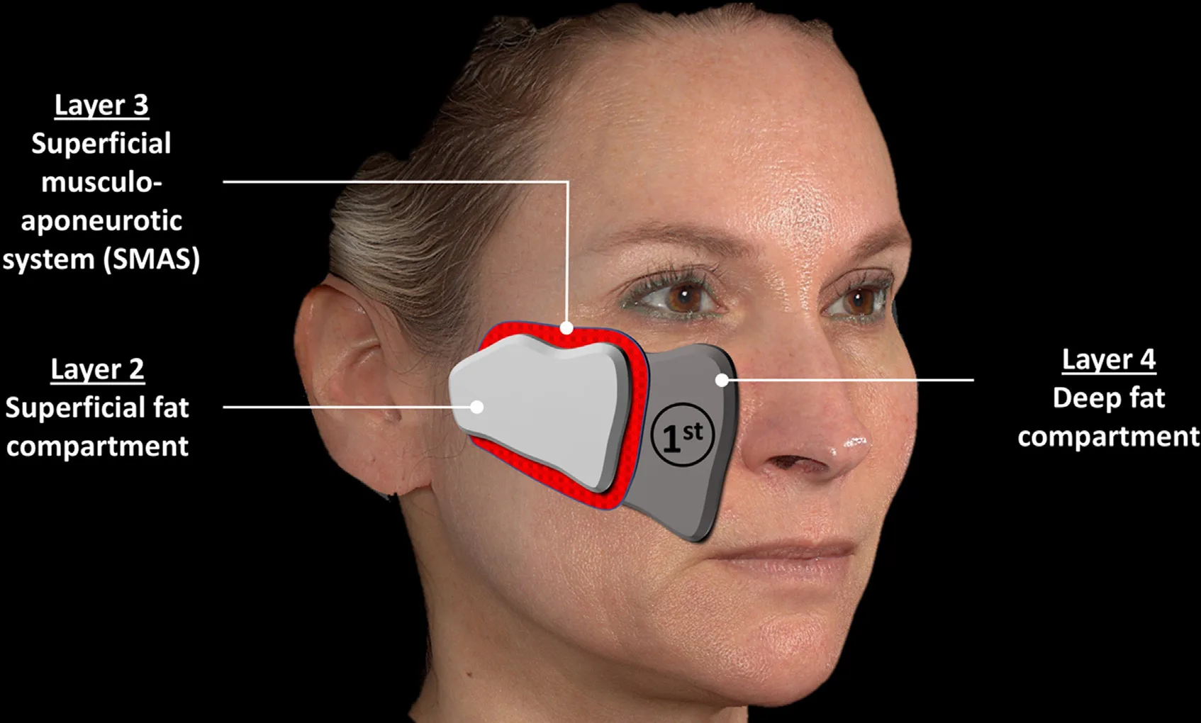 Fig 2. The deep fat compartment (layer 4) is treated first. (Freytag, 2022, p.6).
> Fuentes: Freytag, Understanding Facial Aging Through Facial Biomechanics [2022], p.6
Fig 2. The deep fat compartment (layer 4) is treated first. (Freytag, 2022, p.6).
> Fuentes: Freytag, Understanding Facial Aging Through Facial Biomechanics [2022], p.6 [B].
The five-layer scaffold this rests on (skin, subcutaneous fat, SMAS, deep areolar/deep fat, periosteum) is constant across the face; the variability is in the thickness of each layer and in where deep compartments sit (deep medial cheek, deep pyriform space medial to the infraorbital foramen, medial/lateral SOOF) [8][9][10]. Detail in A1 — Facial & Regional Anatomy.en.
Reading resistance: what each change at the tip means
Resistance is the injector's primary feedback, and each kind means something specific [12]: - A firm stop is bone: the supraperiosteal plane, the target for structural depots. - A sudden rise in injection resistance where the product was flowing means the tip has risen a plane (into dermis) or entered fibrosis or prior product; keep injecting and you deposit too superficial (Tyndall) or into a scar. - Free fanning mobility confirms the subcutaneous plane; a tip that will not fan is in dermis or under a septum. - The pop through a fascial layer is felt as a give, and the borders of the superficial cheek compartments are felt as the resistance of the vascularized septa dividing them [8]. - A dull periosteal ache confirms depth; a sharp sting confirms dermis.
The trap is that all five readings are calibrated to virgin tissue. Over prior product and fibrosis the same needle reports a plane it is not in, which is why the touch-up is the setting where an objective check (ultrasound) earns the most and where the operator must most distrust the feel [12]. When resistance and expectation disagree, believe the resistance and stop, rather than pushing through what feels wrong.
Plane by region: the default, and the exception that bites
| Region | Default structural plane | Superficial refinement | Danger-plane exception |
|---|---|---|---|
| Malar / zygoma | Supraperiosteal bolus [9] | Subdermal fan for texture [9] | Transverse facial artery laterally |
| Chin / jaw | Supraperiosteal on bone | Subcutaneous blend | Facial artery at antegonial notch |
| Tear trough | Deep, on the orbital rim, low volume [4] | Avoid superficial (Tyndall, swelling) [14] | Angular vessels; hydrophilic HA swells |
| Temple | Deep supraperiosteal microbolus, or subdermal cannula [33] | Subdermal fan | Sentinel vein; intracranial if force >4 kg [14] |
| Nose | Midline supraperiosteal microdroplets [11] | None (high risk) | Dorsal nasal/lateral nasal to ophthalmic: blindness [26] |
| Glabella | Avoid or dermal microdroplets only | None | Supratrochlear/supraorbital to ophthalmic [13] |
| Lips | Submucosal body, vermilion edge | Serial puncture at the border [15] | Labial arteries in the wet-dry junction |
| Nasolabial fold | Deep at the piriform + subdermal thread | Subdermal retrograde [15] | Facial/angular artery ascending the fold |
The table encodes the governing rule: structural planes are deep on bone or in the deep fat, refinement is intradermal/subdermal, and the named artery is the reason a region has a plane it must not be injected into. The nose and glabela are the two regions where the "intermediate territory" rule is absolute, because their vessels drain to the ophthalmic artery and a wrong plane there blinds [13][26]. Detail per region in the D-series chapters.
Recognizing the plane without seeing it: five tactile signs, in order of reliability
- Resistance to advance. Bone stops the tip; fat offers almost nothing; dermis demands force.
- Resistance to injection. A product that should flow suddenly resists: you have risen a plane or entered fibrosis.
- Tip mobility. In the subcutaneous plane a cannula fans freely; in dermis it does not. During cannula runs the borders of the superficial medial and middle cheek compartments are felt as the resistance of the vascularized septa dividing them [8].
- Skin appearance. Blanching or a papule means superficial. Diffuse elevation means deep.
- Pain. Periosteum gives a characteristic dull ache; dermis, a sharp sting.
⚠ Touch-up over prior product: residual filler and fibrosis change the resistance and falsify all five signs. This is one of the most frequent causes of a deposit in the wrong plane, and it is almost never taught. The objective check is ultrasound (§C1.5): map what is already there before adding [12].
The regional exceptions to "deep is safer"
The supraperiosteal-is-safer rule holds in general because the facial arteries mostly run in more superficial planes, but it has named exceptions where an artery crosses onto or near periosteum, and those exceptions are where deep injection still embolizes [7][9]:
- Glabella / medial brow: the supratrochlear and supraorbital arteries emerge from their foramina and run cephalad in a plane that becomes superficial quickly; a "deep" glabellar injection is still an ophthalmic-artery danger [13].
- Nasal dorsum / tip: the dorsal nasal and lateral nasal arteries and prior-rhinoplasty scarring make even a midline supraperiosteal plane hazardous; the nose is the region where "deep" does not mean "safe" [26].
- Temple: the deep temporal plane is relatively avascular on the bone, but the middle temporal vein (sentinel vein) and the intracranial risk above the pterion (force >4 kg) make it a low-force decision, not a confident bolus [14][33].
- Lateral cheek: the transverse facial artery runs in the mid-lateral cheek and can be reached by a deep lateral pass.
The cadaver evidence closes the argument: intra-arterial injection occurred with the needle tip on periosteum under constant bone contact [1]. "Deep on bone" lowers the average risk; it never zeroes it, and in these regions it barely lowers it. Detailed vessel maps are in A1 — Facial & Regional Anatomy.en and the danger-zone chapter.
Depth by geometry
Plane is also read from needle length and angle. A 13 mm needle held perpendicular reaches bone in the malar eminence; angled shallow it lands in dermis (and injects a wheal). A subdermal wheal that tents and blanches confirms a superficial plane; a deposit that spreads with no surface change confirms a deep one. For the temple, note the intracranial trap: needle force >4 kg may penetrate the pterion into the intracranial space, so the temple is a low-force, defined-access-point plane decision, not a push [14].
Consensus: deep structural volume goes supraperiosteal; skin-quality and fine-line work goes intradermal/subdermal; the intermediate planes are entered deliberately and regionally, never by drift [7][12]. Discrepancy: deep-support-first vs multilayering: the deep-structural school (MD Codes, Beut/Surek prezygomatic, Cotofana/Freytag) builds the supraperiosteal and deep-fat scaffold before any superficial work [7][11]; the multilayering school (Rosso MLT) treats three planes in one session from one port, adding superficial redensification alongside structure. Decide by: skin condition (poor texture favours added subdermal fanning) and the amount of deep deficit [9]. Never average the plane; a bolus split between two planes is a deposit in neither.
Classic pitfall: treating "subcutaneous" as one plane. The superficial and deep fat are separated by SMAS/platysma and behave differently (the deep compartments are ligament-bound and stable; the superficial ones descend). Product meant for deep support placed in superficial fat migrates and can worsen a fold (the nasolabial and jowl compartments are specifically not targeted a priori because filling them worsens the fold) [7][8]. Signature: a result that looked right on the table and slid within weeks, or an accentuated fold. Fix: confirm depth by resistance and, in a touch-up, by ultrasound.
C1.4 · Deposition movements, one by one
Answer first: the movement catalog with indication and risk.
| Movement | How | Where | Risk |
|---|---|---|---|
| Bolus / depot | Single static deposit, on bone | Structural volume: malar, chin, angle | Highest embolic risk per volume at one point [12] |
| Microbolus / microdroplet | Very small repeated deposits (<0.1 mL) | Lower-risk alternative to a bolus; forehead, temple | Low, if per-point volume is truly small [12] |
| Retrograde linear threading | Inject on withdrawal | Folds, lines, contour | Low–moderate; the anterograde version is the dangerous one [15] |
| Anterograde threading | Inject while advancing | Reserved: vermilion border only | Higher; product pushed ahead of the tip [15] |
| Fanning | Several threads from one entry point | Wide surfaces, cheek, temple | Moderate; depends on plane [15][34] |
| Cross-hatching / mesh | Two perpendicular planes of threads | Surface support, body, diffuse volume | Moderate; a lattice for PLLA/biostimulator [15] |
| Serial puncture | Small contiguous depots along a line | Vermilion, fine rhytides, glabella, philtral columns | Low; beading if spacing fails; more bruising [15][34] |
| Cross-tunnel depot | Moderate volume in a deep plane | Biostimulators, cheek mounds, temporal hollows, scars | Moderate; nodule if the plane is superficial [15] |
| Tower / column | Vertical deposit crossing planes on withdrawal | Chin, deep lines | Moderate–high; seeds product along the tract |
Fig 3 is the movement catalog in a single plate: anterograde and retrograde linear threading (top, by arrow direction), a single depot/bolus (middle-left), serial puncture as a row of small deposits with repositioning (middle-right), radial fanning (bottom-left), and cross-hatching (bottom-right).
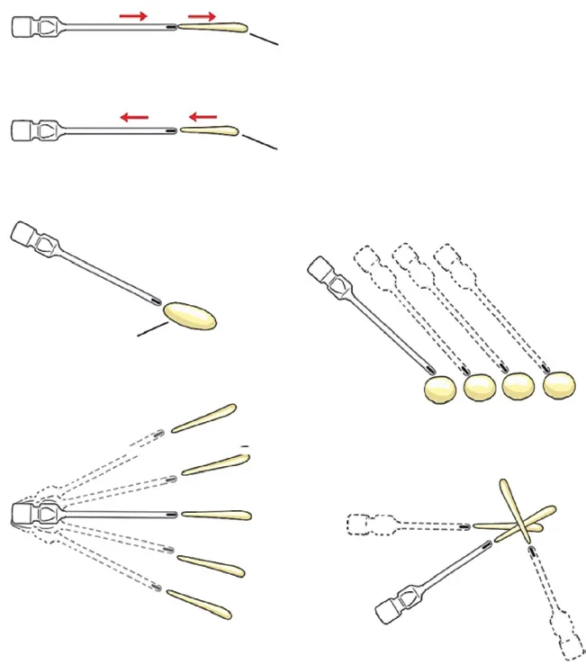 Fig 3. Various injection techniques, panel by panel. (Towne, 2019, p.109).
> Fuentes: Towne, Neurotoxins and Fillers in Facial Esthetic Surgery [2019], p.109
Fig 3. Various injection techniques, panel by panel. (Towne, 2019, p.109).
> Fuentes: Towne, Neurotoxins and Fillers in Facial Esthetic Surgery [2019], p.109 [C] [MEDLIB].
One by one, with the indication
Bolus (depot). A single aliquot placed and left, classically supraperiosteal on bone for structural support (MD Code point work). It carries the most embolic risk of any movement because it concentrates volume and pressure at one point; mitigate with the ≤0.025 mL ceiling per bolus, low pressure, and, in danger zones, manual arterial compression proximal to the point [12]. Andrews' cheek approach illustrates the sequence: if skin is good, multiple small depot injections first in the supraperiosteal plane, then reassess for contour before any subdermal work [9].
Fig 4 shows the bolus in practice: tinted product extruded at the needle tip (top), then a deep depot deposited and molded to shape (bottom, arrows).
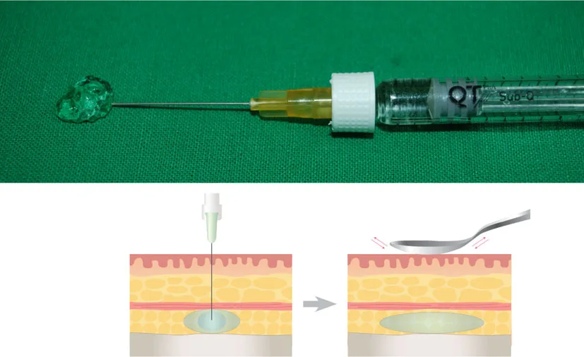 Fig 4. Bolus deposited in the deep plane and then molded. (Hong, 2020, p.97).
> Fuentes: Hong, Art and Science of Filler Injection [2020], p.97
Fig 4. Bolus deposited in the deep plane and then molded. (Hong, 2020, p.97).
> Fuentes: Hong, Art and Science of Filler Injection [2020], p.97 [C] [MEDLIB].
Microbolus / microdroplet (<0.1 mL). The lower-risk substitute for a bolus: the same territory built in many tiny deposits so no single point holds enough volume to embolize a critical vessel. First choice in the forehead and temple, where the plane is thin and the vessels superficial [12].
Retrograde linear threading. The needle or cannula is advanced to the far end of the target and product is laid down only on withdrawal, so the tip is always moving away from the deposit. This is the default safe movement: the preference for retrograde over anterograde is based on lower intravascular risk, because injecting while withdrawing keeps the moving tip from ramming product into a vessel lumen ahead of it [15]. Maximum 0.025 mL per retrograde thread in danger zones [12].
Anterograde threading. Injecting while advancing pushes a bolus of product ahead of the tip and is reserved for a single indication where it is the correct choice: the vermilion border, where a controlled advance defines the lip edge. Everywhere else it is the movement to avoid [15].
Fanning. Multiple threads radiating from one entry point cover a wide surface with one puncture. Useful for the cheek and for diffuse subdermal texture work (overlapping subdermal fans when skin needs texture improvement) [9][15]. It does not lend itself to aspiration, because the tip changes position between passes; this is one reason the aspiration debate is moot for fanning (§C1.8) [6].
Cross-hatching / mesh. Two sets of threads laid perpendicular to each other build a lattice that distributes volume and supports a surface; the technique of choice for diffuse enhancement and for poly-L-lactic acid placed across multiple planes [15]. Like fanning, it cannot be aspirated meaningfully.
Serial puncture. Small depots placed contiguously along a line or fold, so the operator can put an exact volume at exact points; best for fine wrinkles (glabella, philtral columns, nasolabial folds) and the vermilion. The trade-offs: discontinuous "beads" of product that can be palpable or visible if spacing is wrong, and more bruising from the higher number of punctures. Advocates argue a lower chance of entering a vessel because the needle is not passed through long subdermal distances [15][34][41].
Cross-tunnel depot. A moderate depot in a deep plane used to raise depressed scars or fill cheek mounds and temporal hollows; nodule risk if the plane is too superficial [15].
Tower / column. A vertical deposit that crosses planes as the needle withdraws, used for the chin and deep lines. Because it seeds product along the whole tract it is a moderate-to-high-risk movement and is chosen deliberately, not by default.
Precision, bruising and safety by movement
| Movement | Precision | Bruising | Intravascular risk | Aspiration possible |
|---|---|---|---|---|
| Bolus / depot | High at one point | Low (few passes) | Highest per point | Yes (fixed needle) [2] |
| Microbolus | High, distributed | Low | Low | Partly |
| Retrograde thread | Moderate | Moderate | Low–moderate | No (tip moving) [6] |
| Anterograde thread | Moderate | Moderate | Higher (product ahead of tip) | No [6][15] |
| Fanning | Moderate | Moderate–high | Moderate | No (tip repositions) [6] |
| Cross-hatch | Diffuse | Higher | Moderate | No |
| Serial puncture | Very high (point control) | Higher (many sticks) | Low (short passes) | Impractical |
The grid explains why the safest movements (retrograde, microbolus) are also the ones where aspiration is useless: the tip is either moving or the volume is already sub-critical, so the maneuver that "reassures" adds nothing to the movements that actually protect [2][6]. It also explains the serial-puncture trade: maximal point control and low intravascular risk (short passes), paid for in bruising from the number of sticks. Movement is chosen for the deposit shape the region needs, then made safe by volume and pressure, not by adding an aspiration step that the movement cannot support [6][15].
Movement and volume ceiling by region
| Region | Preferred movement | Volume discipline | Why |
|---|---|---|---|
| Nose / glabella | Micro-droplets, retrograde | ≤0.05 mL per point, ≤0.025 mL danger-zone bolus | Ophthalmic-artery embolus blinds at 0.04–0.12 mL [12][13] |
| Temple | Deep supraperiosteal microbolus or subdermal fan | Small per-point; low force (>4 kg penetrates bone) | Sentinel vein, intracranial risk [14][33] |
| Cheek | Supraperiosteal depot + subdermal fan | Staged; reassess contour before adding | Deep-first builds the fundament [9] |
| Tear trough | Retrograde thread on the rim | Very low; avoid superficial | Hydrophilic HA swells, Tyndall [4][14] |
| Lips | Vermilion serial puncture; body retrograde | Small aliquots, symmetric | Beading and labial-artery risk [15] |
| Nasolabial fold | Retrograde subdermal + deep piriform support | Retrograde only | Facial/angular artery ascends the fold [15] |
| Jawline / chin | Supraperiosteal bolus; tower for chin projection | Moderate per point | On-bone, fewer vessels [12] |
The pattern the table encodes: the higher the vascular stakes, the smaller the aliquot and the more the movement shifts from bolus to micro-droplet, until in the nose and glabella the only acceptable delivery is a moving micro-droplet under the 0.025 mL ceiling [12][13]. Volume is a safety parameter, not only an aesthetic one.
The rule that crosses the whole table
> Inject on withdrawal, not on advance; slowly; small volume; low pressure; and moving.
The reasoning is mechanical, not dogmatic. Embolization requires pressure sufficient to overcome arterial pressure retrograde and volume sufficient to reach a critical territory. Injecting while moving, at low pressure, means that even with an intraluminal tip the volume introduced before you notice anything is minimal. A static bolus under pressure does the opposite [12].
The consequence that is routinely forgotten: the plunger of a 1 mL syringe generates far more pressure per unit of thumb force than a 3 mL syringe. Small syringe + dense product + fine needle = high pressure without you feeling it. If a danger-zone injection needs a small syringe for control, it also needs deliberate awareness that the pressure margin has shrunk [12][14].
Classic pitfall: a static high-pressure bolus because it is fast and yields volume. It is the embolic scenario par excellence. Signature: a large depot delivered in one push, often with the operator "finishing the syringe". Fix: microboluses in motion, ≤0.025 mL per point in danger zones, and stop at the first abnormal sign rather than emptying the syringe [12].
C1.5 · Ultrasound guidance: when it changes the decision, and when it is theater
Answer first: what ultrasound is for, ranked by immediate yield.
| Use | What it changes | Yield today |
|---|---|---|
| Locate prior product | Know what is there, where, and what type before adding or dissolving | Highest; easy and immediate [12] |
| Guide hyaluronidase | Deliver the enzyme into the deposit or the occluded vessel, not blind | High; less enzyme, faster resolution [12][24] |
| Diagnose a nodule | Anechoic vs hypoechoic vs hyperechoic vs vascular, with or without flow | High; separates biofilm/granuloma/vascular from HA lump [12][24] |
| Pre-treatment vessel mapping | See this patient's artery in a danger zone before injecting | High in nose/temple/glabella/piriform [24][27] |
| Real-time guided injection | Track the tip and deposition across planes live | Contested as routine; a two-hand skill and the last to learn [29] |
The honest split: ultrasound reduces anatomical uncertainty; it does not remove vascular risk. An operator with a probe and no judgement can still inject into an artery. Detail in G6 — Facial Ultrasound (Diagnostic & Guided).en.
Equipment
High-frequency linear probe: 18–22 MHz resolves dermis and subdermis; 15 MHz reaches deep planes and is what most portable devices provide. Color Doppler is mandatory to identify flow and to distinguish a vessel from a duct or a septum [27][28]. Point-of-care handheld devices ("the stethoscope of the future") have driven adoption because they are small, portable and cheap enough to sit beside the injector [12].
Fig 5 shows the tool the workflow depends on: a portable point-of-care ultrasound console with linear probes, small enough to sit beside the injector.
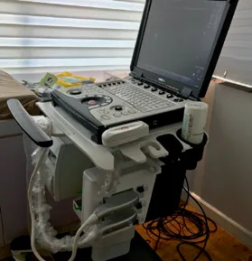 Fig 5. Portable point-of-care ultrasound unit. (Miedany, 2022, p.301).
> Fuentes: Miedany, Musculoskeletal Ultrasound-Guided Regenerative Medicine [2022], p.301
Fig 5. Portable point-of-care ultrasound unit. (Miedany, 2022, p.301).
> Fuentes: Miedany, Musculoskeletal Ultrasound-Guided Regenerative Medicine [2022], p.301 [C] [MEDLIB].
Reading the image: echogenicity
| Echo pattern | Appearance | Structures / products |
|---|---|---|
| Anechoic (black) | No reflection | Water, blood, hydrophilic HA deposits [12][24] |
| Hypoechoic (grey) | Some reflection | Muscle, fat, HA [24] |
| Hyperechoic (white) | Strong reflection, may shadow | Bone, calcium hydroxylapatite, air bubbles, PMMA granules [12][24] |
| Doppler signal | Movement | Blood flow, and probe motion itself [24] |
Fillers split into hydrophilic (HA, polyacrylamide/Aquamid, polyalkylimide/Bio-Alcamid) which read anechoic-to-hypoechoic, and hydrophobic (liquid silicone, PMMA/Artecoll, poly-caprolactone/Ellanse) which read hyperechoic; CaHA (Radiesse) and PLLA (Sculptra) are mixed [12]. A PMMA deposit shows a heterogeneous hyperechoic band with posterior shadowing, distinct from the clean anechoic pockets of HA [12].
Ultrasound-guided hyaluronidase, the highest-value intervention
The workflow van Loghem codifies at the Erasmus MC filler-complication clinic: after identifying the causative HA accumulation on ultrasound, inject hyaluronidase under ultrasound guidance directly into the substance; success in almost every case at 15–75 units per session, because delivering the enzyme exactly into the HA has a far better effect than injecting "here and there" blind [12].
For arterial occlusion, the monitor shows dilated collateral arteries, an image nicknamed the "Medusa head"; the anechoic HA sits at its center. Injecting that spot with 50–100 units, sometimes repeated once or twice after 15 and 30 minutes, almost invariably restores flow; more than one artery is often blocked because fragments travel distally, so the whole downstream course must be followed [12].
The 2025 complications roundtable (Fabi, Desyatnikova, Dayan) frames the practice numbers: ultrasound-guided hyaluronidase uses less time and less enzyme than blind flooding; stock ≥10 vials or >1500 units; a targeted vascular-occlusion injection of 150–200 IU with 0.25 mL of 1–2% lidocaine (lidocaine without epinephrine for vasodilation); a single early ultrasound-guided injection may prevent necrosis [24]. The needle must be thick enough to be seen: 25G or lower is recommended for ultrasound-guided delivery [12]. Cross-reference the vascular emergency protocol in J2 — Vascular Occlusion & Emergency Response.en and the high-dose pulsed protocol (DeLorenzi) in §Safety [25].
Probe technique and the nodule differential
In-plane vs out-of-plane. In-plane, the needle runs along the probe's long axis and the whole shaft is visible as a bright line; out-of-plane, the needle crosses the beam and appears only as a moving bright dot at the point of intersection. In-plane gives shaft-and-tip control for a threading approach; out-of-plane suits a perpendicular depot. Either way the operator confirms the tip, not the shaft, because a shaft echo can be mistaken for a tip one plane too deep [29]. A straight physician-to-monitor-to-patient sightline is a precondition, not a nicety [12].
The nodule differential on ultrasound, the daily reason the probe earns its cost:
| Finding | Echo pattern | Doppler | Reading |
|---|---|---|---|
| HA deposit | Anechoic to hypoechoic pocket | None | Reversible with hyaluronidase [12] |
| CaHA / PMMA | Hyperechoic, posterior shadow / granular band | None | Not HA-reversible [12] |
| Inflammatory / biofilm nodule | Hypoechoic, ill-defined, may be vascularized | Increased peripheral flow | Do not reflex-steroid; treat as infective [24][31][32] |
| Vascular occlusion | Anechoic filler beside a distended artery ("Medusa head") | Collateral flow, blocked segment | Ultrasound-guided hyaluronidase now [12][24] |
| Foreign-body granuloma | Heterogeneous, ill-defined | Variable | High-frequency US separates it from a simple deposit [32] |
High-frequency ultrasound distinguishes a nodular filler deposit from a foreign-body granuloma and maps palpable nodules for targeted treatment, which changes the management (dissolve vs anti-inflammatory vs excise) rather than guessing [31][32].
The learning curve, stated honestly
Adoption is slow because the curve is real. The order that works [12][29]: 1. Normal anatomy on your own face and colleagues' until layers are identified without hesitation. 2. Identify prior product, which is easy and immediately convincing. 3. Doppler and vessels: map the facial and angular arteries, whose course, diameter and depth vary widely between patients [27][30]. 4. Guide hyaluronidase into a known deposit. 5. Real-time guided injection, last, because it demands two coordinated hands and a straight physician-to-monitor-to-patient line of sight [12][29].
Pre-treatment mapping: the workflow that pays off before the needle
The single most transferable ultrasound habit for a general injector is not real-time guidance, it is the five-minute pre-treatment scan in a danger zone [24][27]:
- Map the artery in this patient. Facial-artery course, diameter and depth vary widely between individuals, so the atlas mean is a starting point, not a location; Doppler shows where the vessel actually is today [30][35]. Angiographic series confirm the variability the probe reveals (variant facial-artery anatomy across hundreds of cases) [35].
- Detect prior product. Many patients do not recall or will deny previous filler; the scan converts that into an honest conversation and a real risk assessment before adding or dissolving [12].
- Type the prior product. Anechoic/hypoechoic points to HA (reversible); a hyperechoic granular band points to a non-HA or permanent product (not HA-reversible), which changes the plan entirely [12].
- Plan the plane. With the vessel and any prior deposit located, choose an entry and a plane that keep the tip away from both.
DERMUS-group guidance frames how to perform a dermatologic ultrasound examination reproducibly, which matters because an unstandardized scan is as misleading as no scan [39]. The yield is asymmetric: the scan costs minutes and the miss it prevents is a vascular catastrophe, which is why the four highest-value situations (unknown prior filler, nodule to characterize, danger zone, any hyaluronidase dissolution) are exactly the ones where a pre-treatment scan changes the plan [12][24].
Scan-before vs scan-while: where the field is moving
The corpus documents ultrasound as a complication-diagnosis and pre-treatment-mapping tool [12][24][27][29]. The frontier, formalized in recent upper-face best-practice consensus work, is a codified "scan-while-injecting" workflow: tracking the needle/cannula tip and filler deposition live across subdermal, interfascial and supraperiosteal planes (temple, glabella, forehead, supraorbital) with in-plane versus out-of-plane probe positioning. [MATERIAL GAP] The specific 2024 upper-face scan-while-injecting consensus is not in the own corpus (a declared corpus-acquisition gap from the C1 scope scout); the practice is bounded here by the Doppler-guided-injection literature that is held (Vasconcelos-Berg's "useful tips to integrate ultrasound in daily practice"; Lee Won's Doppler-guided HA injection) [28][29]. Treat routine real-time guidance of every injection as an emerging standard, not an established one.
Cost, availability and the realistic adoption path
The honest barrier is not evidence, it is cost, availability and the learning curve. Portable point-of-care units have lowered the price and the footprint enough to sit in a treatment room, but they are still absent from many practices where HA is injected, so a protocol that mandates real-time guidance for every injection is not yet realistic [24]. The realistic path is staged: acquire the skill on normal anatomy and prior-product detection first (immediate value, low cost in time), reserve the device for the four high-yield situations, and treat real-time guidance as the last competency to build. A clinic that injects danger zones without any access to ultrasound should at minimum know where to refer a suspected occlusion for ultrasound-guided hyaluronidase, because a single early guided injection can prevent necrosis [24][25]. The trajectory is clear even if the present is uneven: imaging is where the technique of this field will change most over the next decade.
Consensus: ultrasound is high-value for prior-product detection, nodule characterization, danger-zone vessel mapping, and guiding hyaluronidase [12][24][27][29]. Discrepancy: whether real-time guidance should be routine for every injection. Pro (Vasconcelos-Berg, Lee Won): integrating Doppler into daily practice raises safety and precision [28][29]. Against (pragmatic): cost, the two-hand learning curve, and device availability make it selective today [24]. Decide by: zone risk and operator skill, not by owning the machine.
Classic pitfall: the probe as reassurance theater. Scanning a low-risk cheek to "look modern" while skipping the map on a nose, or reading anechoic HA as "a vessel" (both are dark) without Doppler to confirm flow. Signature: time spent scanning where nothing is at stake and none where everything is; or a misread static anechoic pocket. Fix: use Doppler to prove flow, map only the danger zones and prior-product cases, and remember the machine informs the decision, it does not make it [24][27].
C1.6 · Anaesthesia and comfort
The real objective is not perfect analgesia: it is a still patient. A patient who moves is a safety problem, not a comfort problem. Everything below serves that first [16][17].
Comfort in filler work is not one drug but a layered sequence, because two different pains must be covered: the needle stick and the tissue expansion of instillation, which topical anaesthetic blunts poorly [16][18]. The layers are chosen by region and by procedure length, and the guiding constraint is to reach a still patient with the least anaesthetic that works, since every added source (topical, product lidocaine, infiltration, block) stacks toward a systemic-toxicity ceiling [16][20].
Answer first: the comfort ladder.
| Method | How | When |
|---|---|---|
| Cold | Ice or cold pack before and after; contact cooling device | Universal, free; also reduces bruising and swelling [16] |
| Topical anaesthetic | EMLA / BLT / Ela-Max / Pliaglis under time (and often occlusion) | Lip, tear trough, intradermal, microneedling [17][19] |
| Lidocaine in the product | Most modern HA carries 0.3% lidocaine | Standard; does not numb the first puncture [14] |
| Nerve block | Infraorbital / mental / supraorbital | Lip, perioral, chin, long procedures [18] |
| Vibration / pressure | Gate-control theory, tapping at or adjacent to site | Effective, no cost [16][17] |
| Communication ("talkesthesia") | Anticipate each step; upright, unhurried | Underrated; the biggest reducer of vasovagal [17] |
Topical agents, with their numbers
| Agent | Composition | Onset / application |
|---|---|---|
| EMLA | 2.5% lidocaine + 2.5% prilocaine | ~60 min, with or without occlusion; prescription [17] |
| Ela-Max / LMX | 4–5% lidocaine | ~30 min onset; over-the-counter [17] |
| BLT | 20% benzocaine / 6% lidocaine / 4% tetracaine (compounded) | ~20 min onset [17] |
| BLT (Garg variant) | 10% benzocaine / 20% lidocaine / 10% tetracaine / 10% DMSO in Lipoderm | ~15 min; DMSO speeds penetration [16] |
| Pliaglis | 7% lidocaine + 7% tetracaine (peel) | FDA-approved; forms a peel [19] |
Topical anaesthetic effectiveness depends on skin penetration depth, surface location, exposure time and ingredient concentration; without occlusion (or without the built-in penetration enhancer) it does not reach depth [16]. The key limitation to state to the patient: topicals blunt the needle-entry pain but do not blunt the pain of HA instillation (tissue expansion), which is why lidocaine-containing product and, for the lip, a block are added [18]. ⚠ Compounded high-concentration formulas over large areas carry a real lidocaine-toxicity risk (case reports); keep the treated surface small and know the pharmacy's concentration [16][19].
The three blocks that cover almost the whole face
| Block | Target | Territory |
|---|---|---|
| Mental | Mental foramen, on the pupillary line, at the level of the 2nd premolar; intraoral (buccal sulcus) or transcutaneous | Lower lip, chin, mucosa [18] |
| Infraorbital | Infraorbital foramen, ~1 cm below the rim, on the same vertical line | Upper lip, nasal ala, medial cheek, lower eyelid [18] |
| Supraorbital / supratrochlear | Supraorbital notch on the pupillary line; supratrochlear ~1 cm medial | Forehead, glabella, anterior scalp [18] |
The three foramina align on a single vertical line through the pupil: that line is the landmark to memorize [18]. The sensory bundles are the supraorbital, supratrochlear/infratrochlear, infraorbital, zygomaticofacial and submental [18]. For lip and nasolabial work, an intraoral gingival/gingivobuccal block (needle above the canine in the gingival sulcus, directed below the infraorbital rim, 1–2 mL) is precise and avoids surface distortion [18]. A lip ring block places 0.5 mL at the minor points and 1.0 mL at the major points of lidocaine-epinephrine [16].
Rules of the block: fine needle, aspirate first (here it is indicated, because the injectate is a liquid anaesthetic and the risk is systemic toxicity, not embolism), small volume, and wait for it to work before starting. ⚠ The block distorts surface anatomy through the infiltrated volume. Mark the plan before blocking, not after; injecting the block first and then marking is a frequent lip error [18].
Adjuncts
- Vibration and tapping (gate-control): flood and then down-regulate the painful stimulus at or adjacent to the site; a vibration device applied concurrently with the anaesthetic injection is effective and cost-free [16][17].
- Contact cooling numbs and reduces post-treatment bruising and swelling [16].
- Shaking the lip as the needle enters the mucosa, or pulling the mucosa onto the tip, minimizes the first-stick discomfort [17].
- Upright 60° positioning for lip work exposes the gingivobuccal margin and reduces the vasovagal tendency of a supine anxious patient [16].
Anaesthesia by region, and the systemic-toxicity ceiling
| Region | First choice | Add if needed |
|---|---|---|
| Forehead / glabella | Topical + supraorbital/supratrochlear block | Vibration [17][18] |
| Temple | Topical + cold | Rarely a block |
| Cheek / midface | Product lidocaine + topical | Infraorbital block for the anteromedial cheek [18] |
| Tear trough | Topical (occluded) + cold | Infraorbital block [18] |
| Nose | Topical + cold; work with minimal anaesthetic distortion | Dorsal nasal infiltration sparingly |
| Upper lip | Infraorbital or gingivobuccal block | Topical first-stick aid [16][18] |
| Lower lip / chin | Mental block (intraoral or transcutaneous) | Ring block for full lip [16][18] |
| Nasolabial fold | Infraorbital block + topical | Gingival block [18] |
Local anaesthetic systemic toxicity (LAST), the ceiling nobody calculates in aesthetics. Compounded high-concentration topicals over large surfaces have caused lidocaine toxicity; keep the treated area small and know the pharmacy's concentration [16][19]. For infiltration, respect the lidocaine dose ceiling (roughly 4.5 mg/kg plain, up to about 7 mg/kg with epinephrine, as a general anaesthesia reference, not a filler-specific licence); a full-face session of multiple blocks plus product lidocaine plus a compounded topical stacks three sources, and the total is what matters. Early LAST is perioral tingling, metallic taste, tinnitus and agitation; the response is to stop, support, and have lipid emulsion available in the emergency kit [20]. This is why "a still patient" is achieved with the least anaesthetic that works, not the most.
Vasovagal: the most frequent complication in all of aesthetic medicine
Prevention: patient supine, not fasting, cool room, warn before each puncture. Recognition: pallor, sweating, yawning, nausea, blurred vision. The rule that matters: before any odd symptom during an injection, rule out the dangerous thing first: a vasovagal and a vascular compromise can begin with the same frightened face [17]. Detail in J2 — Vascular Occlusion & Emergency Response.en and J6 — Emergency Preparedness & Anaphylaxis.en.
Positioning, sequencing and post-care
Comfort is a sequence, not a single agent. Apply the topical early enough to work (EMLA about 60 minutes under occlusion; Ela-Max about 30; BLT about 15 to 20) so the wait is not spent with the patient in the chair; the topical blunts the entry, the product lidocaine and the block cover the instillation and the deep work [16][17]. Position matters: a 60-degree upright posture for lip work exposes the gingivobuccal margin and reduces the vasovagal tendency of an anxious supine patient, while the deep facial work is done reclined [16]. Cooling is the cheapest multi-purpose adjunct: it numbs, reduces bruising and reduces post-treatment swelling, before and after [16]. Vibration and tapping at or adjacent to the site flood and down-regulate the painful stimulus through the gate-control mechanism, and shaking the mucosa onto the needle tip minimizes the first stick [16][17].
Post-care is part of comfort and of safety: written instructions, no make-up for several hours (and washing with wipes rather than at the sink, to avoid tap-water mycobacteria), no saunas or pools for a couple of days, arnica or cold for bruising, and a named emergency contact with a phone number so a delayed vascular or infective sign reaches the clinic fast [19][21]. The patient who knows what a normal bruise looks like and what a blanch looks like is a safety asset.
Consensus: topical + cold + communication for most; add a block for lip/perioral and long cases; aspirate on the block (systemic risk) even though you do not rely on aspiration for filler (embolic risk) [16][17][18]. Discrepancy: epinephrine in the block. Pro: prolongs anaesthesia and reduces bleeding for lip work [16]. Against (danger-zone rule): do not add adrenaline to lidocaine near a filler danger zone because the induced pallor can mask the blanch of vascular compromise [12]. Decide by: proximity to a filler danger zone and whether you need the blanch as a warning sign.
Classic pitfall: marking after the block. The infiltrated volume shifts the very landmarks you are treating, so the plan drawn on distorted tissue is wrong. Signature: asymmetry that was not there before the block, most often in the lip. Fix: mark seated, block second, treat third [18].
C1.7 · Asepsis of the act and skin preparation
Late infection and biofilm are complications of technique, not bad luck, and their prevention happens entirely in the three minutes before the first puncture. Skin decontamination is the single biggest modifiable infection and biofilm factor. Full detail, including biofilm biology and management, in J5 — Infection, Biofilm & Sterilization.en (§J5.5 asepsia/esterilización) [12][21].
Answer first: the sequence.
- Remove make-up completely. Residual make-up is the most frequent and most invisible failure [12].
- Hand hygiene and gloves. Gloves always; sterile when the procedure demands it.
- Antiseptic. Chlorhexidine-alcohol is the default for intact skin (see grid). ⚠ Contraindicated periocular (keratitis, ocular injury; potential ototoxicity if it reaches the ear); use aqueous povidone-iodine or another safe alternative there [19][21].
- Respect the dry time. A wet antiseptic has not acted; alcohol and chlorhexidine reach maximum effect after drying. The most skipped step [20].
- Work centre to periphery, without returning over prepared skin [21].
- Do not re-palpate the prepared field with a contaminated glove. If you palpate, you re-prep.
- Clean field: needle, syringe and product never touch surfaces.
- One needle per zone when changing territory; never reuse between patients.
- Record lot and UDI. Legal traceability (B7 — Spanish Market & Practice Particularities.en), and the first thing requested if there is a problem.
Fig 6 shows the prep motion: the antiseptic applied over a draped field and worked from the centre outward (the spiral), never returning over already-prepared skin.
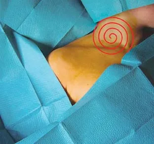 Fig 6. Skin decontamination worked centre to periphery. (Curso Terapia Intravenosa, p.110).
> Fuentes: Curso de Terapia Intravenosa (corpus monograph), p.110
Fig 6. Skin decontamination worked centre to periphery. (Curso Terapia Intravenosa, p.110).
> Fuentes: Curso de Terapia Intravenosa (corpus monograph), p.110 [C] [MEDLIB].
The antiseptic grid
| Agent | Spectrum | Speed | Residual | Cautions |
|---|---|---|---|---|
| Alcohol 60–90% | Gram+/Gram− excellent, virus good | Excellent | None | Flammable; max effect after it dries [20] |
| Povidone-iodine | Gram+ excellent, Gram− good | Moderate | Minimal | Inactivated by blood; shellfish allergy not a contraindication [20] |
| Chlorhexidine gluconate | Gram+ excellent, Gram− good | Moderate | Excellent | Avoid cornea, nerves, meninges [20][21] |
| Chlorhexidine + alcohol | Gram+/Gram− excellent | Excellent | Excellent | The best combination; avoid periocular [20] |
| Benzalkonium chloride | Broad, but can be self-contaminated | Moderate | Moderate | Alone does not protect against mycobacteria [19][37] |
Evidence for the default. No universal guideline exists for pre-filler skin prep; the data are borrowed from surgical practice [21][23]. A prospective study of 849 patients undergoing clean-contaminated surgery found chlorhexidine-alcohol more effective than povidone-iodine; general healthcare-associated-infection guidance is 2% chlorhexidine gluconate in 70% isopropyl alcohol [19]. Chlorhexidine handles resistant Gram-positive strains including S. aureus and some Gram-negative, viral and fungal microbes, and its residual activity is its advantage; but it can cause skin sensitivity, and it is barred periocular [21]. Some authors prefer benzalkonium chloride reinforced with chlorhexidine and benzyl alcohol (synergy against Gram-positive/negative, mycobacteria, fungi and viruses at lower concentrations) precisely because benzalkonium alone can be contaminated by mycobacteria [37]. For intraoral prep before perioral filler, chlorhexidine 0.12% mouth rinse plus 70% alcohol or 2% chlorhexidine on the skin [36].
The tap-water and ice trap. Mycobacterium chelonae infection after cosmetic HA injection has been traced to ice cubes used at the injection site, with clinic-faucet isolates matching the patient's culture; avoid facial skin exposure to tap water peri-procedurally, and instruct patients to wash with cleansing wipes (not at the sink) and to not reapply make-up for several hours [19]. The atypical mycobacteria (M. fortuitum, M. chelonae) and staphylococci are the organisms of poor-asepsis nodules [37].
Why the three minutes before the first puncture decide the infection
Late infection and biofilm are not random: they are seeded at the moment of injection and declare weeks later, which is why prevention is entirely front-loaded [12][21]. The mechanism: skin flora and any surface contaminant (residual make-up, non-dried antiseptic, a re-palpated field, tap-water mycobacteria) are carried into a closed tissue space by the needle, where filler is an ideal scaffold for a biofilm that the immune system cannot clear and antibiotics penetrate poorly [12]. The three-minute window controls every modifiable factor: complete make-up removal, an antiseptic with residual activity (chlorhexidine-alcohol) given time to dry, a no-touch field, and the fewest punctures compatible with the plan. Each skip (a wet antiseptic, a re-touched field, an extra unnecessary pass) is an independent seed, and because more punctures raise biofilm risk exponentially, the count itself is a variable [12][21]. The biofilm's late, sterile-appearing, steroid-worsened presentation is the reason a late nodule is treated as biofilm until proven otherwise, and the reason the prevention is worth more than any treatment (J5 — Infection, Biofilm & Sterilization.en).
No-touch handling and prophylaxis by scenario
No-touch technique is the operational core of asepsis: after the skin is prepped and dry, the needle, the product hub and the injection field never contact a non-sterile surface or a re-contaminated glove. More punctures raise biofilm risk (rising exponentially with penetration count), so the fewer-entry cannula approach is an asepsis argument as much as a vascular one, provided the single entry point is handled no-touch [12]. Practical rules: change the needle when moving to a new territory, never recap by hand into a used cap, keep the product cap sterile until the moment of loading, and re-prep any point that is re-palpated with a working glove.
Prophylaxis is targeted, not blanket:
| Scenario | Action | Evidence status |
|---|---|---|
| Recurrent perioral HSV + lip/perioral filler | Antiviral prophylaxis (start before, continue after) | Standard of care; trigger is known [21] |
| Semipermanent / permanent filler | Antibiotic prophylaxis sometimes used | Not validated; case-by-case [21] |
| Recent or upcoming dental work | Separate procedures in time | Transient bacteraemia rationale [21] |
| Active acne / folliculitis in field | Postpone | Late infection starts here [21] |
| Active periodontal disease | Address the focus first | Underrated late-nodule factor [21] |
| Prior unexplained late nodule | Image first; treat as biofilm | Do not reflex-steroid [12] |
Routine antibiotic prophylaxis for standard HA filler is not supported; the protective measures that are supported are skin decontamination, appropriate depth for the agent, avoiding filler stacking and large-volume boluses, and not injecting through active infection [21]. Institutional-SOP structure (activation criteria, roles, checklist order) belongs in the clinic protocol and is detailed for emergencies in J6 — Emergency Preparedness & Anaphylaxis.en.
Risk factors that change the decision to treat today
- Active infection in or near the field (inflammatory acne, folliculitis, herpes): postpone [21].
- Recurrent labial herpes + perioral treatment: antiviral prophylaxis; lip manipulation is a known trigger [21].
- Recent or upcoming dental work: transient bacteraemia; separate the two in time [21].
- Active periodontal disease: a chronic focus, an underrated factor in late nodules.
- Filler stacking (layering multiple filler types over each other at one session), large-volume boluses, and injecting through active acne all raise infective risk; keep depths appropriate to the agent and location [21].
The biofilm rule, because it explains the stakes: a late inflammatory nodule over filler is biofilm until proven otherwise, not an allergic reaction. The corollary is uncomfortable and must be known: the reflex of infiltrating corticosteroid can worsen it. More punctures raise biofilm risk (exponentially with penetration count), which is one of the real arguments for fewer-entry cannula work with strict no-touch handling [12]. Management in J5 — Infection, Biofilm & Sterilization.en.
Consensus: clean skin thoroughly, remove make-up, chlorhexidine-alcohol on intact non-periocular skin, respect the dry time, no-touch, record lot/UDI [19][20][21]. Discrepancy: periocular antiseptic choice. Chlorhexidine is barred (keratitis); options are aqueous povidone-iodine or careful alcohol. Decide by: distance to the eye and whether the field can be kept out of the tear film. This is a change-the-gesture discrepancy, so it stays.
Classic pitfall: not respecting the antiseptic dry time because of haste. A wet antiseptic has not acted, so the skin was never decontaminated; this is a direct biofilm cause. Signature: a delayed nodule weeks to months later with no other explanation. Fix: prep, wait for visible drying, then inject [20][21].
C1.8 · Aspiration, plunger pressure, and the safety maneuvers: what evidence each has
Answer first: every maneuver graded by what the evidence actually shows.
| Maneuver | What it claims | Evidence | Verdict |
|---|---|---|---|
| Aspirate-wait-inject | A negative aspirate means extravascular | 33% true positive ≤1 s; 128/340 false negatives after 10 s [2][3] | Not a safety test. A negative authorizes nothing |
| Slow injection, low extrusion force | Limits volume entering a vessel and retrograde spread | Mechanistic; endorsed by consensus [12] | Effective. First-line |
| Micro-aliquots (≤0.025 mL / <0.1 mL) | Below the blinding threshold per point | Blinding needs 0.04–0.12 mL; JVL bolus 0.025 mL [12] | Effective. First-line |
| Keep the needle/cannula moving | No static intraluminal delivery | Mechanistic [12][15] | Effective |
| Smallest needle that still injects | Less retrograde flow once intraluminal | >27G reached retrograde flow faster than 30G [12] | Rational, choose smallest workable bore |
| Inject away from / not perpendicular to axial vessels | Avoids the perforation geometry | Tansatit: perpendicular angle favours wall injury [6] | Rational |
| Digital compression of the arterial path | Blocks retrograde travel past the finger | Van Loghem "manual arterial closure" [12] | Adjunct in danger zones |
| No adrenaline in the block near danger zones | Preserves the blanch warning | Consensus caution [12] | Effective safety rule |
Aspiration: what the evidence demonstrates, uncomfortable as it is
The most disputed gesture in the specialty, and the evidence is clear if inconvenient.
Van Loghem, Fouché and Thuis tested a bench of 24 different fillers and 11 needle gauges, pressurized to 150 mmHg to simulate arterial pressure, against anticoagulated blood and tinted lactated Ringer, over 340 aspiration tests [2]:
| Result | Count |
|---|---|
| True positives within ≤1 second | 112 / 340 (33 %) |
| Positives between 1 and 10 seconds | 101 |
| False negatives after a full 10 seconds | 128 / 340 |
The outcome was governed by needle diameter, needle length and product rheology; the authors' conclusion, in substance: additional safety measures are required, aspiration alone does not suffice [2]. Goodman and colleagues state the corollary in their title: neither a positive nor a negative aspiration should be relied upon as a safety maneuver [3]. Hong's in-vitro image makes it visible: a 27G/13 mm needle aspirated blood in 1 second, while a 25G/40 mm cannula did not aspirate even after 10 seconds [13].
Why it fails, mechanically: four independent reasons, and one is enough [3][6][13]: 1. Viscosity. A cross-linked HA will not move up the needle in the time anyone aspirates; the denser the product and the finer the needle, the longer the required time, and nobody aspirates 10 seconds. Torbeck argues rheology is the main determinant of a true positive [6]. 2. Vessel collapse. Suction pulls the arterial wall against the tip and seals the port. 3. The tip moves. Between aspirating and injecting, a millimetre of displacement invalidates the test; with a 1 mL syringe, withdrawing the plunger moves the hand [13]. 4. A negative reassures. The most dangerous effect: the maneuver transfers confidence without transferring safety. 5. It is impossible for whole categories of technique. Linear threading and fanning do not lend themselves to aspiration at all, because the tip is moving through tissue [6].
Documentation, because the maneuver you skip must be defensible
Whatever the clinic decides about aspiration, the decision must be written into the protocol and applied consistently, because the medicolegal exposure is in the inconsistency, not in the choice. Omitting aspiration systematically is defensible with this literature; omitting it ad hoc, without being able to say why, is not [3]. The record for every injection carries the product, lot and UDI, volume per zone, plane, tool and any incident; that record is the traceability a regulator or a responding clinician asks for first, and it is what lets a delayed complication be worked backward to a cause [21]. The consent names the vascular risk explicitly [12]. None of this is separate from technique: a maneuver you cannot document and defend is a maneuver you have not really adopted, and a chapter that recommends abandoning a taught safety step owes the reader the reasoning to put in the chart, which is exactly what §C1.8 provides.
The position of this atlas, said without ambiguity
- Aspiration is not a safety test and must not be used as one. A negative does not authorize a bolus.
- A positive does inform, and mandates repositioning. Its value is asymmetric: it only helps when it fires.
- It replaces nothing. What lowers risk is the correct plane, low volume, low pressure, motion, microboluses instead of boluses, and knowledge of the arterial course [12].
- If you aspirate, do it right or not at all: tip still, hand unchanged, and knowing that two times in three it will not warn you [2][3].
⚠ Medicolegal reservation, and it is real: aspiration is part of the taught standard in many settings and appears in some consent forms and protocols. Systematically omitting it is defensible with this literature; omitting it without being able to explain why is not. Record the reasoning in the clinic protocol [3].
Plunger pressure: the physics nobody feels
Embolization requires injection pressure to exceed arterial pressure retrograde. The 1 mL syringe generates far more pressure per unit of thumb force than a 3 mL syringe; small syringe + dense product + fine needle = high pressure you do not perceive [12][14]. Tejero's good-practice directives: inject slowly at low pressure (this limits how much can inadvertently enter a vessel, limits retrograde flow, and limits the extent of any embolus); consider a cannula ≥25G; inject in small increments per site; if you aspirate, understand it is not fail-safe and is unreliable even done correctly; use targeted digital compression of the arterial paths to prevent retrograde filler travel; and do not use adrenaline with lidocaine, because it can mask the pallor of vascular compromise [12]. Van Loghem's manual-arterial-closure maneuver: while the injecting hand delivers a maximum of 0.025 mL per retrograde thread, the fingers of the other hand press on the nasion to compress and temporarily stop flow in the supratrochlear and dorsal nasal arteries, so a potential embolus cannot be pushed past that point [12].
Extrusion force and injection rate: the numbers behind "slow and low"
The reason "slow and low" works is quantitative. Embolization has two requirements that must both be met: injection pressure above arterial pressure (so product can be driven retrograde into and up a vessel) and a delivered volume large enough to reach a critical territory [12]. Both are directly controlled at the thumb. Extrusion force rises with product G-prime, with narrower needle bore, and with narrower syringe barrel; the 1 mL syringe's small plunger area converts a modest thumb force into a high tip pressure, which is why a dense product through a fine needle on a 1 mL syringe is the maximum-pressure configuration [12][14]. Injection rate sets how fast that pressure delivers volume: a slow rate means that even with an intraluminal tip, the volume introduced before a warning sign (pain, blanch) appears is sub-critical, whereas a fast bolus can deliver the blinding volume (0.04 to 0.12 mL) before the operator reacts [12].
The experimental model makes the trade-off concrete: in a bifurcated-tube system, needles larger than 27G reached retrograde flow and the bifurcation faster and more often than 30G needles, so a larger bore lowers the chance of puncture but raises the ease of retrograde occlusion once intraluminal, because larger aliquots inject more easily [12]. The synthesis is the smallest bore that still lets you inject the product at low, controlled force. This is also why threading and fanning, which keep the tip moving through tissue, are inherently lower-pressure-at-a-point than a static bolus: the pressure is never sustained against one spot [15].
The safety maneuvers, step by step
Because no single maneuver is protective, they are layered. The sequence Tejero's good-practice directives encode, in order of weight [12]:
- Know the arterial course of the region before the first puncture; rehearse it aloud if learning.
- Choose the plane that keeps the tip away from the named artery (deep on bone or superficial in dermis).
- Low extrusion force, slow rate. This limits inadvertent intravascular volume, limits retrograde flow, and limits the extent of any embolus that does form.
- Small aliquots per site (micro-droplets; ≤0.025 mL per danger-zone bolus or thread), so the volume delivered before a warning sign is sub-critical.
- Keep the tip moving on withdrawal, so no static intraluminal delivery is possible.
- Manual arterial closure where anatomy allows: while the injecting hand delivers ≤0.025 mL, the other hand's fingers compress the proximal artery (for example on the nasion for the supratrochlear and dorsal nasal system), so an embolus cannot be pushed past the finger [12].
- No adrenaline in the block near a filler danger zone, so the blanch of a compromise is not masked [12].
- Watch the skin continuously and stop at the first abnormal sign; do not finish the syringe.
Aspiration, if it is done at all, done correctly: a fixed needle that does not move between test and injection, an unfilled needle (a primed needle cannot aspirate blood into the barrel in the time available), and sustained negative pressure held long enough for the product's rheology, knowing that even then two of three intravascular placements will not signal [2][3]. This is the only setting (a fixed supraperiosteal needle bolus) where a positive still changes the decision; a negative never authorizes the bolus [3][12].
Needle size and retrograde occlusion
The trade-off is not one-directional. Larger needles (>27G) reached retrograde flow and the arterial bifurcation faster and more often than 30G needles in a bifurcated-tube model; so larger needles may lower the chance of puncturing a vessel, but once intraluminal they cause retrograde occlusion more readily because larger aliquots inject more easily. The synthesis: choose the smallest needle that still permits the injection to limit retrograde-flow risk [12].
Consensus: low volume, low pressure, motion, microaliquots, danger-zone compression, smallest workable needle, and hyaluronidase ready [2][3][12]. Discrepancy: aspirate or not: the aspirate-as-one-check school (keep it for supraperiosteal needle boluses, done correctly with a fixed unfilled needle and sustained negative pressure it can still catch some intravascular placements) versus the abandon-aspiration school (currently no evidence supports it as a safety measure, it is impossible with cohesive fillers and with threading/fanning; replace with motion + microaliquots + low pressure + compression) [3][12]. Decide by: local protocol and whether the specific injection is a fixed supraperiosteal needle bolus (the only scenario where a positive can still inform). Never present it to the patient as protection.
Classic pitfall: trusting a negative aspirate. Signature: a bolus delivered "safely" after a clean aspiration that was a false negative (128/340), ending in blanch or necrosis. Fix: treat aspiration as, at best, a one-way check that never authorizes a bolus, and rely on plane, volume, pressure and motion [2][3].
C1.9 · Nomenclature: which MD Code and which eponym maps to which gesture
This block is an index, not a technique chapter. It cross-references code ↔ proper name ↔ gesture so that a region chapter can name a point without redefining it. The full point-by-point system, volumes and sequences live in C2 — MD Codes Systematic Approach.en and the region chapters (D-series). [MODELO] The codes are a structural naming convention; a code never carries a dose here.
A shared vocabulary is a safety instrument as much as a teaching one: when a treatment is recorded and handed off as coded points, a second clinician managing a complication knows what was placed and where, in what plane and by what movement, which is exactly what a freehand note loses [11][21]. The two naming systems in daily use are the MD Codes anatomic point system (de Maio), which labels supraperiosteal and deep entry points region by region, and the eponymous/school techniques (deep-injections-first, prezygomatic deep-cannula, multilayering, the six temple techniques), which name a whole gesture rather than a point [7][11][33]. This block indexes both against the plane and the movement this chapter defines, so a region chapter can invoke a code or a name without redefining it and without importing a volume the label never carried.
MD Codes: the de Maio anatomic point system
De Maio's MD Codes are a methodological, anatomy-anchored point system: each facial region gets a letter-number label tied to a specific supraperiosteal or deep entry point and a specific gesture, so that a treatment can be prescribed and reproduced as a sequence of coded points rather than a freehand impression [11]. The point is standardization: same label, same anatomic target, same plane, same movement, across operators. Representative index (framework labels; targets and volumes in C2 and the region chapters):
| Code family | Region | Typical point / gesture (index only) |
|---|---|---|
| Ck1–Ck5 | Cheek | Zygomatic/malar and mid-cheek points; supraperiosteal bolus + subcutaneous support [11] |
| TT | Tear trough | Deep medial supraperiosteal depot; low-volume [11] |
| C1–C4 (chin) | Chin | Central/paramedian supraperiosteal boluses; tower for projection [11] |
| Jw1–Jw4 | Jawline | Angle, body and pre-jowl supraperiosteal/subcutaneous points [11] |
| NL / M | Nasolabial / marionette | Retrograde subdermal threads plus deep support [11] |
| Lp | Lips | Vermilion serial puncture; body retrograde threading; 8-point lip framework [11] |
| T / F | Temple / forehead | Deep temporal supraperiosteal or subdermal microbolus [11][33] |
| Rh | Nose | Midline supraperiosteal microdroplets, a high-risk index (danger zone) [11] |
Aggregate frameworks that ride on the points: the 8-point lip reshape and 5-point cheek reshape are named sequences of these coded points, not new anatomy [11].
Eponymous and named techniques mapped to their gesture
| Name | Region / gesture | Plane / tool |
|---|---|---|
| Deep injections first (Cotofana/Freytag) | Build the bony fundament before superficial work | Supraperiosteal first [7] |
| Six temple techniques (Cotofana) | Six discrete ways to augment the temple, each with a defined plane | Deep temporal to subdermal [33] |
| Prezygomatic deep-cannula (Beut / Surek) | Deep prezygomatic-space support of the midface | Deep cannula, prezygomatic space |
| MLT multilayering (Rosso) | One entry port, three planes in one session: structural support, dynamic volume, superficial redensification | Cannula across planes |
| Andrews cheek approach | Depot supraperiosteal first, reassess contour, then subdermal fans | Needle depot + subdermal fan [9] |
| JVL bolus (van Loghem) | Retrograde thread capped at 0.025 mL for danger-zone safety | Needle/cannula, retrograde [12] |
| Manual arterial closure (van Loghem) | Finger compression proximal to the injection to block retrograde embolus travel | Adjunct maneuver [12] |
| HDPH (DeLorenzi) | High-dose pulsed hyaluronidase for a vascular event, hourly to resolution | Rescue protocol [25] |
| Serial puncture / linear threading / fanning / cross-hatch | Deposition movements (see §C1.4) | Generic, not eponymous [15] |
Brand names, generic gestures, and the safety handoff
Nomenclature is not pedantry: it is the handoff. When a complication presents to a second clinician, the record must say what was placed, where, in what plane, by what movement, so the responder can plan a dissolution or a rescue. A note reading "1 mL Ck1" is legible only if "Ck1" resolves to a zygomatic supraperiosteal bolus of a named product, which is why this chapter insists a code resolve to a plane and a movement, and why the lot/UDI record is a technique item, not just a legal one [11][21]. Brand names carry rheology information a generic label loses (a high-G' volumizer versus a soft-tissue soft HA implies a plane and a bore), so this private wiki names products where the name is the datum, while the generic gesture (supraperiosteal bolus, retrograde subdermal thread) is what the technique chapter standardizes [14]. The failure mode to avoid is a record that names a region and a volume but not a plane or a product, which tells a future responder almost nothing when it matters most.
Region-to-code quick cross-reference
| Region | MD Code family | Named/school gesture | Plane + movement (this chapter) |
|---|---|---|---|
| Zygoma / malar | Ck1, Ck2 | Deep-injections-first; Andrews depot | Supraperiosteal bolus, then subdermal fan (§C1.3/§C1.4) [9][11] |
| Anteromedial cheek | Ck3–Ck5 | Prezygomatic deep-cannula (Beut/Surek); MLT (Rosso) | Deep fat depot + subcutaneous support [11] |
| Chin | C1–C4 (chin) | Tower for projection | Supraperiosteal bolus + tower/column (§C1.4) [11] |
| Jawline | Jw1–Jw4 | Angle/body/pre-jowl | Supraperiosteal + subcutaneous retrograde [11] |
| Tear trough | TT | Cannula deep on rim (Spada) | Low-volume retrograde on the orbital rim [4][11] |
| Lips | Lp; 8-point lip | Vermilion serial puncture | Serial puncture at the edge, retrograde in the body [11][15] |
| Temple | T | Six temple techniques (Cotofana) | Deep supraperiosteal microbolus or subdermal fan [33] |
| Nose | Rh | Midline micro-droplet | Supraperiosteal micro-droplets, ≤0.025 mL (danger zone) [11][12] |
The table resolves the promise of §C1.9: a region chapter can write "Ck1" and this index returns the plane and the movement, so a code never travels without its gesture. The full sequences and product volumes stay in C2 — MD Codes Systematic Approach.en and the region chapters.
The naming clash worth knowing
The same gesture carries different names in different schools, and the region chapters must not treat them as different techniques. A supraperiosteal malar bolus is "Ck1" in MD Codes, a "deep injection first" in the biomechanics school, and simply "on-bone depot" in a generic manual; they are the same gesture in the same plane [7][11]. Conversely, one label can hide a plane choice: "temple augmentation" spans a deep supraperiosteal microbolus and a subdermal cannula fan, with completely different risk profiles [33]. When a region chapter cites a code, it must resolve to a plane and a movement here, or it is decoration.
Aggregate frameworks are sequences of points, not new anatomy
The named "reshape" frameworks are ordered recipes built from the single-point codes, and they belong in this index so a region chapter can invoke one without re-teaching it [11]: - 8-point lip: a fixed sequence of coded vermilion and body points that reshapes and projects the lip, each point a serial-puncture or retrograde gesture at a defined depth. - 5-point cheek: a sequence of zygomatic and mid-cheek points (the Ck family) that restores the cheek's convexity, deep-first then subdermal. - Deep-injections-first, upper-face-first, lateral-face-first: not point codes but sequencing rules that order whichever points a plan uses [7].
The safety point these frameworks carry: a "reshape" is a total volume distributed over several points, and the danger is reading the framework's total as a single-point dose. Each coded point still obeys the per-point volume ceilings of §C1.4, and in a danger-zone framework (a nose or a glabella has no reshape framework precisely because the volume discipline forbids it) the sequence never overrides the ≤0.025 mL micro-droplet rule [11][12]. The framework tells you where and in what order; §C1.3 and §C1.4 tell you in what plane and how much, and the region chapter binds the product.
Consensus: a shared point-and-name system improves reproducibility and teaching; MD Codes is the most widely used [11]. Discrepancy: none that changes the gesture. The schools disagree on sequence and philosophy (deep-support-first vs multilayering, §C1.3), not on what a given code names. Averaging is not at issue here because this block assigns no volumes.
Classic pitfall: treating a code as a dose. A code names a point, a plane and a movement, never a millilitre; reading "Ck1" as "inject X mL" imports a volume the label never carried and that varies by product, patient and school. Signature: over- or under-correction from a copied number. Fix: resolve every code to plane + movement here, and take the volume from the product and the region chapter, not from the label [11].
C1.10 · Technique errors and their clinical signature
Answer first: read the sign backwards to the error.
| Error | Why it happens | Clinical signature | Fix |
|---|---|---|---|
| Too superficial (HA) | Plane not verified; touch-up over fibrosis falsifies resistance | Bluish Tyndall discoloration, beading, visible ridge | Deeper next time; dissolve with hyaluronidase [14] |
| Too superficial (non-HA) | Same, with an irreversible product | Persistent papule/nodule, no easy reversal | Prevent; place below deep reticular dermis [15] |
| Intramuscular placement | Aiming for deep plane, entering muscle | Product displacement/lumping on contraction, migration | Confirm plane; supraperiosteal or subcutaneous, not intramuscular [12] |
| Overcorrection | Volume by feel, not by plan | Visible/palpable excess, unnatural contour | Hyaluronidase (HA); conservative staging [14] |
| Wrong plane in a danger zone | Anatomy not respected; static bolus | Vascular occlusion: immediate disproportionate pain, blanch, mottling, then dusky skin; visual symptoms if periorbital | Emergency protocol; hyaluronidase now [12][26] |
| Large static bolus | Speed and volume yield | Migration; palpable mass; embolic risk realized | Microboluses, moving, ≤0.025 mL danger zones [12] |
| Poor asepsis / wet antiseptic | Haste, skipped dry time | Late inflammatory nodule (biofilm until proven otherwise) | Prevent; do not reflex-inject steroid [12][21] |
| Filler stacking | Layering product types in one session | Delayed nodule, harder to diagnose | Separate agents and sessions [21] |
| Hydrophilic HA in tear trough | Wrong product for the site | Sausage-like delayed swelling, months later | Low-hydrophilicity HA; Restylane/Belotero/Volbella class [14] |
| Cannula too thin for product | "Less traumatic" belief | Off-plane deposit, raised pressure, shallow result | Match bore to product [13] |
| Trusting a negative aspirate | Taught as a safety test | A false-negative bolus into a vessel | Never authorize a bolus on aspiration [2][3] |
| Bone-contact false security | Intuitive | Embolus despite tip on periosteum | Bone contact is not protection [1] |
| Marking after the block | "Saving time" | Post-block asymmetry, especially lip | Mark seated, block second [18] |
| Finishing the syringe past a warning sign | Sense of waste | Progression of an occlusion | Stop at the first abnormal sign [12] |
| No hyaluronidase available | Assumed in the drawer | An untreatable HA occlusion | No HA without hyaluronidase stocked [24] |
| No lot/UDI record | Forgotten | No traceability if a problem arises | Record every lot/UDI [21] |
Timing of the signature: immediate, early, late
The clock is a diagnostic axis: the same nodule means different things at 5 minutes, 5 days and 5 weeks.
| Onset | Signature | Most likely error / cause | First move |
|---|---|---|---|
| Seconds to minutes | Disproportionate pain, blanch, mottling | Intravascular / wrong plane in danger zone | Stop; hyaluronidase; occlusion protocol [12][26] |
| Seconds to minutes | Pallor, sweating, yawning, nausea | Vasovagal (rule out occlusion first) | Supine, legs up, reassure [17] |
| Minutes to hours | Bluish hue over a superficial deposit | Too-superficial HA (Tyndall) | Hyaluronidase; place deeper next time [14] |
| Hours to days | Spreading erythema, warmth, tenderness | Acute infection / poor asepsis | Antibiotics; culture; image [21] |
| Hours to days | Grouped vesicles, perioral | HSV reactivation from lip manipulation | Antivirals [21] |
| Days | Firm swelling, delayed | Early hypersensitivity / immune | Anti-inflammatory pathway (J1) |
| Weeks to months | Inflammatory nodule | Biofilm until proven otherwise | Image; treat as infective; do not reflex-steroid [12][21] |
| Weeks to months | Sausage-like swelling, tear trough | Hydrophilic HA in the wrong site | Hyaluronidase [14] |
| Months | Contour migration | Large bolus / wrong plane | Assess; dissolve if HA [12] |
Reversibility is part of the error's cost. HA errors are reversible with hyaluronidase (Tyndall, overcorrection, malposition, occlusion); CaHA, PLLA, PMMA and silicone are not, so a plane error with a non-HA product is a permanent one, which is why plane discipline matters most with the irreversible agents [12][14]. A persistent small non-emergent nodule has resolved with as little as 15 to 30 U of hyaluronidase, a different setting from the 200 IU-and-up used for impending necrosis [22][24].
The three signatures that must never be missed
- The Tyndall effect is pathognomonic of superficial HA: a blue-grey hue from Rayleigh scattering over a superficial HA deposit. It is reversible with hyaluronidase and is a plane error, not a product defect [14].
- Displacement on animation points to intramuscular or wrong-plane placement: a deposit that looks fine at rest and lumps or shifts when the patient contracts. The deep, ligament-bound compartments move little; a product that moves a lot is in the wrong layer [8][12].
- Disproportionate immediate pain plus blanch is vascular compromise until proven otherwise: stop, do not finish the syringe, and run the occlusion protocol; a vasovagal and an occlusion can start identically, so rule out the dangerous one first [12][26]. Full management in J2 — Vascular Occlusion & Emergency Response.en.
Reversal: what an error costs by product
The correctable errors are the HA ones. Hyaluronidase reverses HA-specific technique errors: Tyndall from superficial placement, overcorrection, malposition, delayed hydrophilic swelling, and vascular occlusion [14][24]. The dose scales with the setting: a small persistent non-emergent nodule has resolved with as little as 15 to 30 U, whereas impending necrosis is treated with doses not below 200 IU, and a vascular event with the high-dose pulsed protocol (hourly high-dose hyaluronidase until resolution, scaled to the ischaemic tissue) [22][24][25]. Ultrasound-guided delivery uses less enzyme than blind flooding because it puts the hyaluronidase into the deposit or the occluded segment (15 to 75 U per session for a malposition, 50 to 100 U into the "Medusa head" for an occlusion) [12][24].
The non-HA products have no equivalent rescue: calcium hydroxylapatite, poly-L-lactic acid, polymethylmethacrylate and silicone are not dissolvable, so a plane error with any of them is permanent and may require intralesional steroid, laser, or excision. This asymmetry is a technique rule in disguise: the irreversible agents demand the strictest plane discipline, because the safety net that catches an HA mistake does not exist for them [12][14].
Why the touch-up is the highest-error setting
Residual product and fibrosis change tissue resistance and falsify the five tactile plane signs (§C1.3), so a plane that reads "subdermal" by feel may be deeper or shallower than it is. This is why the objective check (ultrasound) earns its highest yield exactly here: map what is present before adding or dissolving [12]. An operator who treats a touch-up as identical to a first session is injecting into an anatomy that no longer matches the atlas.
The five habits that prevent most of these errors
Nearly every row of the error table above is prevented by five habits, which is why the chapter reduces to them [1][3][12]: 1. Verify the plane before injecting, by the five tactile signs and, in a touch-up, by ultrasound; most placement errors are plane errors read from falsified resistance [12]. 2. Inject low-and-slow-and-moving, on withdrawal, with micro-aliquots; this converts the physics of embolization against the operator's mistakes [12][15]. 3. Keep hyaluronidase stocked and imaging accessible, so an HA error is recoverable and an occlusion is treatable early [24][25]. 4. Do not trust the reassuring gestures: a negative aspirate, bone contact and the cannula all fail, and treating any of them as protection is itself the error [1][3][5]. 5. Document plane, product and lot, so a delayed sign is traceable and the next clinician can act [21].
The errors that survive these habits are the irreversible-product plane errors, which is why the discipline is strictest with CaHA, PLLA, PMMA and silicone: there is no hyaluronidase to catch them [14]. Prevention is not a separate skill from technique; it is the technique, and every region chapter inherits these five habits without restating them.
Consensus: most technique errors are plane errors or pressure/volume errors, and both are preventable by verifying the plane and injecting low-and-slow-and-moving [12][15]. Discrepancy: management of the late nodule (biofilm vs immune) shapes the fix (antibiotics/anti-inflammatory vs hyaluronidase vs steroid) and is adjudicated in J5 — Infection, Biofilm & Sterilization.en and J1 — Delayed Nodules & Immune Reactions.en; it is a change-the-management discrepancy, kept there, not averaged here.
Classic pitfall: reading a late nodule as allergy and reflexively injecting corticosteroid, which can worsen a biofilm. Signature: a nodule weeks to months out that flares after steroid. Fix: treat a late nodule as biofilm until proven otherwise, image it, and follow J5 — Infection, Biofilm & Sterilization.en [12][21].
Coverage vs UPO
| UPO topic (Máster ME) | State in this chapter | What the atlas adds |
|---|---|---|
| Needle vs cannula: geometry, gauge, indications | Covered, expanded (§C1.2) | The 25G watershed; the safety conflict printed whole (Zhou, Tansatit vs cadaver), not averaged [1][5][6] |
| Tissue planes: supraperiosteal, deep fat, SMAS, subdermal, intradermal | Covered, expanded (§C1.3) | Deep-injections-first biomechanics; five tactile signs; touch-up falsification [7][8] |
| Injection techniques: bolus, retrograde, fan, cross-hatch, serial puncture, depot | Covered, expanded (§C1.4) | Anterograde reserved to vermilion; the low-volume/low-pressure/moving triad; 1 mL vs 3 mL pressure [12][15] |
| Aspiration: evidence, limitations, false negatives | Covered, expanded (§C1.8) | The 340-test numbers; four mechanisms of failure; the medicolegal reservation [2][3] |
| Aseptic technique, skin prep, infection control | Covered, expanded (§C1.7) | Antiseptic grid; tap-water/ice mycobacteria; biofilm-until-proven-otherwise [19][21][38] |
| Pain management: topical, blocks, ice | Covered, expanded (§C1.6) | Agent table with onsets; the pupillary-line landmark; aspirate-on-the-block distinction [16][17][18] |
| Ultrasound-guided injection: principles, adoption | Covered, expanded (§C1.5) | Echogenicity grid; Medusa head; scan-while-injecting frontier declared as a corpus gap [12][24][29] |
| Instrumentation by product rheology | Added (§C1.2) | UPO teaches needle vs cannula, not bore-to-G' matching; added [13][14] |
| Deposition movements one by one with volume ceilings | Added (§C1.4) | UPO lists techniques; the atlas assigns each an indication and a danger-zone volume cap [12][15] |
| Nomenclature index (MD Codes + eponyms) | Added (§C1.9) | UPO does not index code ↔ name ↔ gesture; added, cross-referenced to C2 [11][33] |
| Technique-error signatures | Added (§C1.10) | UPO teaches complications by cause; the atlas reads the sign backwards to the error [12][26] |
UPO is the fastest-ageing lane: a dose resting only on a UPO slide is never_sufficient_alone. The UPO complications decks (Tejero, M2/T10) are used here as corroboration for the aspiration and cannula-conflict facts, never as the sole support for a number [38].
Self-assessment
- What is the JVL bolus ceiling, and why that number?
answer
0.025 mL per bolus or retrograde thread. The volume that blinds sits at 0.04 to 0.12 mL, so staying below 0.025 mL drives the chance of blinding "next to zero" [12]. - Aspiration true-positive rate at one second, and false-negatives after ten?
answer
33% true positive within ≤1 s (112/340); 128/340 false negatives after a full 10 s. Governed by needle diameter, needle length and product rheology [2]. - In Zhou's series, how many of the severe embolism cases were injected with a cannula?
answer
25 of 28 (cannula 22 to 27G). The board consensus that followed: avoid cannulas smaller than 25G [5]. - What did the cadaver study find with the needle tip on periosteum?
answer
Intra-arterial injection occurred with the tip resting on periosteum under constant bone contact; the needle also seeded product retrograde up its tract into multiple layers [1]. - Name the three governing biomechanical principles for filler placement.
answer
Upper face first, lateral face first, deep injections first. The supraperiosteal plane is generally safer because the arteries mostly run more superficially [7]. - Which movement is reserved for the vermilion, and why is it otherwise avoided?
answer
Anterograde threading; it pushes product ahead of the tip. Everywhere else retrograde (inject on withdrawal) is preferred for lower intravascular risk [15]. - Why does a 1 mL syringe raise embolic risk versus a 3 mL?
answer
It generates far more pressure per unit of thumb force; small syringe + dense product + fine needle = high pressure you do not feel [12][14]. - On ultrasound, how do hydrophilic and hydrophobic fillers read, and what is the Medusa head?
answer
Hydrophilic (HA) reads anechoic to hypoechoic; hydrophobic (silicone, PMMA) reads hyperechoic. The Medusa head is dilated collateral arteries around a central anechoic HA deposit in arterial occlusion; inject 50 to 100 units there [12][24]. - Which antiseptic is default, and where is it contraindicated?
answer
Chlorhexidine-alcohol (2% CHG in 70% IPA), more effective than povidone-iodine; contraindicated periocular (keratitis) [19][20]. - What is a late inflammatory nodule over filler until proven otherwise, and what must you not reflexively do?
answer
Biofilm until proven otherwise, not allergy. Do not reflexively infiltrate corticosteroid, which can worsen it [12][21].
What's new and trends
| Year | Change | Maturity | Reference |
|---|---|---|---|
| 2025 | Consensus review consolidating ultrasound-guided hyaluronidase (150 to 200 IU + lidocaine), stock ≥10 vials / >1500 U, echogenicity-based nodule triage | clinically actionable now | [24] |
| 2023 | Split-face needle-vs-cannula tear-trough data (25G vs 31G) with ultrasound/MRI to 365 days: cannula less bruising but more superficial; needle more precise | promising but not validated (n=10) | [4] |
| 2023 | Doppler-guided HA injection formalized as integrable into daily practice | promising but not validated | [28][29] |
| 2022 | Facial vascular/neural ultrasound pictorial standard; deep-injections-first biomechanics distilled to three named principles | clinically actionable now | [7][27] |
| 2021 | Zhou "false sense of safety" reframes the cannula as the tool in the majority of severe embolism cases | clinically actionable now | [5] |
| 2021 | Goodman consensus: neither positive nor negative aspiration is a safety maneuver | clinically actionable now | [3] |
| 2018–2021 | High-frequency ultrasound differentiates filler nodules from granulomas and maps palpable nodules | clinically actionable now | [31][32] |
| 2024→ | Real-time "scan-while-injecting" ultrasound guidance of every injection (upper-face consensus, corpus-acquisition gap) | preclinical/speculative as a routine standard | [29] |
| Ongoing | Retrobulbar hyaluronidase to reverse filler-induced blindness | preclinical/speculative (animal-model evidence) | [13] |
| Ongoing | Marketing of the blunt cannula as an inherently safe device | unsupported commercial claim | [5][6] |
What did NOT change, and why the older references still stand. The mechanical core is stable: embolization still requires pressure above arterial pressure and a critical volume, so low-volume, low-pressure, moving technique remains first-line, and the 2018 aspiration and cadaver data [1][2] are not superseded because no newer study has overturned the physics; they have only been reinforced (Goodman 2021, Zhou 2021) [3][5]. The plane taxonomy (five layers, deep compartments ligament-bound) is anatomy and does not date [8][9][10]. The genuine movement is in imaging: ultrasound has shifted from a complication-diagnosis tool toward pre-treatment mapping and, at the frontier, real-time guidance; that is where the field will change over the next decade, and where the own corpus is thinnest (the 2024 scan-while-injecting consensus is a declared acquisition gap, §C1.5). Anatomical variability data (facial-artery course and depth) continue to accumulate and argue for patient-specific mapping over atlas averages [30][35].
Unexplored directions (AI speculation)
> Disclaimer. The following are model-generated research directions, not clinical recommendations. Every item is tagged [IA-ESPEC], carries no dose, product or actionable protocol, and states what would settle it. They are hypotheses the chapter's own evidence exposes, nothing more.
[IA-ESPEC] A rheology-indexed nomogram for the narrow case where aspiration still informs.
Anchor: aspiration true-positive rate is 33% at one second and is governed by needle diameter, needle length and product rheology [2].
Proposal: characterize, across product G-prime and needle bore, the specific combination (fixed supraperiosteal needle bolus, low-viscosity product, adequate dwell) in which a positive aspirate retains predictive value, so the maneuver is neither universally taught nor universally abandoned.
Expected effect: a defined, small subset of injections where a positive result changes the decision, and an explicit larger set where it does not.
Confounder: tip displacement between the test and the injection, which invalidates the test independently of rheology [13].
What would settle it: a prospective in-vivo study pairing aspiration outcome with an objective intravascular marker across stratified product-and-gauge cells.
[IA-ESPEC] A bore-and-stiffness threshold that separates vessel displacement from wall penetration.
Anchor: the cannula displaces vessels in some settings yet penetrates the arterial wall in others, and below 25G it behaves like a needle [5][6].
Proposal: test whether a measurable stiffness-and-gauge threshold predicts, per region, whether a cannula tip displaces or perforates an artery of a given depth.
Expected effect: a bore rule grounded in mechanics rather than in the current binary "cannula safer / cannula not safe".
Confounder: regional variability in artery depth and in fibrous-septum density, which changes tip behaviour independently of the cannula [30].
What would settle it: a cadaver perforation-force study across gauges and regions with matched artery-depth measurement.
[IA-ESPEC] Whether routine pre-injection Doppler mapping lowers occlusion incidence at population scale.
Anchor: facial-artery course, diameter and depth vary widely between patients, and ultrasound can map them before injecting [27][30][35].
Proposal: test whether making pre-injection Doppler mapping routine in danger zones reduces the population incidence of vascular occlusion, versus the current selective use.
Expected effect: a measurable reduction in vascular events attributable to mapping rather than to operator selection.
Confounder: operator-skill confounding and the low base rate of catastrophic events, which demand very large samples.
What would settle it: a large multi-centre registry comparing mapped versus unmapped danger-zone injections with prospective event capture.
Safety
The governing thesis, restated because everything else depends on it: no maneuver turns an unsafe injection into a safe one. Not aspiration, not the cannula, not bone contact. What lowers risk is anatomical knowledge, the correct plane, and low-volume, low-pressure, moving technique [1][3][5][12].
Before every injection, without exception: 1. Signed consent naming the vascular risk (B3 — Ethics, Consent & Medicolegal.en). 2. Hyaluronidase on the trolley, in date and in quantity (danger-zone practice: ≥10 vials / >1500 U). If there is none, do not inject HA [24]. 3. Mark the plan seated, before blocking; rehearse the regional arterial course [18]. 4. Remove make-up, antiseptic, respect the dry time [19][21]. 5. Choose plane, tool and gauge for that product and zone, in that order [12][14].
During: inject on withdrawal, slowly, small volume, low pressure, moving; ≤0.025 mL per bolus/thread in danger zones; watch the skin continuously for blanch, mottling and disproportionate pain; at the first abnormal sign, stop, do not finish the syringe [12][15].
Step-by-step protocol for any injection, no exception
- Signed consent naming the vascular risk (B3 — Ethics, Consent & Medicolegal.en).
- Standardized pre-treatment photograph (B1 — Facial Assessment & Aesthetic Analysis.en).
- Check the emergency trolley: hyaluronidase in date and in quantity. If there is none, do not inject HA [24].
- Mark the plan with the patient seated, before reclining and before blocking [18].
- Rehearse the regional arterial course, aloud if you are learning [12].
- Remove make-up, apply antiseptic, respect the dry time [19][21].
- Choose plane, tool and gauge for that product and that zone, in that order.
- Verify the plane with the five tactile signs (§C1.3) before injecting anything.
- Inject on withdrawal, slowly, low volume, low pressure, moving [12][15].
- Watch the skin continuously: blanch, mottling, disproportionate pain.
- At any sign, stop. Do not finish the syringe.
- Massage and mold as the product allows.
- Reassess with the patient seated, talking and animating.
- Record product, lot/UDI, volume per zone, plane, tool and any incident [21].
- Give written post-care and a named emergency contact with a phone number.
- Review at 2 to 4 weeks with comparable photography.
This is the salvaged operational spine of the technique: every region chapter assumes these sixteen steps and adds only what its anatomy changes.
Vascular occlusion, the event that defines the specialty's floor. The mechanism: product injected against flow at pressure above arterial pressure reverses up the artery; from the supratrochlear or dorsal nasal artery it can reach the ophthalmic artery and the central retinal artery, and a small volume can occlude the retinal circulation and blind the patient; more pressure can carry product into the internal carotid and the cerebral circulation (stroke) [13][40]. Recognition: immediate disproportionate pain, blanch, then dusky mottling; periorbital cases add visual symptoms. Response is a protocol, not improvisation: stop, high-dose hyaluronidase into the ischaemic territory, and follow the emergency chain in J2 — Vascular Occlusion & Emergency Response.en. The high-dose pulsed hyaluronidase protocol (DeLorenzi): high-dose hyaluronidase hourly until the adverse event resolves, dose scaled to the ischaemic tissue volume [25]. Consensus doses for impending necrosis are not below 200 IU, with lidocaine for vasodilation; small persistent nodules have resolved with as little as 15 to 30 U in non-emergent cases, which is a different setting from an occlusion [22][24].
Danger zones (highest ophthalmic-anastomosis risk): glabella, nose, nasolabial fold, forehead, temple, periorbital region; the internal-to-external carotid anastomoses mean a facial injection elsewhere can still embolize the eye [13]. In these zones: cannula ≥25G when used, trajectory away from the named artery, microdroplets, manual arterial compression, and the lowest workable pressure [12].
Do not, ever: - Rely on a negative aspirate to authorize a bolus [2][3]. - Treat the cannula or bone contact as a guarantee [1][5][6]. - Use adrenaline with lidocaine near a filler danger zone (it masks the blanch) [12]. - Inject HA without stocked hyaluronidase, or inject over active infection/herpes [21][24]. - Reflexively infiltrate corticosteroid into a late nodule before excluding biofilm [12][21].
Ultrasound reduces uncertainty, not risk: an operator with a probe and poor judgement can still inject into an artery [24][27].
References
- van Loghem JAJ, Humzah D, Kerscher M. Cannula Versus Sharp Needle for Placement of Soft Tissue Fillers: An Observational Cadaver Study. Aesthet Surg J. 2018;38(1):73-88.
[B]https://doi.org/10.1093/asj/sjw220 · PMID 27986754 - van Loghem JAJ, Fouché JJ, Thuis J. Sensitivity of aspiration as a safety test before injection of soft tissue fillers. J Cosmet Dermatol. 2018;17(1):39-46.
[B]https://doi.org/10.1111/jocd.12437 · PMID 28987016 - Goodman GJ, Magnusson MR, Callan P, et al. Neither Positive Nor Negative Aspiration Before Filler Injection Should Be Relied Upon as a Safety Maneuver. Aesthet Surg J. 2021;41(4):NP134-NP136.
[A]https://doi.org/10.1093/asj/sjaa215 · PMID 33236760 - Spada J, Ravera K, Schneider C. Needle versus Cannula to Treat Tear Trough: A Prospective Study Comparing both Methods. Plast Reconstr Surg Glob Open. 2023;11(11):e5327.
[B]https://doi.org/10.1097/GOX.0000000000005327 · PMID 37936826 - Zhou SB, Chiang CA, Liu K. False Sense of Safety: Blunt Cannulas Cause the Majority of Severe Vascular Complications in Hyaluronic Acid Injection. Plast Reconstr Surg. 2021;147(4):240e-241e.
[B]https://doi.org/10.1097/PRS.0000000000007714 - Tansatit T, Apinuntrum P, Phetudom T. A Dark Side of the Cannula Injections: How Arterial Wall Perforations and Emboli Occur. Aesthetic Plast Surg. 2017;41(1):221-227.
[B]https://doi.org/10.1007/s00266-016-0725-7 - Freytag L, Alfertshofer MG, Frank K, et al. Understanding Facial Aging Through Facial Biomechanics: A Clinically Applicable Guide for Improved Outcomes. Facial Plast Surg Clin North Am. 2022;30(2):125-133.
[B]https://doi.org/10.1016/j.fsc.2022.01.001 - Cotofana S, Gotkin RH, Frank K, et al. The Anatomy of the Facial Fat Compartments and their Relevance in Aesthetic Surgery. J Dtsch Dermatol Ges. 2019.
[B]https://doi.org/10.1111/ddg.13737 - Cotofana S, Schenck TL, Trevidic P, et al. Midface: Clinical Anatomy and Regional Approaches with Injectable Fillers. Plast Reconstr Surg. 2015;136(5 Suppl):204S-218S.
[B]https://doi.org/10.1097/PRS.0000000000001837 - Sykes JM, Cotofana S, Trevidic P, et al. Upper Face: Clinical Anatomy and Regional Approaches with Injectable Fillers. Plast Reconstr Surg. 2015;136(5 Suppl):204S-218S.
[B]https://doi.org/10.1097/PRS.0000000000001830 - de Maio M. MD Codes: A Methodological Approach to Facial Aesthetic Treatment with Injectable Hyaluronic Acid Fillers. Aesthetic Plast Surg. 2021;45(2):690-709.
[B]https://doi.org/10.1007/s00266-020-01762-7 - van Loghem J. Soft Tissue Filler Complications: Prevention and Management. CRC Press; 2023. ISBN 9781032440460.
[C][MEDLIB] - Hong KH. The Art and Science of Filler Injection. Springer; 2020. ISBN 9789811306105.
[C][MEDLIB] - Jones DH, Fitzgerald R, eds. Injectable Fillers: Principles and Practice. 2nd ed. Wiley-Blackwell; 2019. ISBN 9781119046967.
[C][MEDLIB] - Osborn T, Towne BM, eds. Neurotoxins and Fillers in Facial Esthetic Surgery. Wiley-Blackwell; 2019. ISBN 9781119294283.
[C][MEDLIB] - Garg S. Dermal Fillers for Dental Professionals. Quintessence; 2021. ISBN 9780867158304.
[C][MEDLIB] - Kontis TC, Lacombe VG. Cosmetic Injection Techniques: A Text and Video Guide to Neurotoxins and Fillers. 2nd ed. Thieme; 2019. ISBN 9781626234574.
[C][MEDLIB] - Lipham WJ. Cosmetic and Clinical Applications of Botox and Dermal Fillers. 2015.
[C][MEDLIB] - Alam M, Nguyen TH, Dover JS, et al, eds. Evidence-Based Procedural Dermatology. 2nd ed. Springer; 2019. ISBN 9783030020224.
[C][MEDLIB] - Benzon HT, Rathmell JP, Wu CL, et al. Essentials of Pain Medicine. 3rd ed. Elsevier; 2011. ISBN 9781437722420.
[C][MEDLIB] - Emer J, Waldorf H, Cohen JL. Illustrated Manual of Injectable Fillers. Informa Healthcare; 2011.
[C][MEDLIB] - Carruthers J, Carruthers A. Soft Tissue Augmentation. 3rd ed. Elsevier; 2013.
[C][MEDLIB] - Sclafani AP, ed. Master Techniques in Facial Rejuvenation. 2nd ed. Elsevier; 2018. ISBN 9780323358767.
[C][MEDLIB] - Fabi SG, Desyatnikova S, Dayan SH. Prevention and Management of Dermal Filler Complications: A Review. Facial Plast Surg Aesthet Med. 2025.
[A]https://doi.org/10.1089/fpsam.2024.0020 - DeLorenzi C. New High Dose Pulsed Hyaluronidase Protocol for Hyaluronic Acid Filler Vascular Adverse Events. Aesthet Surg J. 2017;37(7):814-825.
[B]https://doi.org/10.1093/asj/sjw251 - Yang Q, Lu B, Guo N, et al. Fatal Cerebral Infarction and Ophthalmic Artery Occlusion After Nasal Augmentation with Hyaluronic Acid: A Case Report and Review of Literature. Aesthetic Plast Surg. 2019.
[B]https://doi.org/10.1007/s00266-019-01589-x - Wu WT, Chang KV, Chang HC, et al. Ultrasound Imaging of Facial Vascular Neural Structures and Relevance to Aesthetic Injections: A Pictorial Essay. Diagnostics (Basel). 2022;12(7):1766.
[B]https://doi.org/10.3390/diagnostics12071766 - Lee W. Hyaluronic Acid Filler Injection Guided by Doppler Ultrasound. Arch Plast Surg. 2023;50(4):348-353.
[B]https://doi.org/10.1055/s-0043-1770078 - Vasconcelos-Berg R, Goldie K, Cotofana S, et al. Doppler Ultrasound-Guided Filler Injections: Useful Tips to Integrate Ultrasound in Daily Practice. Aesthet Surg J. 2023.
[B]https://doi.org/10.1093/asj/sjac353 - Ten B, Kara T, Kaya Tİ, et al. Evaluation of facial artery course variations and depth by Doppler ultrasonography. J Cosmet Dermatol. 2021;20(7):2247-2258.
[B]https://doi.org/10.1111/jocd.13838 - Mlosek RK, Migda B, Skrzypek E, et al. The use of high-frequency ultrasonography for the diagnosis of palpable nodules after the administration of dermal fillers. J Ultrason. 2021;20(83):e248-e253.
[B]https://doi.org/10.15557/JoU.2020.0044 - Mlosek RK, Skrzypek E, Skrzypek DM, Malinowska S. High-frequency ultrasound-based differentiation between nodular dermal filler deposits and foreign body granulomas. Skin Res Technol. 2018;24(3):417-422.
[B]https://doi.org/10.1111/srt.12444 - Cotofana S, Gaymard A, Alfertshofer M, et al. Six Different Injection Techniques for the Temple Relevant for Soft Tissue Filler Augmentation Procedures. J Cosmet Dermatol.
[B]https://doi.org/10.1111/jocd.13491 - Cohen JL, ed. Advances in Cosmetic Surgery, Vol 1. Elsevier; 2018.
[C][MEDLIB] - Hong SJ, Park SE, Jo JW, et al. Variant facial artery anatomy revisited: Conventional angiography performed in 284 cases. Medicine (Baltimore). 2020;99(28):e21048.
[B]https://doi.org/10.1097/MD.0000000000021048 - Paulos. Riesgos y Complicaciones en Estética Facial.
[C][MEDLIB] - Ordiz I. Tratado de Mesoterapia.
[C][MEDLIB] - UPO Sorted (Máster ME): Tejero P. Complicaciones de los materiales de relleno (M2/T10), presentation decks.
[D][MEDLIB](never sufficient alone) - Wortsman X, Alfageme F, Roustan G, et al. Guidelines for Performing Dermatologic Ultrasound Examinations by the DERMUS Group. J Ultrasound Med. 2016;35(3):577-580.
[A]https://doi.org/10.7863/ultra.15.06046 - Vieira Braz A. Atlas de Anatomia e Preenchimento Global da Face. 2017. ISBN 9788527732482.
[C][MEDLIB] - Ordiz I. Cosmetic Medicine & Surgery (corpus monograph, general injection technique).
[C][MEDLIB]
Verification: 2026-08-24. Author: atlas-chapter agent (fresh context). New EN canonical chapter, cross-reference C1 (technique fundamentals), salvaged in full from the retired ES C1 — Injection Planes, Tools & Technique Fundamentals.es.md.
Corpus lane [MEDLIB]: retrieval ran per subchapter, evaluation/runs/C1.{1..7}.jsonl on disk (7/7, all exit 0, ~139 facet answers); scoped WIDE for planes/ultrasound/anaesthesia and TIGHT for products/schools per the brief.
Primary lane [B]/[A] verification: DOIs and PMIDs written as resolvable URLs and passed to the research-methodology refverify (Crossref existence + Retraction-Watch) at close; Zhou 2021 [5] and Tansatit 2017 [6] were verified live via NCBI during writing (identifiers resolved as links in the reference list). The four salvage identifiers (van Loghem cadaver, van Loghem aspiration, Goodman, Spada) were retained from the prior verified chapter and re-checked.
Conflicts preserved, not averaged (⚠): cannula vs needle (cadaver plane-confinement vs Zhou/Tansatit false-safety, §C1.2/§C1.8); aspirate-as-one-check vs abandon-aspiration (§C1.8); routine real-time ultrasound vs selective (§C1.5); periocular antiseptic choice (§C1.7); epinephrine in the block near danger zones (§C1.6).
Declared gaps: the 2024 upper-face "scan-while-injecting" real-time ultrasound consensus is a corpus-acquisition gap ([MATERIAL GAP], §C1.5), tagged, not filled from memory.
Coverage additions declared: this chapter maps to 10 blocks (C1.1–C1.10), expanding the retired 7-subchapter ES version; the added blocks (instrumentation by rheology, movements with volume ceilings, the MD-Codes/eponym nomenclature index, and the error-signature reader) are new ground the theme required and are named in §Coverage vs UPO. No content from the prior version was dropped.