D12 · Línea mandibular y ángulo (jawline and jaw angle)
> Currency and provenance — 44 references · median 2018, range 2007-2025, 27 % from 2022 on · provenance: verified external 50 % (22) · MEDLIB corpus 50 % (22, of which 1 from the UPO master's) · 1 flagged [D] never_sufficient_alone.
> Tags: [A] label, guideline or dated consensus · [B] primary literature with PMID/DOI · [C] monograph · [D] slide or opinion, never sufficient alone · [MEDLIB] own corpus · [MODELO] structure, never a figure · (P) model reasoning, never a dose · ⚠ disputed or stale number.
Chapter blocks: - [x] D12.1 In 30 seconds - [x] D12.2 Layered anatomy, skin to bone - [x] D12.3 Vessels, nerves and the danger zone - [x] D12.4 Regional ageing - [x] D12.5 Assessment - [x] D12.6 Goal and patient selection - [x] D12.7 Technique, full grid - [x] D12.8 Toxin of the region - [x] D12.9 Combination and sequence - [x] D12.10 Region-specific complications
D12.1 · In 30 seconds
The lower-border rule. The jawline is a line, not a volume: definition is a continuous shadow-casting border from the gonial angle to the chin, not the amount of gel deposited. Two planes carry that line, and they map to two products and two instruments [4][6][14].
| Segment | Target | Plane | Instrument | Product | Volume/side (start) | Ceiling/side |
|---|---|---|---|---|---|---|
| Gonial angle (posterior) | width, angularity, ramus height | supraperiosteal bolus on the angle/ramus | 25-27G needle, bone contact | high-G′ HA (Volux/VYC-25L) or CaHA supraperiosteal | 0.4-0.8 mL | ~1.2 mL |
| Body / border (middle) | the shadow line | subdermal / supra-SMAS retrograde | 25G 50 mm or 27G cannula | high-G′ HA or CaHA subdermal | 0.4-0.8 mL | ~1.0 mL |
| Prejowl sulcus (anterior) | close the wave in front of the jowl | supraperiosteal microbolus | 27G needle or cannula | high-G′ HA | 0.2-0.5 mL | ~0.6 mL |
Session ceiling: VYC-25L jawline pivotal used a mean of roughly 3-4 mL total for both sides with an optional day-30 touch-up [12]; Voluma-type gels are labelled ≤2 mL per area per session [10]. Above the ceiling the border stops defining and starts weighing: added mass drags a tissue that is already descending.
The toxin half of the region (subtractive, not additive):
| Muscle | Goal | Points/side | Dose/side (ona) | Plane | Do NOT touch |
|---|---|---|---|---|---|
| Masseter | slim a wide lower face (hypertrophy) | 3 (lower posterior third) | 20-30 U ona (≈48-72 U in prabo/abo-equivalent dose-finding) | deep, bone-contact then withdraw 2-3 mm | stay ≥1 cm above the mandibular border; keep anterior to the parotid/risorius zone [21][22] |
| Platysma (Nefertiti vector) | release the downward pull on the border | 4-6 along the posterolateral band + subauricular | ~15-20 U total, 2 U/point | superficial | do not chase the depressors of the lower lip along the border [26] |
| DAO | lift the drooping commissure / marionette | 1 | 4-8 U | deep, at the mandibular border below the commissure | mentalis and orbicularis oris [26] |
| Mentalis | soften pebble chin, support the anterior line | 1-2 | 4-8 U | deep, central | do not inject superficially into the muscle belly |
Red lines of this region (memorise these four):
- ⚠ Antegonial (premasseteric) notch = the facial artery crossing the lower border, anterior to the masseter, in deep fat. It is palpable. Palpate the pulse before any bolus there [6][29]. Occlusion here embolises the chin, lower lip and (via the submental branch) the floor of mouth.
- ⚠ Marginal mandibular nerve (VII): on or just above the border in most, but up to ~19-32 % of sides it runs below the inferior border [27][28]. Injury = an asymmetric smile with an incompetent lower lip.
- ⚠ Mental foramen (mental nerve, terminal branch of the inferior alveolar): on the mid-body, roughly below the second premolar. A depot in the foramen gives lower-lip and chin numbness [4][6].
- ⚠ Facial vein, posterior and deep to the artery, and the submental artery branch: additional embolic routes when a body cannula is driven blindly [6][29].
The plane rule that neutralises three of the four red lines: on the body, either stay superficial in the subdermal plane (above the marginal mandibular nerve and above the facial vessels) with a cannula, or go onto bone in the supraperiosteal plane (deep to the artery) with an aspirated needle bolus. The intermediate deep-fat plane, between platysma and periosteum, is where the facial artery and the motor nerve both live; it is the plane to cross quickly and never to deposit in. The mental foramen is avoided by marking it. Four structures, one plane discipline.
The one diagnostic that flips the whole plan: pinch the border skin and let go. If there is true laxity, filler will not define; it will add weight to what is falling. In that phenotype the order is energy or threads first, then less volume, or surgery. And "I want a sharper jaw" means add in the thin face with a lost border, but subtract (masseter toxin) in the wide face from masseteric hypertrophy: the same complaint, opposite gestures [21].
What defines versus what weighs (the mental model for the whole region):
| Defines (do) | Weighs (avoid) |
|---|---|
| a continuous shadow-casting border, angle to chin [9] | volume dumped into the jowl or a lax body [1] |
| high-G′ on bone at the angle/prejowl [11][12] | soft low-G′ gel spread to "build" the border [11] |
| toxin to subtract width and release the depressor vector [21][26] | filler added to a masseteric-hypertrophy width [21] |
| energy/threads first on a positive pinch [1] | volume onto a failing suspension [1][3] |
| less product, better line | more product, heavier face |
Emergency kit on the tray, not in the cupboard: hyaluronidase for high-dose pulsed rescue of an HA occlusion (≈450-1500 IU in repeated hourly pulses until refill returns) [31]; the CaHA/biostimulator equivalent has no dissolver, which is why the body/border of a lax face is a bad place for an irreversible product [14][16].
Step-by-step regional consult (the 12 points): 1. Hear the complaint and classify it: width, lost definition, jowl, small chin, heavy neck. Each starts a different tree. 2. Assess in profile (chin projection, cervicomental angle, ramus) and treat from the front; the region is mis-diagnosed because it is diagnosed frontally [6]. 3. Ask the patient to bite: microgenia (normal occlusion) versus retrognathia (class-II) changes the whole indication [6]. 4. Pinch the border and submental skin: laxity versus volume, and subcutaneous fat versus everything else [1]. 5. Clench: masseter bulk and dominance; decide additive versus subtractive [21]. 6. Full-smile photograph: baseline lower-lip symmetry before any border work [27]. 7. Palpate the antegonial notch pulse and locate the mental foramen [6]. 8. Where available, ultrasound-map the facial artery and vein at the mid-body [29][30]. 9. Standardised photographs: frontal, both profiles, both obliques, neutral head; grade on a validated scale (ALJDS/Merz) [12][17]. 10. Plan by sequence: subtract/reposition before adding; deep before superficial; masseter before contour in hypertrophy [9][21]. 11. State the cost and the ceiling, and name any referral (surgery, orthodontics) before opening product. 12. Inject on bone at the angle/prejowl (needle, aspirate) and subdermally on the body (cannula); review at 2-4 weeks, optional touch-up at day 30 [12].
Classic pitfall: starting at the gonial angle "for character". Without the intermediate border the face reads wider, not sharper: this is the pattern that manufactures square young faces. Continuous border first, angle last and only if the ramus is genuinely short [4][9].
D12.2 · Layered anatomy, skin to bone
The six layers, in the fixed order, read for the mandibular body and angle. The lower face is the one region where layer 4 (the SMAS) is a named contractile muscle, the platysma, and where layer 5 (deep fat) hides the artery. This changes what "deep" means on the jaw versus the cheek [4][6].
JAWLINE / GONIAL ANGLE: 6 layers, superficial → deep
┌───────────────────────────────────────────────────────────────┐
│ 1 SKIN          thin over body, thicker over the masseter;      │
│                 mandibular skin is mobile except where a         │
│                 ligament tethers it (jowl border)                │
│ 2 DERMIS        anchor for skin boosters / intradermal HA;       │
│                 mentalis & platysma insert INTO dermis here      │
│ 3 SUPERFICIAL   3 mandibular/jowl compartments (Schenck);        │
│   FAT           SUPERIOR & INFERIOR jowl fat + a mid segment,     │
│                 each a discrete pocket, NOT buccal (Bichat) fat  │
│ 4 SMAS =        the SMAS of the lower face IS the PLATYSMA;       │
│   PLATYSMA      a depressor that pulls the border down; the       │
│                 marginal mandibular nerve runs deep to it        │
│ 5 DEEP FAT      thin premasseteric / prejowl deep fat; the        │
│                 FACIAL ARTERY sits in this plane at the notch,    │
│                 deep to platysma, superficial to periosteum      │
│ 6 PERIOSTEUM    the supraperiosteal target for structural bolus; │
│                 mandibular bone resorbs at the angle with age    │
└───────────────────────────────────────────────────────────────┘
| Layer | At the jawline it contains | Injection relevance |
|---|---|---|
| 1 Skin | thin anteriorly, thick over masseter | quality lane: skin boosters, PN, microneedling; not where the border is built [20] |
| 2 Dermis | insertions of mentalis and platysma into dermis | superficial toxin/product here becomes visible on animation; keep product out of the muscle belly [26] |
| 3 Superficial fat | superior + inferior jowl compartments and a mid segment, each bounded by septa [1][2] | descent of these pockets over an attenuated ligament is the jowl; do not fill them |
| 4 SMAS / platysma | platysma = the lower-face SMAS; marginal mandibular nerve deep to it | plane you cross with a cannula in the body; the nerve lives here [4][7] |
| 5 Deep fat | thin premasseteric/prejowl fat; facial artery and vein at the notch | vascular plane; supraperiosteal bolus should be deeper than this, on bone [6][29] |
| 6 Periosteum | mandibular cortex, resorbing at the gonial angle | structural supraperiosteal target for angle and prejowl [3][14] |
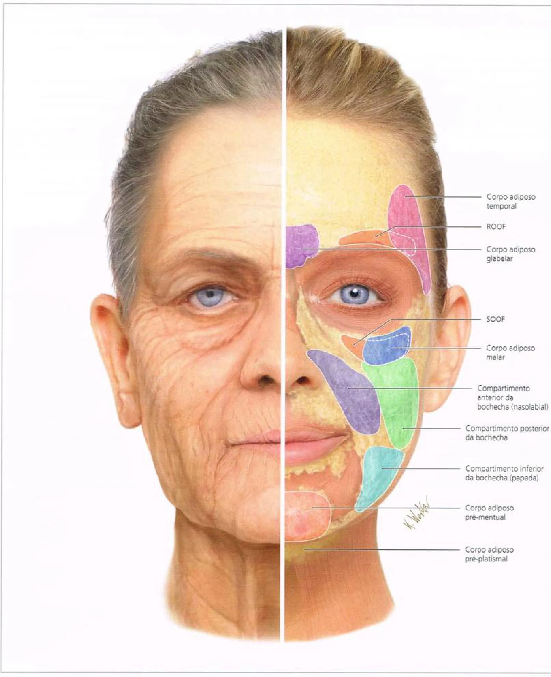 Fig 1. Superficial fat compartments of the face, split young (right) versus aged (left). The lower-third pockets that build the jowl are the inferior cheek compartment (compartimento inferior da bochecha / papada), the pre-mental and the pre-platysmal fat (Radlansky, 2017, p. 58).
> Fuentes: Radlansky, Atlas Ilustrado de Anatomia Clínica da Face [2017].
Fig 1. Superficial fat compartments of the face, split young (right) versus aged (left). The lower-third pockets that build the jowl are the inferior cheek compartment (compartimento inferior da bochecha / papada), the pre-mental and the pre-platysmal fat (Radlansky, 2017, p. 58).
> Fuentes: Radlansky, Atlas Ilustrado de Anatomia Clínica da Face [2017].
Skin and dermis, the quality lane that filler does not fix [20]. The skin over the body is thin anteriorly and thicker over the masseter; the dermis is where the mentalis and platysma insert, which is why superficial product or toxin in the wrong plane becomes visible on animation. Crepe and thinning of the jaw and upper-neck skin are a quality deficit treated with skin boosters, polynucleotides, microneedling and energy, never with structural volume; confusing the two (filling a skin-quality problem) adds weight without improving the surface. The skin layer is the last domino of ageing (§D12.4) and is treated on its own lane.
The jowl is a compartment problem, not a fat surplus [1][2]. Schenck 2018 and Cotofana 2019 map the superficial fat of the lower face as discrete, septated compartments; the jowl is the inferior part of the superficial cheek fat that has descended over an attenuated mandibular retaining ligament and now sits above the border. The Fig 1 panels document, compartment by compartment, that the fat which becomes the jowl (inferior cheek / pre-platysmal pockets) is separate from the buccal (Bichat) deep fat and from the pre-mental fat [5]. Consequence at the chairside: adding volume into the jowl enlarges a descended pocket; the region is treated in front of and below it.
The two ligaments that build the wave [1][3][4]:
| Ligament | Runs | What it does | Failure sign |
|---|---|---|---|
| Mandibular (retaining) ligament / mandibular septum | osteocutaneous, from the anterior mandibular body to the dermis | tethers skin to bone in front of the jowl; the descending tissue stops here | the jowl piles up behind it, the prejowl sulcus appears in front of it |
| Masseteric-cutaneous ligaments | from the masseter fascia to the dermis, along the anterior masseter border | hold the lateral cheek up; caudal fibres are the danger-zone landmark for the cervical/marginal branch | attenuation lets the lateral cheek and jowl descend [7] |
Consensus: every school treats the prejowl sulcus and the border in front of the jowl, not the jowl itself [1][3][4][9]. Discrepancy: whether the mandibular "ligament" is a true osteocutaneous ligament (Cotofana, Standring [1][4]) or a septum forming the caudal wall of the premasseteric space (Mendelson school, cited in [4]): both agree it is a fixed osteocutaneous barrier, so the gesture is unchanged (support in front, do not chase behind).
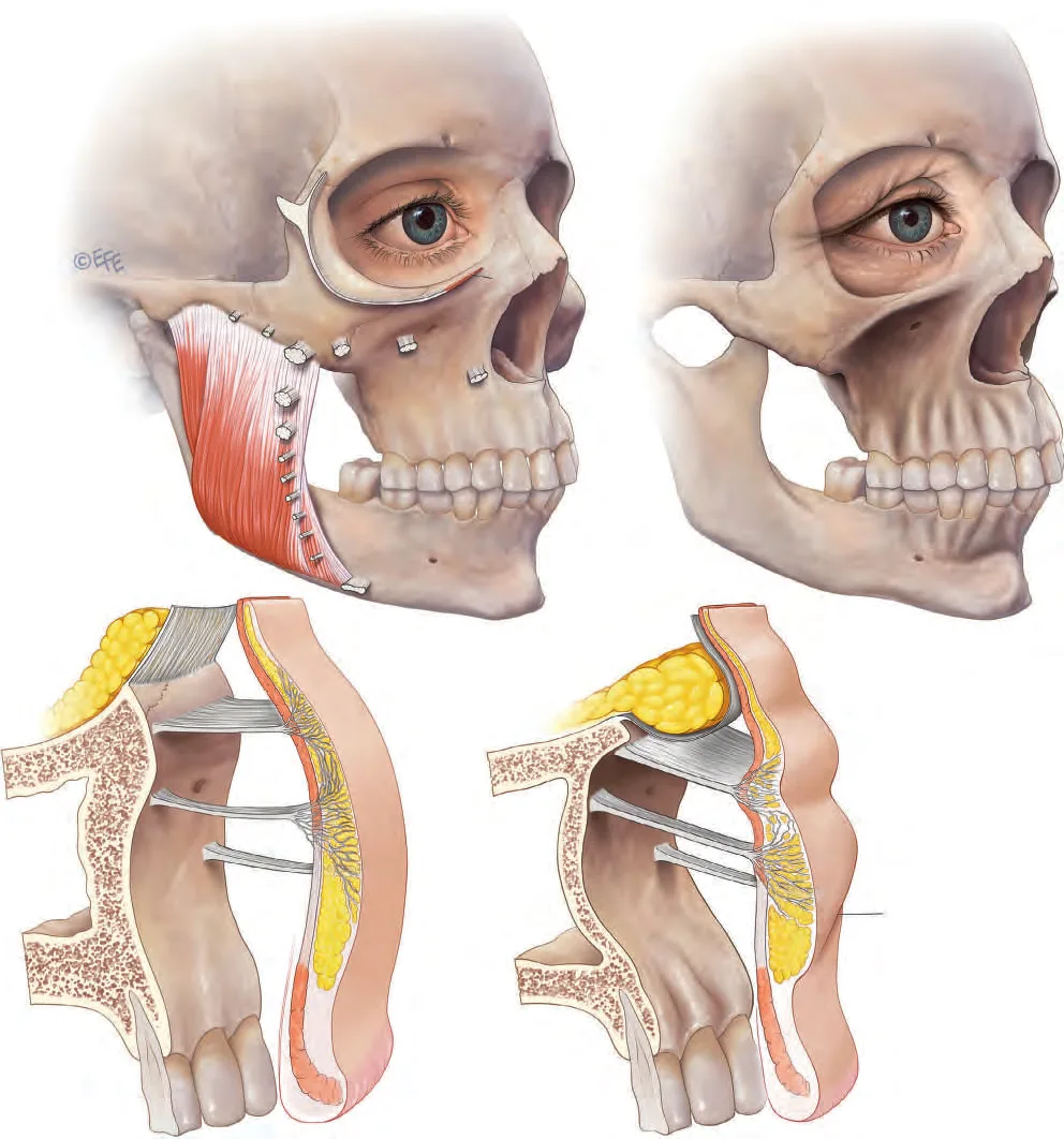 Fig 2. Linking of ageing changes through the tissue layers. Top: youthful (left) with tight retaining ligaments and a full masseter versus aged (right) with an atrophied masseter and a jowl. Bottom: cross-sections in youth (left, ligaments taut, fat held up) versus age (right, ligaments attenuated, fat descended, skin redundant) (Standring, 2015, p. 960).
> Fuentes: Standring, Gray's Anatomy [2016].
Fig 2. Linking of ageing changes through the tissue layers. Top: youthful (left) with tight retaining ligaments and a full masseter versus aged (right) with an atrophied masseter and a jowl. Bottom: cross-sections in youth (left, ligaments taut, fat held up) versus age (right, ligaments attenuated, fat descended, skin redundant) (Standring, 2015, p. 960).
> Fuentes: Standring, Gray's Anatomy [2016].
The Fig 2 cross-sections document the mechanism panel by panel: the retaining ligaments attenuate, the superficial fat compartment slides forward and down, and the skin follows. It is the anatomical reason the border becomes a wave. The platysma, the SMAS of this region, is drawn on the same figure as the roof of the lower premasseteric space; when it loses tone it stops holding the border and becomes a net depressor, which is what the Nefertiti toxin vector addresses (§D12.8).
Why "deep" means something different here than on the cheek [4][6]. On the cheek, deep injection means the deep medial or lateral fat, safely away from named arteries in most planes; on the jawline, the deep-fat plane between platysma and periosteum is exactly where the facial artery and the marginal mandibular nerve run. So a technique taught for the midface ("go deep, on bone") is safe on the jaw only if it is truly on bone; stopping in the deep fat that would be safe elsewhere is, here, the danger plane. The layer diagram is not academic: it is the reason the same word carries a different risk in this region.
Premasseteric space (Standring, [4]). Between the platysma (roof) and the masseter fascia (floor) is a glide space crossed by the masseteric-cutaneous ligaments. Its lower, anterior wall is the mandibular ligament: the exact spot where descent stops and the jowl forms. Knowing the space explains why a cannula in the body plane glides until it snags a ligament: that snag is the anatomical border of the jowl.
The platysma is the anatomy that makes the lower face different [4][26]. In the cheek, layer 4 (SMAS) is a fibrous sheet; in the lower face it is a contractile muscle, the platysma, continuous with the SMAS above. Its upper-lateral fibres and its bands act as depressors of the border, and its medial fibres interdigitate with the DAO and the lip depressors. Three consequences follow: the border has a muscular downward vector that toxin can release (Nefertiti, §D12.8); the marginal mandibular nerve runs deep to it and is the plane hazard; and deep injection in the neck below it reaches the swallowing musculature (dysphagia). The platysma is why the lower third is treated with toxin as much as with filler.
Compartment count, stated for the record [1][2]. The superficial fat of the lower face is not one pad: Schenck and Cotofana describe discrete jowl segments (a superior and an inferior jowl compartment with a mid segment) plus the pre-mental and pre-platysmal fat, each bounded by septa. This is why the jowl descends as a defined bulge with a defined leading edge (the ligament), not as a diffuse sag, and why the correction is geometric (rebuild the line in front) rather than volumetric (fill the sag).
Classic pitfall: treating the jowl as buccal-fat excess and reaching for deep-fat reduction. The jowl is superficial fat that descended over a ligament [1][2]; deoxycholate or buccal-fat removal there is the wrong compartment and sits on the marginal mandibular nerve. Support the prejowl and border; do not deflate the jowl.
D12.3 · Vessels, nerves and the danger zone
Four structures own this border. Two are vascular (necrosis routes), two are neural (functional palsy). Full map with course, depth, variant frequency and consequence; cross-linked to the vascular emergency chapter (§J2) and the danger-zone/anatomy chapter set (§J1-J8) [36]. The current framing prefers naming the vascular safe zones (where the large vessels are not) over memorising danger points, because the lower face is supplied by a dense, variable network [39].
| Structure | Course at the jawline | Depth | Variant / frequency | What fails if you hit it |
|---|---|---|---|---|
| Facial artery | crosses the inferior border at the antegonial (premasseteric) notch, just anterior to the anteroinferior masseter border, then ascends tortuously toward the labial commissure | deep at the border: Doppler mean ≈ 6.3 mm at the lower-border level, artery narrows and deepens as it ascends (≈8.0 mm, diameter 2.1→1.5 mm) [29] | palpable pulse at the notch in most; a premasseteric course and duplicated/absent variants occur; unilateral dominance common | ischaemia/necrosis of chin, lower lip and (via submental branch) the perioral skin; retrograde embolus a theoretical route to deeper territory [6][29][30] |
| Submental artery (facial branch) | leaves the facial artery near the border and runs medially along the mandibular body, deep, toward the chin and floor of mouth | deep, on/near periosteum of the body | present in essentially all; caliber variable | chin and floor-of-mouth ischaemia; mucosal necrosis reported with low-volume chin/border filler [31] |
| Facial vein | posterior and deep/lateral to the artery over the border, running to the retromandibular/angle region | deeper and more lateral than the artery [6] | constant | venous compromise, haematoma; a landmark, not usually the primary embolic risk |
| Marginal mandibular nerve (VII) | runs deep to the platysma, superficial to the facial vessels, along/above the border; loops down below it posteriorly | between platysma and deep fascia | above the border in ≈81 %, below the inferior border in ≈19 % (Dingman-Grabb) [28]; a cadaver series found it along the angle/lower border in 52 % and below the border in 32 % [27]; measured up to 13-40 mm below the border (mean ≈22 mm) [28] | incompetent lower lip, asymmetric smile (loss of depressor labii/DAO); the region's signature functional injury |
| Mental nerve (V3, via mental foramen) | exits the mental foramen on the mid-body, roughly below the second premolar, ~ mid-way between border and alveolar crest | intraosseous then subcutaneous | foramen position varies with dentition and resorption; may sit higher on an edentulous, resorbed mandible | lower-lip and chin numbness/dysaesthesia; a depot in the foramen is the mechanism [4][6] |
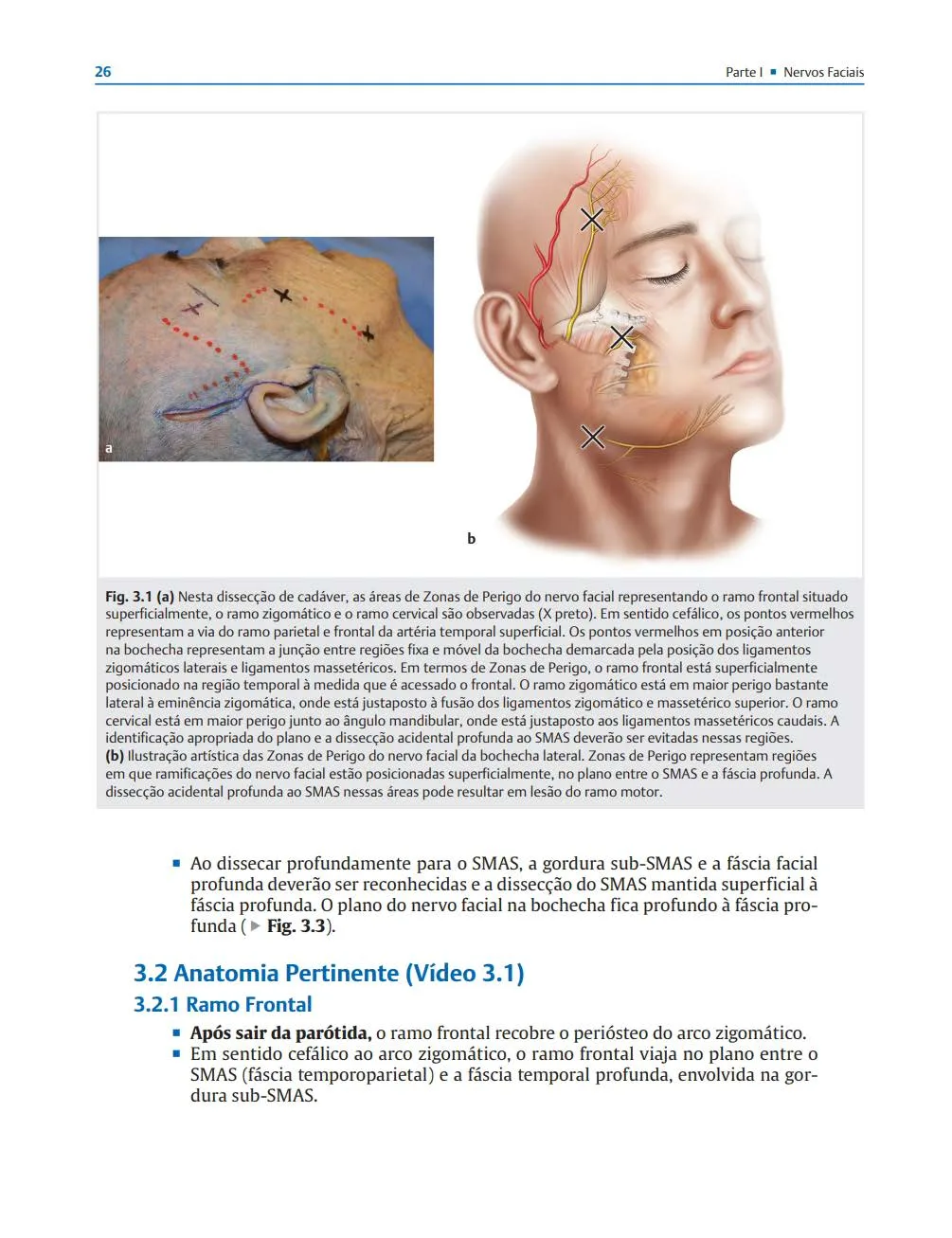 Fig 3. Facial-nerve danger zones (marked X). The branches lie in the plane between the SMAS and the deep fascia; the cervical branch is most exposed at the mandibular angle, juxtaposed to the caudal masseteric ligaments, and accidental dissection deep to the SMAS there risks motor injury (Rohrich, 2020, p. 33).
> Fuentes: Rohrich, Facial Danger Zones / Zonas Faciais de Perigo [2020].
Fig 3. Facial-nerve danger zones (marked X). The branches lie in the plane between the SMAS and the deep fascia; the cervical branch is most exposed at the mandibular angle, juxtaposed to the caudal masseteric ligaments, and accidental dissection deep to the SMAS there risks motor injury (Rohrich, 2020, p. 33).
> Fuentes: Rohrich, Facial Danger Zones / Zonas Faciais de Perigo [2020].
The cervical branch and the marginal mandibular branch are the two that matter at the jawline: the cervical branch is most exposed at the angle, and the marginal mandibular branch governs the lower-lip depressors along the border. Both sit in the same superficial SMAS-to-deep-fascia plane, which is why a single plane discipline protects both.
The Fig 3 dissection documents the operative point for this region: the motor branches of VII sit superficially, in the SMAS-to-deep-fascia plane, and the cervical/marginal territory is most exposed at the angle. For an injector this translates into a plane rule, not a depth-to-avoid: on the border, either stay superficial (subdermal cannula) above the nerve, or on bone (supraperiosteal) below it; the dangerous plane is the intermediate one where the nerve and vessels live [7].
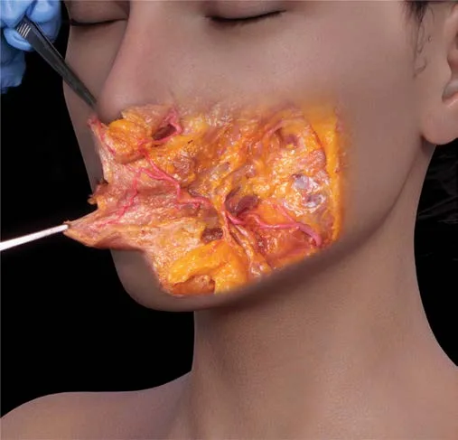 Fig 4. The facial artery ascends over the mandible, deep to the platysma, and runs a tortuous course into the lower face; a live vessel at the antegonial notch (Pirayesh, 2020, p. 232).
> Fuentes: Pirayesh, Aesthetic Facial Anatomy Essentials for Injections [2020].
Fig 4. The facial artery ascends over the mandible, deep to the platysma, and runs a tortuous course into the lower face; a live vessel at the antegonial notch (Pirayesh, 2020, p. 232).
> Fuentes: Pirayesh, Aesthetic Facial Anatomy Essentials for Injections [2020].
The red line, in five seconds: the antegonial notch is where the artery is most accessible to a needle, and it is palpable. The Fig 4 dissection shows why the notch matters: the vessel is right there, deep to platysma, crossing the bone. Palpate the pulse; if a structural bolus is planned near the notch, place it on bone, deep to the artery, aspirate, and inject low and slow, or move the entry to a cannula in the subdermal plane [6][29][30]. Palpating the facial-artery pulse over the jaw before injecting is a long-standing procedural safeguard, not a new one [38].
Depth reconciliation, values kept separate, not averaged: the Doppler meta-analysis reports the facial artery at ≈6.3 mm at the lower-border level and deeper above it [29]; the danger-zone texts describe it as "deep to platysma, on periosteum at the notch" [6]. Both are compatible: at the border the artery is deep (near bone), and a supraperiosteal bolus is only safe when it is truly on bone and the notch is respected. The disagreement to preserve is the marginal mandibular nerve frequency below the border: 19 % (Dingman-Grabb, posterior to the facial artery) [28] versus 32 % (Batra cadaver series) [27]. Plan for the higher figure: assume the nerve can be below the border in up to a third of sides, and keep border toxin superficial and away from the depressors.
Marginal mandibular nerve, variants that matter [27][28]. The nerve arises as one, two or three branches (roughly 37 %, 43 %, 20 % in one cadaver series [27]) and dips furthest below the border posteriorly, near the angle and the antegonial notch, before rising anteriorly toward the depressors it supplies. Measured excursions below the border reach 13-40 mm in some series [28]. Practical reading: the posterior border near the angle and notch is where a low or deep point is most likely to reach the nerve, which coincides with the artery zone; the same "on bone or superficial, never the intermediate plane" rule protects both.
Mental nerve, the underused landmark [4][6]. The mental foramen lies on the mid-body, about below the second premolar, roughly midway between the alveolar crest and the inferior border in a dentate mandible, and higher on a resorbed, edentulous mandible. A body bolus placed without locating it risks a depot in or on the foramen, giving lower-lip and chin numbness. Marking it before mid-body work is a five-second safeguard.
Ultrasound is the disambiguator (§D12.5): a linear-probe scan at the mid-body localises the facial artery and vein before a body bolus and confirms the plane of a cannula; the meta-analysis found the artery detectable at the lower border in almost all cases [29][30].
Facial-artery depth by level (Doppler meta-analysis) [29]:
| Level | Location | Mean depth | Mean diameter |
|---|---|---|---|
| 1 | lower border of the mandible (the notch) | ≈6.3 mm | ≈2.1 mm |
| 2 | cheilion (mouth corner) | intermediate | intermediate |
| 3 | lateral nasal ala | ≈8.0 mm | ≈1.5 mm |
The artery deepens and narrows as it ascends: widest and most superficial at the border, deep and thin near the ala [29]. For the jaw this means the border-level vessel is a wide, relatively superficial target that a body cannula must respect, and the safe supraperiosteal bolus at the notch must be deeper than the artery, on bone. The submental branch adds a medial deep route along the body toward the chin [6][29].
Venous and glandular landmarks: the facial vein sits posterior and deeper than the artery; posteriorly, at the angle, the retromandibular vein and the tail of the parotid are the deep structures, which is why aggressive deep work at the angle risks bleeding and why masseter toxin is kept in the muscle, not driven to the deep angle [4][6].
Classic pitfall: a "supraperiosteal" angle or notch bolus that is actually in deep fat, not on bone. That is precisely the plane of the facial artery. Bone contact is not optional at the notch: no bone, no bolus [6][29].
D12.4 · Regional ageing
The lower third ages in a fixed order, and the order dictates the treatment order. Bone goes first and skin goes last; treating the skin of a face whose bone has resorbed is treating the last domino [3][43].
AGEING SEQUENCE: mandibular line (what fails, in order)
① BONE          gonial angle opens (more obtuse), ramus & body lose
                height, prejowl & pre-angular cortex resorb  [44]
      ↓
② DEEP FAT      thin premasseteric/prejowl deep fat depletes; loss of
                deep support under the border  [3]
      ↓
③ LIGAMENT      mandibular ligament & masseteric-cutaneous ligaments
                attenuate → the tether that stopped descent loosens  [1][3]
      ↓
④ SUPERFICIAL   jowl (inferior cheek) compartment slides forward & down
   FAT DESCENT  over the loosened ligament → the border becomes a wave  [1][2]
      ↓
⑤ SKIN          dermal thinning, elastosis, redundancy; platysma tone
                loss adds a downward vector  [3][4]
| Domain | What is lost / what changes | Clinical read | Where it is treated |
|---|---|---|---|
| Bone | gonial angle more obtuse, ramus + body height fall, prejowl cortex resorbs, mental foramen relatively rises | shortened, posteriorly-rotated lower face; loss of the platform the soft tissue sits on | supraperiosteal structural HA/CaHA at angle and prejowl [12][14][44] |
| Deep fat | premasseteric/prejowl deep fat depletes | loss of deep projection at the border | deep/supraperiosteal volume [3] |
| Ligament | mandibular + masseteric-cutaneous ligaments attenuate | the jowl is released to descend; prejowl sulcus deepens | support in front (prejowl), energy/threads for the ligament laxity [1] |
| Superficial fat | jowl compartment descends over the border | the wave: convexity (jowl) then concavity (prejowl) | never filled directly; border and prejowl rebuilt around it [1][2] |
| Skin + platysma | dermal thinning, elastosis, platysmal downward pull | crepe, redundancy, marionette deepening | skin boosters/EBD for quality; toxin for the platysma vector [26] |
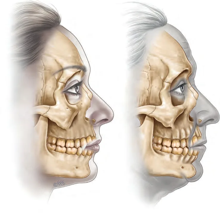 Fig 5. Youthful (left) and aged (right) facial skeleton with the soft-tissue profile overlaid. Skeletal resorption of the mid and lower face changes the profile independent of any soft-tissue change (Standring, 2015, p. 959).
> Fuentes: Standring, Gray's Anatomy [2016].
Fig 5. Youthful (left) and aged (right) facial skeleton with the soft-tissue profile overlaid. Skeletal resorption of the mid and lower face changes the profile independent of any soft-tissue change (Standring, 2015, p. 959).
> Fuentes: Standring, Gray's Anatomy [2016].
Bone first, and it is measurable. Shaw 2010, on 3D CT of 120 subjects across three age bands, found the bony elements of the mandible change significantly with age in both sexes: the gonial angle becomes more obtuse and the ramus and body lose height, which shortens and posteriorly rotates the lower face [44]. Mendelson and Wong 2012 place the prejowl area of the mandible among the resorption-prone regions and argue that failing to rebuild the skeletal platform caps what any soft-tissue procedure can achieve [43]. The Fig 5 profile documents the point: the aged skeleton (right) has changed the outline before a single soft-tissue manoeuvre. This is why the structural HA and CaHA lane exists for the jaw: it substitutes for lost bone platform, not for lost fat.
Then the ligament and the fat, in that order [1][3]. The Cotofana aging review describes the sequence as deep support failing before superficial descent: the deep fat and the ligaments give way, and only then does the superficial jowl compartment slide forward. The Fig 2 cross-sections (§D12.2) are the same event drawn through the layers. This is the anatomical justification for "the jowl is not filled": by the time the jowl is visible, the fault is upstream (bone, ligament, deep fat), and that is where the correction goes.
Sex differences that change the plan [44]: men start with a squarer, wider angle and lose it toward a female-pattern narrowing; women show greater ramus remodelling. A wide gonial angle is a masculine trait, so widening a female angle masculinises the lower third, and if it was done with an irreversible biostimulator there is no way back (§D12.7).
Male versus female ageing targets, concretely [44]. The male lower third starts squarer and wider (a wide gonial angle, a tall ramus, a broad chin) and ages toward a female-pattern narrowing; the female lower third is narrower and softer from the start. So the male restoration goal is often width and angularity (rebuild the angle and the ramus platform), while the female goal is almost always line and shadow without width. Applying the male recipe (angle widening) to a female face masculinises it, and doing so with an irreversible biostimulator makes the error permanent. This is why the sex target is a documented decision at assessment, not an assumption.
The platysma vector. Beyond bone and fat, the platysma (the SMAS of the region) loses tone and, together with its bands, becomes a net downward pull on the border, which is why a toxin that releases that pull (Nefertiti vector, §D12.8) produces a real, if modest, lift without adding weight [26].
Consensus: correct the deepest failed layer first (bone/deep support), soft tissue after [3][43][44]. Discrepancy: the "skeletal-first" school (Mendelson [43]) treats bone platform as the primary target, while a "ligament-and-descent" reading emphasises re-supporting the attenuated ligament with energy/threads before volume; both agree the jowl fat itself is never the target, so at the chairside the gesture converges.
Where the bone resorbs, specifically [43][44]. Mendelson and Wong list the prejowl area of the mandible among the resorption-prone zones, alongside the maxilla and orbital rim [43]; Shaw's 3D-CT series shows the mandibular body and ramus lose height and the angle opens [44]. Two clinical signs follow: the prejowl sulcus deepens because the bone in front of the mandibular ligament recedes, and the mental foramen sits relatively higher on a resorbed, edentulous mandible, which moves the nerve into the field of a body injection. The chin resorbs too, which is why chin and jaw are planned together (§D13).
The skin and the platysma close the sequence. Dermal thinning and elastosis reduce the skin's ability to drape over a rebuilt border, and platysmal tone loss adds the downward vector; in a face where the skin has already failed, volume under it does not re-tension it and can make it hang. This is the anatomical statement of the pinch-test rule: when the skin is the failed layer, the answer is energy, threads or surgery, not more volume [1][3].
Classic pitfall: reading the aged jawline as "needs filler in the jowl". The jowl is the last visible step of an upstream failure [1][3]. Filling it chases the symptom, adds descending weight, and skips the bone/ligament fault that actually changed the border.
D12.5 · Assessment
Measure the bone, photograph the border, test it moving, then scan the artery. Four lanes, each answering a different treatment question.
| Lane | What | Tool / metric | Decides |
|---|---|---|---|
| Measure (static) | gonial angle, ramus + body height, bigonial width, chin projection, cervicomental angle, ogee/contour | goniometry on photo or 3D; E-line / Riedel for chin projection; ALJDS (5-point jawline definition) or Merz jawline scale for grading | is the deficit skeletal (angle/ramus) or contour (border), and how severe [8][12][17] |
| Photograph | frontal, right/left profile, both obliques, neutral head + Frankfort plane, plus a profile with the neck relaxed | standardised distance/lighting; 3D photogrammetry (VECTRA) for gonial-angle quantification | baseline, side asymmetry, and the profile the patient never sees themselves in [6] |
| Test dynamically | clench (masseter bulk + dominance, paradoxical bulge risk), full smile (baseline lower-lip symmetry before any border toxin), pinch test of border skin, submental palpation | clinician exam | additive vs subtractive plan; masseter anterior/posterior dominance; laxity vs volume [21][24] |
| Scan (ultrasound) | facial artery/vein at mid-body; masseter thickness and layer; product/plane after treatment | linear probe, lower-face vascular-mapping protocol | pre-bolus vessel map; masseter dose planning [29][30] |
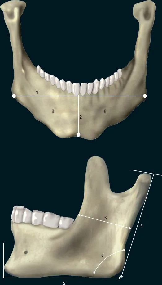 Fig 6. Mandibular measurements used to plan structural contouring: (1) bigonial width, (2) mandibular body height, (3-5) ramus dimensions and length, (6) gonial angle (Vieira Braz, 2017, p. 129).
> Fuentes: Vieira Braz, Atlas de Anatomia e Preenchimento Global da Face [2017].
Fig 6. Mandibular measurements used to plan structural contouring: (1) bigonial width, (2) mandibular body height, (3-5) ramus dimensions and length, (6) gonial angle (Vieira Braz, 2017, p. 129).
> Fuentes: Vieira Braz, Atlas de Anatomia e Preenchimento Global da Face [2017].
The two measurements that route the whole plan [8][44]. The Fig 6 landmarks separate the two deficits the region presents with: an open gonial angle with a short ramus (measurements 4-6) is a skeletal problem, corrected with structural supraperiosteal volume at the angle; a preserved skeleton with a lost border shadow is a contour problem, corrected along the body and prejowl. Quantifying the gonial angle (goniometry on a standardised profile, or 3D VECTRA) turns "the jaw looks weak" into a targeted plan and a before/after that can be graded.
Chin projection references, used as guides not targets [6][43]. The E-line (Ricketts, nose-tip to chin-pommel) and the Riedel plane describe where a balanced chin sits relative to the lips; a chin behind these lines reads as retruded and steepens the cervicomental angle, weakening the whole jaw. These are population-derived and vary by sex and ethnicity, so they orient the plan rather than set a number; the point for the jaw injector is that an under-projected chin makes any border work look weaker and should be addressed with the jaw (§D13).
Grading scales, and why the trial ones matter [12][17]. The jawline pivotal literature grades with a validated 5-point jawline definition scale (ALJDS) and requires a ≥1-grade improvement per side as the response threshold; the CaHA and combination literature uses the Merz Aesthetics jawline scale [17]. Using a validated scale, not a subjective impression, is what lets a clinic decide whether a touch-up is warranted at day 30 (the trial rule) rather than over-filling on the day.
The dynamic exam is where additive vs subtractive is decided [21][24]: - Clench. A wide lower face that widens further on clenching is masseteric hypertrophy: the correct treatment is toxin (subtract), not filler. Palpating and (ideally) scanning the masseter also flags anterior versus posterior dominance, which predicts paradoxical bulge and changes injection points (§D12.8). - Full smile before any border work. Document lower-lip symmetry first: a pre-existing marginal-mandibular asymmetry must be on record before border toxin or a body bolus, or a baseline finding becomes an alleged complication. - Pinch test of the border skin. True laxity means filler will add descending weight, not definition: the order becomes energy/threads first, or surgery (§D12.6). - Submental palpation. A firm, non-pinchable bulge under the border is not fat (submandibular gland ptosis, node, or mass): it is not injected, it is worked up.
Ultrasound has moved from optional to protocolised for the border [29][30]. A linear-probe scan at the mid-body localises the facial artery and vein before a body bolus; the meta-analysis detected the artery at the lower border in almost all cases and quantified its depth and diameter along the body [29]. Post-injection, the same probe confirms the plane and locates a nodule as product versus not. (P) In a region where the artery is deep and palpable but variable, pre-procedural mapping converts an anatomical average into this patient's actual vessel.
Ultrasound of the masseter is the newest assessment lever [24]. Beyond bulk, a static and dynamic scan classifies anterior versus posterior functional dominance: an anterior-dominant masseter is the one prone to the paradoxical bulge, because the DIT tendon leaves the anteroinferior portion under-treated by a standard posterior three-point injection. Pre-assessing dominance lets the injector shift or add a point to the dominant unit and pre-empt the bulge, turning a reactive complication into a planned adjustment (§D12.8, §D12.10). The same probe measures thickness for dose planning and can document the reduction at follow-up.
Referral triggers to record at assessment (detailed in §D12.6): frank cervical skin excess, an established platysmal band with redundant skin, a true jowl with excess skin, a low hyoid, or a class-II skeletal pattern. Each is a surgical or orthodontic problem that inject-only treatment will bill for without solving.
The five-view photographic standard for this region [6][12]: frontal at rest, both true profiles, both obliques (three-quarter), all at a fixed distance and lighting with the head in the Frankfort horizontal, plus a profile with the neck deliberately relaxed to expose the true cervicomental angle and any submental component. The oblique views are where jawline definition and the jowl step are best judged and where the ALJDS is graded; the profile is where chin projection and the cervicomental angle are read. A frontal-only record cannot grade the border and cannot document the profile that the treatment is actually aimed at.
Angles worth putting a number on [8][44]: the gonial angle (obtuse with age and in the descended face), the cervicomental angle (obtuse with a low hyoid or a heavy submentum), and chin projection against the E-line or Riedel plane. Quantifying them separates a skeletal deficit (short ramus, open angle) from a soft-tissue one (lost border shadow) and gives a defensible before/after. 3D photogrammetry (VECTRA) automates the gonial-angle measurement and the volume estimate for structural planning.
Classic pitfall: grading the jaw from the frontal photo alone. The lower third is diagnosed in profile (chin projection, cervicomental angle, ramus) and treated from the front, which is exactly why it is mis-assessed [6]. No profile, no plan.
D12.6 · Goal and patient selection
Who benefits, who does not, what leaves the room for a surgeon. The non-surgical jaw has a lower and stiffer ceiling than the midface: it defines and camouflages, it does not remove skin or lift a true jowl [35].
| Candidate | Deficit | Non-surgical plan | Verdict |
|---|---|---|---|
| Young, skeletally weak angle, good skin | short ramus / open angle, no laxity | structural HA/CaHA supraperiosteal at angle + border | ✅ ideal responder |
| Mild-moderate jowl, border still readable | early descent, prejowl sulcus | support prejowl + border, toxin for platysma/DAO | ✅ camouflage, set expectations |
| Masculinising a defined but under-projected male jaw | contour/projection | high-G′ structural volume, angle + border | ✅ with the gender target stated |
| Wide lower face on clench | masseteric hypertrophy | toxin, not filler | ✅ subtractive plan (§D12.8) |
| True border laxity (positive pinch) | skin/ligament laxity | energy/threads first, then less volume | ⚠ volume alone worsens it |
| Frank cervical skin excess / established platysmal band with redundant skin | skin surplus | none | ❌ refer: lower rhytidectomy / neck lift |
| True jowl with excess skin | descended fat + skin surplus | none injectable removes skin | ❌ refer: facelift |
| Low hyoid, obtuse cervicomental angle | constitutional | none changes the hyoid | ❌ declare prognosis before charging |
| Class-II retrognathia, abnormal occlusion | skeletal + functional | filler only camouflages the profile | ❌ refer: orthodontics / orthognathic surgery |
| Body dysmorphia / unrealistic "snatched" demand | psychological | none | ❌ decline, safeguard |
The bite test is the cheapest and most-skipped step [6]. Ask the patient to occlude and look at the bite. Microgenia (small chin, normal occlusion) is a filler indication; retrognathia (whole mandible retruded, class-II malocclusion) is an orthodontic and orthognathic problem that filler only camouflages. Distinguishing them in thirty seconds redirects the entire plan and is absent from most filler courses.
The masculinisation trap, stated explicitly [44]. A wide, square gonial angle is a masculine trait. Widening a female angle with structural volume masculinises the lower third, and if it was done with an irreversible biostimulator (PLLA, PCL, CaHA past its dissolvable window) there is no reversal. In the female jaw the usual target is line and shadow, not width; angle widening is a deliberate, consented decision for a specific aesthetic goal, not a default.
The weight principle governs selection [1][3]. Everything added to the lower third pulls down on a tissue that is already descending. In the midface, over-volumising makes an odd face; in the lower third it also accelerates descent. The positive pinch test is therefore a hard gate: laxity means the correct first move is energy, threads or surgery, and volume, if any, comes second and smaller.
What is referred, and to whom: - Frank skin excess (cervical or jowl): lower-face/neck rhytidectomy. No injectable removes skin. - Established platysmal bands with redundant skin: platysmaplasty / neck lift, not only toxin. - Subplatysmal fat, submandibular gland ptosis, a firm non-pinchable mass: surgical or diagnostic work-up, never injected. - Class-II / retrognathia: orthodontics and orthognathic surgery. - Low hyoid: counselled that the prognosis for any lower-face treatment is limited, before any fee.
A firm submental bulge is not fat until proven otherwise [6]. Under the border, a soft pinchable pad is subcutaneous fat; a firm, delimited, non-pinchable swelling is a ptotic submandibular gland, a lymph node, or a mass, and injecting or lipolysing it is an error with risk. Palpation that distinguishes the two is a required step, and any unexplained firm mass is investigated, not treated. The submental fat itself (deoxycholate territory) belongs to §D6/§D13; here it matters only as a differential the jaw injector must clear before working near the border.
The chin is part of the plan, not a separate region for the patient [43]. A retruded or short chin makes any jawline look weaker and steepens the cervicomental angle; projecting the chin (§D13) often does more for the perceived jaw than border filler, and the two are assessed together. Selling border volume while ignoring an under-projected chin treats the symptom and misses the lever.
Consensus: in a lax or skin-excess lower third, energy/threads/surgery precede volume, and volume is reduced or omitted [1][3]. Discrepancy: "preventive" jaw definition in the young, defined jaw. One school offers early structural augmentation to pre-empt descent; the counter-school warns that adding weight and an irreversible product to a jaw that has not yet aged manufactures the square young face and the future descent it was meant to prevent [9][44]. The decision variable is the pinch test and the gonial-angle measurement, not the request.
The demand-driven overtreatment pattern of this region [1][9]. Because a sharp jawline is currently a high-demand feature, the region attracts repeat structural volume, session after session, chasing an ideal the patient's anatomy cannot hold. The result is a heavy, over-projected lower face on a descending tissue: the opposite of the sharp line requested. The defence is the ceiling conversation at assessment, a validated grading scale to show that a result was achieved, and a willingness to say the next syringe will worsen, not sharpen, the line. This region punishes "a little more" more than any other.
(P) The most valuable sentence in this consultation is often "this is a surgical problem", said before any product is opened: it protects the patient from paying for a result an injectable cannot deliver, and it protects the clinic from an unfixable dissatisfaction.
A phenotype decision, in one pass: - Thin face, lost border, good skin, normal bite: structural filler (angle if the ramus is short, border and prejowl for the line). The ideal responder. - Wide face, widens on clench: masseter toxin. Re-measure before any filler [21]. - Positive pinch, lax border: energy/threads first; volume second and smaller, or not at all [1]. - Frank skin excess, true jowl, established bands: surgery. No injectable removes skin. - Small chin, normal bite: chin projection (§D13), which also opens the cervicomental angle. - Retruded chin, class-II bite: orthodontics/orthognathic referral; filler only camouflages [6]. - Low hyoid: counsel limited prognosis before any fee.
The psychological gate [42]. A demand for an extreme, "snatched" jawline out of proportion to the anatomy, a fixation on a filtered image, or serial dissatisfaction after technically good results should trigger a pause for body-dysmorphia screening, not more product. Injecting a dysmorphic concern deepens it. This is a decline-and-safeguard decision, and it belongs in patient selection.
The realistic ceiling, said out loud [1][12]. Non-surgical jaw definition can build a border, sharpen an angle and camouflage a mild-to-moderate jowl; it cannot remove skin, lift a true descended jowl, or change a class-II skeleton. Half a defined jawline is worse than none (it reads as heaviness), so the volume, the staging and the cost are agreed before the first depot, and the patient is told what the ceiling is for their phenotype.
Classic pitfall: accepting "I want a sharper jaw" as a filler order. In the wide, hypertrophic face the answer is toxin (subtract); in the lax face it is energy/surgery; only in the thin face with a lost border and good skin is it filler [21][44]. The complaint is identical; the correct gesture is opposite.
D12.7 · Technique, the full grid
The jawline is built with one of five product classes, through one of two instruments, in one of two safe planes, with one of three movements, per one of two schools. The grid below is the whole option space; a row with no alternative is a region left half-treated [9][11][12].
The region has two sub-targets that demand different tools. The gonial angle and prejowl are a structural, on-bone problem, solved with a high-G′ product placed supraperiosteal by needle bolus; the body and border are a shadow-line problem, solved with an equal or softer product laid subdermally by retrograde cannula [11][12][14]. The toxin half of the region (masseter, platysma, DAO, mentalis) is treated separately and usually first, because it changes the resting shape the filler must match (§D12.8, §D12.9) [21]. Everything below enumerates the full option space for those decisions: product by rheology and role, instrument by gauge and length, plane, movement, school, volume with ceilings, and the point at which the correct answer is not an injectable at all [1][14]. Read it as a grid, not a recipe: the phenotype (§D12.6) selects the row, and the pinch test and the gonial-angle measurement decide between the structural and the support-first reading [1][44].
Product grid (by rheology and role)
| Product | Rheology / property | Role at the jawline | Plane | Duration | Reversible? |
|---|---|---|---|---|---|
| High-G′ HA (Volux / VYC-25L, VYCROSS) | very high G′, low-medium cohesivity, high projection capacity, "mimics bone" | structural angle + border + prejowl; the reference product for on-label jaw [12] | supraperiosteal bolus (angle), subdermal (body) | ~18-24 mo; jaw pivotal sustained to 12 mo with maintenance [12] | yes (hyaluronidase) |
| High-G′ HA (Voluma / VYC-20L) | high G′, medium cohesivity | structural, where Volux unavailable; midface crossover | supraperiosteal / subdermal | ~12-18 mo | yes |
| Cohesive HA (CPM / Belotero-type, Restylane Lyft) | lower G′, higher cohesivity | softer border blending, transitions | subdermal / supra-SMAS | ~9-12 mo | yes |
| CaHA normal (Radiesse / Radiesse+) | high elasticity + biostimulation (dual: fill now, collagen later); FDA on-label jaw 2023 | angle + border structural, skin tightening bonus | ala-tragus rule: supraperiosteal above the line, subdermal below [14][16] | ~12-18 mo (biostimulation longer) | no dissolver |
| CaHA hyperdiluted (1:1 / 1:2) | biostimulatory, minimal volumising | skin quality + light tightening of the border/upper neck | subdermal | progressive, months | no dissolver |
| PLLA (poly-L-lactic acid) | collagen biostimulator, no immediate volume | slow structural support + skin quality at the angle | deep/supraperiosteal, diluted | 18-24 mo, gradual | no dissolver |
| PCL (polycaprolactone, Ellansé) | biostimulator with immediate gel carrier | regenerative border support, longer scaffold | subdermal / supraperiosteal | ~24-48 mo | no dissolver |
| PMMA (Bellafill) | permanent microspheres in collagen | permanent contour; high threshold, expert only | subdermal | permanent | no (surgical only) |
| Autologous fat | living graft, variable take | large-volume structural restoration | supraperiosteal / deep | long, unpredictable take | partial |
| Polynucleotides | regenerative, skin quality | dermal quality of the border/upper neck; not a volumiser | intradermal / subdermal | months, repeated | n/a |
| Skin booster (low-G′ HA) | hydration, not structure | crepe skin of the jaw/neck | intradermal | months | yes |
The rheology decision, stated once [11][12]. Fagien 2019 differentiates fillers by G′ (elastic modulus, resistance to deformation) and cohesivity (resistance to fragmentation); the two do not track together (VYCROSS gels are high-G′, low-medium cohesivity). For a load-bearing, shadow-casting border on mobile bone, the target is high G′ with enough cohesivity to stay a discrete projection: that is the VYCROSS/Volux profile, which is why it is the on-label jaw product. Soft, high-cohesivity, low-G′ gels blend transitions but do not build a border; using them for structure wastes product and defines nothing.
CaHA and the ala-tragus rule [14][16][17]. Radiesse+ is the first FDA-approved filler on-label for jawline (supraperiosteal and/or subdermal), building on an earlier jawline-rejuvenation consensus for the material [15], and Moradi and Green 2023 codify best practice: draw the ala-tragus line, place CaHA supraperiosteal above it (angle, posterior body, on bone) and subdermal below it (anterior body, border), where the tissue is thinner. CaHA is dual: immediate volume plus collagen biostimulation, so it both contours and tightens. The price is irreversibility: there is no CaHA dissolver, so it does not belong in the lax anterior border of a face that may need adjustment.
Named products and where they sit on the jaw
| Product (class) | Relative G′ | Role at the jaw | Typical target |
|---|---|---|---|
| Juvéderm Volux / VYC-25L (VYCROSS HA) | very high | on-label structural jaw; angle + border + prejowl | supraperiosteal bolus (angle), subdermal (body) [12] |
| Juvéderm Voluma / VYC-20L (VYCROSS HA) | high | structural where Volux is unavailable | supraperiosteal / subdermal |
| Restylane Lyft / Defyne (NASHA / XpresHAn) | high / medium | border projection and flexible contour | supraperiosteal / subdermal |
| Belotero Volume / Intense (CPM HA) | medium, high cohesivity | blending the border, softer transitions | subdermal |
| Radiesse / Radiesse+ (CaHA) | high + biostimulation | angle + posterior body, skin tightening | ala-tragus rule: supraperiosteal above, subdermal below [14][16] |
| Sculptra (PLLA) | biostimulator | slow structural support + skin quality | deep / supraperiosteal, diluted, staged |
| Ellansé (PCL) | biostimulator + gel | longer scaffold border support | subdermal / supraperiosteal |
| Bellafill (PMMA) | permanent | permanent contour, expert-only, high threshold | subdermal |
G′ ordering, kept practical [11]. For the load-bearing jaw the working order from most-to-least structural is roughly VYCROSS high-G′ (Volux/Voluma) ≥ NASHA high-G′ (Lyft) > CPM cohesive gels; the softer, high-cohesivity gels are for transitions, not for the border itself. Fagien's point stands: G′ and cohesivity are separate axes, so a gel can be high-G′ and only medium-cohesive (VYCROSS), which is exactly the profile that holds a discrete projection on mobile bone [11].
Biostimulator specifics for the jaw [16][20]. PLLA (poly-L-lactic acid) provides no immediate volume; it drives gradual collagen deposition over months and is placed deep/diluted for slow structural support and skin quality at the angle, in a staged series. PCL (polycaprolactone) pairs an immediate gel carrier with a longer-lasting collagen scaffold, giving both instant contour and durability at the border. Hyperdiluted CaHA (1:1 or 1:2) is used subdermally for skin tightening and quality of the border and upper neck rather than for projection. All three share one hard limitation on this region: no enzymatic reversal, so they are kept off the mobile, thin-skinned anterior border and reserved for deeper, on-bone or skin-quality roles.
Autologous fat is the large-volume structural option when the deficit is global (a whole lost lower-face platform), not a border line: it restores volume as a living graft but with unpredictable take and no fine-line precision, so it is a different tool from a border filler, used for a different problem [37]. PMMA (permanent microspheres) can give a permanent contour but demands a bovine-collagen allergy test and expert placement, and its permanence makes any error permanent; it is a high-threshold, last-choice product on a mobile border. Polynucleotides and skin boosters treat the quality of the border and upper-neck skin (crepe, thinning), never the structure, and are layered in on their own schedule [20].
Threads and energy are the non-injectable structural lane [1]. Suspension threads reposition a descended jowl over a lax ligament (a mechanical lift filler cannot give); monopolar/bipolar radiofrequency and microfocused ultrasound tighten the lax border without adding weight. They are the correct first move on a positive pinch test, where filler would only add descending load. Surgery (lower rhytidectomy, submentoplasty, genioplasty) is the ceiling for frank skin excess, a true jowl, or a class-II chin (§D12.6).
Instrument grid
| Instrument | Gauge / length | Where | Why |
|---|---|---|---|
| Sharp needle | 25-27G, ~13-25 mm | supraperiosteal bolus at angle, prejowl, on bone | precision on bone, controlled depot; aspirate before injecting |
| Microcannula | 25G / 50 mm or 27G / 40-50 mm | body / border subdermal retrograde | pushes vessels aside; the safer instrument over the artery zone |
| Needle or cannula + ultrasound | as above, with linear probe | anywhere near the notch | live vessel map; confirm plane and avoid the facial artery [29][30] |
Plane × movement matrix
| Plane | Bolus / microbolus | Retrograde linear | Fan | Cross-hatch |
|---|---|---|---|---|
| Supraperiosteal (angle, prejowl) | ✅ structural bolus on bone (needle) | rarely | rarely | no |
| Deep fat | ⚠ avoid: the facial artery plane at the notch | no | no | no |
| SMAS / platysma | no (nerve plane) | no | no | no |
| Subdermal / supra-SMAS (body, border) | microbolus (cannula) | ✅ retrograde linear (cannula) | ✅ fan (cannula) | ✅ light cross-hatch for skin support |
| Intradermal | no | no | no | serial puncture / booster only |
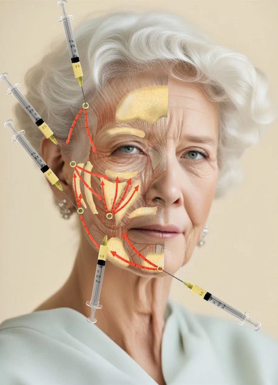 Fig 7. Cannula entry points at the preauricular and mandibular level with fan/retrograde vectors over the lower-face superficial compartments; the movement pattern used for border and body contouring (Piccolo, 2025, p. 60).
> Fuentes: Piccolo, A.R.T. Autologous Regenerative Therapy in Aesthetic Medicine [2025].
Fig 7. Cannula entry points at the preauricular and mandibular level with fan/retrograde vectors over the lower-face superficial compartments; the movement pattern used for border and body contouring (Piccolo, 2025, p. 60).
> Fuentes: Piccolo, A.R.T. Autologous Regenerative Therapy in Aesthetic Medicine [2025].
The Fig 7 vectors document the body technique: a cannula entered at the border or preauricular point, advanced subdermally, and withdrawn in retrograde linear and fan passes to lay a continuous shelf under the border. Contrast this with the angle, where a needle places a supraperiosteal bolus on bone: two instruments, two planes, two movements, for two sub-targets of the same line.
Movements, matched to sub-target: - Bolus / microbolus (supraperiosteal, needle): the structural depot at the angle and prejowl, on bone, aspirated. Small aliquots, single slow depots. - Retrograde linear (subdermal, cannula): the border shelf, laid on withdrawal to build a continuous line. - Fan (subdermal, cannula): distributing product across the body from one entry to cover a segment without multiple punctures. - Cross-hatch (subdermal, cannula): light, for diffuse support or skin scaffolding, not for a sharp border. - Serial puncture / intradermal: skin-quality products (boosters, PN) only; never for structure. - Tower / column: rarely used on the jaw; a vertical stacked depot is a chin-projection manoeuvre (§D13), not a border technique.
The border is a line built by continuous retrograde/fan on the body plus discrete supraperiosteal boluses at the angle and prejowl; it is not a field to be blanket-filled. Blanket-filling widens and weighs.
Schools, kept side by side, never averaged
MD Codes (de Maio) [9]: the jaw and chin are treated with the Jw codes (jawline, from angle to prejowl) and the C codes (chin), each specifying depth, tool and a minimum active volume: - Jw1 (mandibular angle): supraperiosteal bolus on bone, structural, needle. - Jw2-Jw3 (along the body toward the prejowl): subdermal, cannula, retrograde. - Jw4 / prejowl and C (chin) points: supraperiosteal at the prejowl, blending to the chin (§D13 chin). - Product: high-G′ VYCROSS (Voluma/Volux); the method is structural, angle-and-border first.
Rosso mandibular-line technique / structural-line schools: a continuous bolus (angle) + retrograde linear (body) to draw one shadow line, prioritising continuity over posterior width.
A second axis of disagreement: structural volumising versus support-first. The high-load structural school treats the jaw as a bone-substitution problem and places substantial high-G′ volume to rebuild the platform; the support-first school argues that in any face with even early laxity, re-tensioning (energy, threads) before volume gives a better and longer result with less product, because volume on a lax suspension partly spends itself as dead weight [1][3]. Both are correct in their phenotype: high-load structural for the young, tight, skeletally-deficient jaw; support-first for the descending, lax one. The error is applying the high-load recipe to a lax face, which produces heaviness and accelerated descent. The pinch test decides, not the request.
Discrepancy (preserved, not averaged): the structural / angle-forward school (build the angle for character, then the border [9]) versus the continuity-first school (the intermediate border first, the angle last and only if the ramus is short [4][44]). Both use the same products and planes; they disagree on order and on whether to widen the angle at all. The decision variable is the gonial-angle measurement and the sex target: a genuinely short ramus in a male face justifies angle-forward; a female face with a lost border does not, because angle width there masculinises. Never split the difference into a half-wide angle: that is the square-young-face result neither school wants.
The region in one recipe (structural jaw, good skin)
For the common case (a skeletally-deficient or under-defined jaw with acceptable skin and a normal bite): high-G′ HA, needle bolus supraperiosteal on the gonial angle and prejowl for the platform; then a microcannula subdermal retrograde/fan along the body to draw the continuous border shadow; assess in profile mid-procedure; stay under the per-side and per-session ceilings; palpate the notch and keep bone contact for every bolus. Add masseter toxin first (separate visit) if the face is wide from hypertrophy, and platysma/DAO toxin to release the border vector. Reserve CaHA and biostimulators for the posterior, on-bone, or skin-quality roles, and keep the mobile anterior border in a reversible HA. This is the default; every deviation (angle widening, biostimulator on the border, single-visit combining with a first masseter treatment) is a deliberate, phenotype-justified departure, not a shortcut [9][11][12][14][21].
Volume ladder (start low, know the ceiling)
| Level | Volume | Note |
|---|---|---|
| Per point (supraperiosteal bolus) | 0.1-0.2 mL | on bone, aspirate, single slow depot |
| Per side, angle | 0.4-0.8 mL start, ~1.2 mL ceiling | more than this widens rather than defines |
| Per side, body + border | 0.4-0.8 mL | retrograde subdermal, continuous shelf |
| Per side, prejowl | 0.2-0.5 mL | high-yield, small volume |
| Per session, per area | ≤2 mL/area (Voluma-type labelling); jaw pivotal ≈3-4 mL both sides + optional day-30 touch-up [10][12] | stage large corrections; review at 2-4 weeks |
| Ceiling before overfill | when the border stops sharpening and the face starts widening/heavying | stop; reassess in 2-4 weeks, do not keep adding on the day |
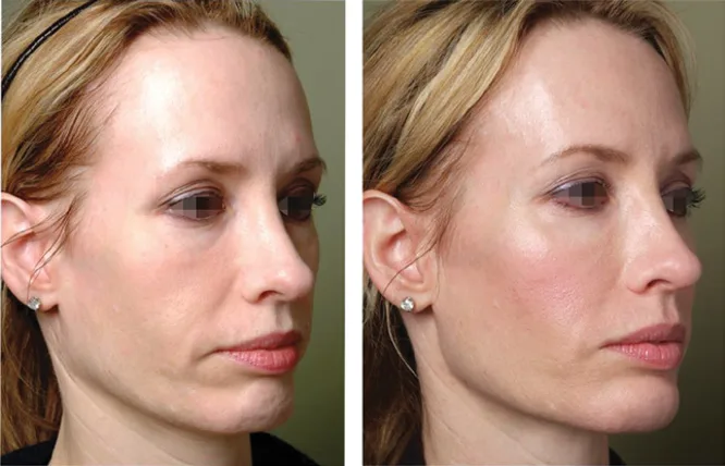 Fig 8. Oblique before (left) and after (right) of jawline contouring, gonial angle to chin: a continuous, shadow-casting border and a less prominent jowl (Jones, 2019, p. 69).
> Fuentes: Jones, Injectable Fillers [2019].
Fig 8. Oblique before (left) and after (right) of jawline contouring, gonial angle to chin: a continuous, shadow-casting border and a less prominent jowl (Jones, 2019, p. 69).
> Fuentes: Jones, Injectable Fillers [2019].
The Fig 8 result is the goal restated visually: a continuous line, not a widened angle [18]. The correction is along the border and prejowl, and the jowl reads less as a step because the line in front of it was rebuilt, exactly the principle of §D12.2 and §D12.4. Illustrated atlases of jawline ageing show the same staged progression from an early prejowl deficit to a frank jowl, and match the correction to the stage [19].
Cannula versus needle on the body, the practical case [29][30]. A blunt microcannula entered through a single port and advanced in the subdermal plane pushes the facial artery and vein aside rather than piercing them, which is why it is the preferred instrument over the body and the notch zone; the trade-off is less precise depth control than a needle. The needle keeps its place for the supraperiosteal bolus on bone, where precision and bone contact matter and where the depot is deliberately deeper than the artery. The two are not interchangeable: needle on bone at the angle/prejowl, cannula subdermal on the body. Aspiration before a needle bolus is a reasonable adjunct but is not a substitute for correct plane and slow, low-pressure delivery.
When the correct answer is NOT an injectable
| Finding | Correct modality | Why not filler |
|---|---|---|
| Border/skin laxity (positive pinch) | monopolar/bipolar RF, microfocused ultrasound (HIFU), fractional laser | volume adds descending weight; energy tightens without weight [1] |
| Descended jowl with a lax ligament | suspension threads (± energy) | threads reposition; filler only pads |
| Frank cervical/jowl skin excess | lower-face rhytidectomy / neck lift | no injectable removes skin |
| Retruded chin with class-II occlusion | orthognathic surgery / genioplasty | filler camouflages, does not correct the skeleton |
| Subplatysmal fat, submandibular gland ptosis | submentoplasty / surgical work-up | wrong compartment, high-risk to inject |
| Skin quality only (crepe, no volume loss) | skin boosters, PN, microneedling ± RF | structure is not the deficit |
(P) The single most protective habit on the jaw is to name, out loud, the modality that is NOT filler when it is the right one: it is the difference between an aesthetic result and an expensive, weight-adding disappointment on a descending tissue.
Classic pitfall: using a soft, high-cohesivity, low-G′ gel to "build" the border, or filling the body in the deep-fat plane "to be supraperiosteal". The first defines nothing and migrates; the second is the facial-artery plane [11][29]. High G′ on bone at the angle, cannula subdermal on the body: match the product and plane to the sub-target.
D12.8 · Toxin of the region
Four muscles shape the lower third with toxin, and the region's whole subtractive plan lives here. Masseter (width), platysma (the downward vector on the border), DAO (the drooping commissure), mentalis (chin support). Doses are onabotulinum-equivalent unless stated; convert for other products, never carry a memorised cross-brand number.
| Muscle | Goal | Points/side | Dose/side (ona) | Per point | Plane / depth | Antagonist / structure NOT to touch | Safety distance |
|---|---|---|---|---|---|---|---|
| Masseter | slim a wide lower face (hypertrophy) | 3 (lower posterior third, in a triangle above the angle) | 20-30 U (dose-finding equivalent ≈48-72 U prabo/abo) [21][22] | ~6-10 U | deep, to bone then withdraw 2-3 mm | anterior masseter / risorius & zygomaticus major (asymmetric smile); do not over-dose both heads unevenly (paradoxical bulge) | stay ≥1 cm above the inferior border (marginal mandibular nerve) and posterior to a tragus-to-commissure line |
| Platysma (Nefertiti vector) | release the downward pull on the border | 4-6 (posterolateral band + subauricular + along inferior border) | ~15-20 U total, 2 U/point | 2 U | superficial (intradermal/subdermal) | depressors of the lower lip along the border; deep neck muscles | small aliquots, low dilution; do not cross deep in the neck (dysphagia) [26] |
| DAO | lift a drooped commissure / soften marionette | 1 | 4-8 U | 4-8 U | deep, at the mandibular border ~1 cm below/lateral to the commissure | depressor labii inferioris & orbicularis oris (too medial → lip asymmetry) | stay at the border, lateral to the modiolus |
| Mentalis | soften pebble chin, support the anterior line | 1-2 | 4-8 U | 4-8 U | deep, central chin | do not inject the belly superficially (visible on animation); avoid DLI | keep central, low on the chin |
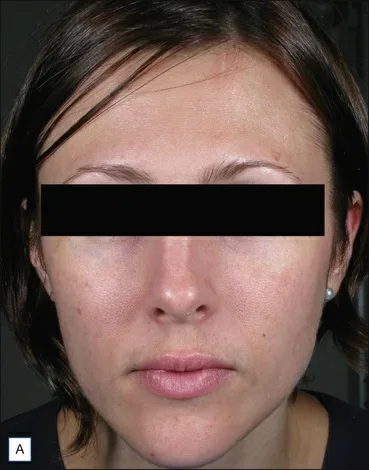 Fig 9. Masseteric hypertrophy widening the lower-facial third: the indication for masseter toxin, where the treatment is subtractive and filler would enlarge the very width the patient wants reduced (Carruthers, 2018, p. 200).
> Fuentes: Carruthers, Botulinum Toxin, Procedures in Cosmetic Dermatology [2018].
Fig 9. Masseteric hypertrophy widening the lower-facial third: the indication for masseter toxin, where the treatment is subtractive and filler would enlarge the very width the patient wants reduced (Carruthers, 2018, p. 200).
> Fuentes: Carruthers, Botulinum Toxin, Procedures in Cosmetic Dermatology [2018].
Masseter, in detail
The injection zone is a triangle, not the whole muscle [21][33]. Ask the patient to clench and mark the inferior-posterior third of the masseter, a triangle bounded by the anterior masseter border, the inferior mandibular border, and a tragus-to-commissure line. The Fig 9 baseline shows why the indication is subtractive: the muscle is the width [34]. Three deep points in that triangle, to bone then withdrawn slightly, keep the toxin in the muscle and away from the neighbours. Named-technique atlases of facial-harmony toxin use the same posterior-inferior triangle and the same risorius-sparing line [41].
Marking the triangle, concretely [33][42]. With the patient clenching, the masseter's anterior and posterior borders and its inferior border become palpable. The safe injection field is the inferior-posterior triangle bounded by: the anterior masseter border (in front, to spare risorius/zygomaticus and the anterior fibres that overlie the facial vessels), a line from the tragus to the mouth corner (above, to spare the risorius), and a line about 1 cm above the inferior mandibular border (below, to spare the marginal mandibular nerve). Three points are spaced within that triangle, each taken deep to bone and withdrawn 2-3 mm so the depot is in muscle, not on periosteum or in the overlying fat. The lowest point drives most of the visible slimming, and it is also the one closest to the nerve, so the 1 cm margin is not negotiable.
Dose, kept as a range with the conflict preserved [21][22][23]. The onabotulinum-equivalent working range is 20-30 U per side, higher in a very hypertrophic male masseter. The prabotulinum dose-finding RCT (24/48/72/96 U groups) found 48-72 U optimal, with 24 U insufficient and 96 U causing chewing discomfort, and re-injection at ~12 weeks [21]; an abobotulinum triple-blind RCT used 75 U per side [22]. These are different toxins on different unit scales: do not average them into a single number, and do not carry an ona dose over to abo or prabo without converting. Effect onset over 2-4 weeks, peak slimming at 2-3 months, duration 4-6 months. A first cycle in a heavy masseter is sometimes repeated at a shorter interval to consolidate the reduction, after which the interval lengthens as the muscle atrophies; the slimming is graded on the same clench assessment used at baseline (§D12.5), not on the patient's impression alone.
Toxin product equivalence (do not carry a memorised cross-brand number)
| Product | INN | Unit relationship to ona | Note for the masseter |
|---|---|---|---|
| Botox / Vistabel | onabotulinumtoxinA | 1 (reference) | the 20-30 U/side working range is quoted in these units |
| Xeomin / Bocouture | incobotulinumtoxinA | ≈1:1 | treat as ona-equivalent |
| Jeuveau / Nabota | prabotulinumtoxinA | ≈1:1 | dose-finding optimum 48-72 U/side [21] |
| Dysport / Azzalure | abobotulinumtoxinA | ≈2.5-3:1 (higher unit count) | RCT used 75 U/side [22]; UPO quotes 12-25 U/point on this scale [42] |
| Myobloc / Neurobloc | rimabotulinumtoxinB | different scale entirely | rarely used cosmetically; not interchangeable |
The single most common dosing error in the region is treating an abobotulinum unit as an ona unit or vice versa: the masseter's high absolute unit count magnifies the error. Convert deliberately, titrate to muscle bulk, and record the product and the units, not just "masseter toxin" [21][22][42].
Microbotox / mesobotox of the lower face and neck [42] is a distinct technique: many superficial intradermal microdroplets of dilute toxin over the lower face and platysma to soften the platysmal pull and refine skin texture, rather than the deep three-point masseter block. It targets the dermal insertion of the platysma and is a skin-and-vector tool, not a muscle-bulk tool; the neck rule (superficial only) still governs.
The two structures that produce the classic complications: - ⚠ Anterior migration to risorius / zygomaticus major → an asymmetric or awkward smile. Keep the points posterior and deep; do not inject the anterior masseter border. - ⚠ The inferior border and the marginal mandibular nerve → stay ≥1 cm above the border; a low point risks lip-depressor weakness.
Paradoxical masseteric bulge [24][25]. In some patients a bulge appears on clenching after treatment, at the anteroinferior masseter. The anatomical basis is an inferior tendon (DIT) within the superficial masseter that divides it into superficial and deep bellies and blocks toxin spread, so an untreated deeper/anterior portion contracts and bulges [25]. Ultrasound reframes it as anterior versus posterior functional dominance: pre-assessing dominance lets the injector adjust points to the dominant unit [24]. Management is a small, superficial touch-up into the bulging portion; it resolves in about ten days [23][24].
Antagonist rule, stated plainly: the masseter's antagonists (the suprahyoid depressors that open the jaw) are not injected; over-weakening or grossly uneven dosing of the two masseter heads is what produces functional chewing complaints and the paradoxical bulge. The target is symmetric partial weakening of the elevator bulk, not paralysis.
The other three, briefly
Platysma / Nefertiti vector [26]. Levy 2007 described draping the jawline by relaxing the platysma's downward pull: deposits along the posterolateral platysmal band and the inferior mandibular border, superficial, ~15-20 U per side in his series of 130 patients (up to 20 U), average duration ~6 months. It is balance, not traction: a modest, real lift by removing a depressor, not a mechanical pull. Two cautions: border deposits can diffuse to the lip depressors (asymmetric smile for weeks), and neck injections must stay superficial (deep injection risks dysphagia/dysphonia).
DAO [26][33]. 4-8 U at the mandibular border, lateral to the modiolus, lifts a down-turned commissure and softens the marionette vector from above. Too medial reaches depressor labii inferioris and orbicularis oris and produces lip asymmetry: stay at the border, lateral.
Mentalis [26]. 4-8 U deep in the central chin softens pebble/orange-peel chin and supports the anterior line, complementing prejowl volume. Keep it deep and central; a superficial belly injection is visible on animation. Over-relaxing the mentalis in a patient with an already short chin can worsen lower-lip incompetence, so it is dosed conservatively and paired with structural chin support where projection is deficient (§D13).
Two under-recognised masseter sequelae [23]. First-bite discomfort and transient dry mouth can follow when toxin reaches the deep angle near the parotid; keeping the injection in the muscle bulk and away from the deep posterior angle limits both. Bruxism overlap: many masseter-hypertrophy patients also clench or brux, and the same treatment addresses both, but the cosmetic dose is titrated to slimming, not to a therapeutic bruxism target, and the two goals are documented separately.
The DAO-platysma-mentalis unit as one lower-face vector [26][42]. These three muscles and the platysma form the depressor complex of the lower third; treating them together (low-dose, superficial where the platysma is involved) resets the resting border upward and softens the marionette region, which is why the toxin plan for this region is rarely a single muscle. The filler then fills the shape this relaxed complex settles into (§D12.9).
Onset, review and expectation-setting [21][23]. Patients should be told the masseter effect is not immediate: slimming appears over weeks and peaks at two to three months, so the review and any judgement of result belongs at that point, not on the day. They should also know that a very hypertrophic muscle may need a consolidating second cycle, that duration is months rather than permanent, and that stopping treatment allows gradual re-hypertrophy. Setting this at the first visit prevents the "it did nothing" call at week one and the "it wore off" surprise at month five.
Consensus: the masseter is treated in the posterior-inferior triangle, deep, 3 points, with a safety margin above the border [21][33]. Discrepancy: total dose and product. The clinical range (20-30 U ona) sits below the prabo/abo dose-finding optima (48-72 U, 75 U) because of unit scale and target thickness; preserve both, convert deliberately, and titrate to muscle bulk, not to a number [21][22].
Classic pitfall: treating the masseter too high, too anterior, or too superficially. High and anterior reaches the risorius (smile asymmetry) and the border nerve; superficial leaves the DIT-bounded portion untreated and invites the paradoxical bulge [24][25]. Deep, posterior-inferior, three points, above the border.
D12.9 · Combination and sequence
The lower third is rarely one procedure. It is a sequence, and the order is set by one rule: subtract and reposition before you add, because volume is committed to whatever shape the muscle and the skin settle into. Cross-linked to the layered-protocol chapter (§L2).
| Step | Procedure | Timing | Why in this position |
|---|---|---|---|
| 0 | Energy / threads if the pinch test is positive | weeks before volume | tighten/reposition the lax border first; volume on laxity adds weight [1] |
| 1 | Masseter toxin (if hypertrophy) | 2-4 weeks before contour filler | the resting width changes; fill the shape the muscle settles into, not the pre-treatment one [21] |
| 2 | Platysma / Nefertiti + DAO toxin | same visit as masseter, or with filler | sets the resting border vector before volume |
| 3 | Structural volume: supraperiosteal angle + prejowl (needle) | after toxin settles | build the platform on the settled shape [12][14] |
| 4 | Border/body contour: subdermal retrograde (cannula) | same session as step 3 or staged | continuous shadow line on top of the platform |
| 5 | Skin quality: boosters / PN / hyperdiluted CaHA / EBD | spaced, own schedule | crepe and skin quality, not structure [16][20] |
| 6 | Review | 2-4 weeks; optional touch-up at day 30; maintenance ~12 months | grade on the scale, top up rather than over-fill on the day [12] |
Masseter before contour, always [21]. Reducing a hypertrophic masseter changes the lower-face width over 2-4 weeks; volumising the border before that resolves means shaping to a width that is about to shrink. Treat the muscle, review at 2-4 weeks, then decide the filler volume against the new resting shape. In a wide, hypertrophic face this frequently means less filler than planned, or none: the width the patient disliked was muscle, not a missing border.
Toxin-first for the resting vector, then volume [24][26]. Beyond the masseter, relaxing the platysma and DAO sets the border's resting position; filling first and then relaxing the depressors can subtly shift a just-placed structural result. The workable rule: toxin sets the shape, filler fills it. DAO and mentalis are low-dose and can go same-session with border filler when the masseter is not part of the plan.
When a single visit is fine, and when it is not [21][26]. DAO, mentalis and low-dose platysma toxin can share a visit with border filler: they are low-dose, their effect on the resting shape is small and predictable, and the filler can be placed accordingly. The visit that should be split is the first masseter treatment in a genuinely hypertrophic face, where the width will change substantially over the following month; filling that border before the width settles wastes product and risks over-correction. The rule is proportional: the more the resting shape will change, the stronger the case for sequencing over combining.
Within a single filler session, deep to superficial [9][14]. Place the supraperiosteal structural bolus (angle, prejowl) first, then the subdermal border shelf, then any intradermal skin-quality product. Building the platform before the surface means the border is laid on a stable base, and it keeps the higher-risk deep work first when the field is clean.
CaHA base plus HA contour [17]. Fakih-Gómez 2021 combines CaHA for biostimulatory tightening and structural base with HA for immediate, mouldable contour: the CaHA does the slow skin-and-platform work, the HA does the reversible, precise border. The pairing is common on the jaw because it covers both the tissue-quality and the shadow-line goals in one plan, while keeping the anterior, adjustable border in a reversible product.
Full-face order matters here [43][44]. The jaw sits on the midface: an unsupported midface lets the lower face read heavier, so structural midface work (§D11 malar) often precedes final jaw contouring [40]. And the neck and jaw age together and are seen together: treating the jaw and leaving the neck is the mismatch that most betrays a treatment, so skin-quality and platysmal work on the upper neck are planned with the jaw (§D6 neck / §L2).
Why "reposition before you add" is not just a preference [1][3][43]. The layered-ageing anatomy (§D12.4) is the reason: adding volume onto a lax ligament and descending fat loads a failing suspension, so any structural gain is partly spent holding up the added mass. Repositioning (threads) or tightening (energy) restores some of the suspension first, so the subsequent volume does structural work instead of dead-weight work. The sequence is anatomy, not fashion.
Intervals between fillers of different classes [14][17]. When CaHA (biostimulatory, progressive) and HA (immediate, reversible) are both used, the CaHA base is often placed first and allowed to begin its collagen effect, with HA border refinement layered at the same or a later visit; stacking multiple biostimulators in one region at once is avoided because it raises the nodule and inflammatory risk without a proportional gain. The reversible product stays on the mobile anterior border.
Intervals to hand the patient: - Masseter: onset 2-4 weeks, peak 2-3 months, repeat ~4-6 months (earlier for the first cycle in heavy hypertrophy) [21]. - Filler: review 2-4 weeks, optional touch-up at day 30, maintenance at ~12 months for high-G′ HA (longer for CaHA/biostimulators) [12][14]. - Skin quality: PN/boosters in a short series, then maintenance; EBD on its own device schedule.
Documenting the sequence for the patient and the record [32]. Each step is recorded with its product, units or volume, plane and side, and the review dates are booked at the time, not left to the patient to request. Because the region often blends a reversible HA, an irreversible CaHA and a toxin across two or more visits, the running record is what lets any subsequent clinician know what is in the tissue and what was placed when, which matters most when the tissue that needs managing is an occlusion at the border.
Consensus: subtract and reposition before adding; deep before superficial within a session [1][9][21]. Discrepancy: whether masseter toxin and structural filler can share one visit. The efficiency school combines them; the sequencing school separates them by 2-4 weeks so the filler is placed on the settled width [21]. The decision variable is how much the width is expected to change: a markedly hypertrophic masseter argues for separating the visits.
A worked sequence (wide, jowly, moderate-laxity lower third): - Visit 1: masseter toxin (subtract the width); platysma/DAO toxin to set the resting border vector; if the pinch test is positive, start energy or plan threads. No filler yet [1][21][26]. - Weeks 2-4: review. The width has changed; re-measure the gonial angle and re-grade the border. Decide the filler volume against the new resting shape, often less than first planned. - Visit 2: structural supraperiosteal volume at the angle/prejowl (needle, on bone, aspirate), then the subdermal border shelf (cannula). Stay under the per-side and per-session ceilings [12]. - Weeks 2-4: review; optional day-30 touch-up if a side is under a 1-grade improvement (the trial rule) [12]. - Ongoing: skin quality (boosters/PN/EBD) on its own schedule; masseter repeat at ~4-6 months; filler maintenance at ~12 months [12][14].
This ordering is not aesthetic preference: it is the direct consequence of the weight principle (§D12.4) and the fact that toxin changes the shape the filler must match. A single-visit "do everything" plan in this phenotype violates both.
Classic pitfall: contouring the border in the same session as a first masseter treatment in a hypertrophic face. The width will shrink over the next month, and the filler was placed on the wrong shape [21]. Toxin, wait, measure, then fill.
D12.10 · Region-specific complications
The generic complications (bruising, swelling, generic occlusion) are in §J1-J8. These are the ones that only happen because of this region's anatomy. Each is tied to a structure from §D12.3 or a muscle from §D12.8.
| Complication | Mechanism (region-specific) | Prevention | Management |
|---|---|---|---|
| Facial / submental artery occlusion at the antegonial notch | structural bolus in deep fat instead of on bone, at the exact plane of the artery [6][29] | palpate the notch pulse; bone contact + aspirate for the bolus; cannula subdermal on the body; ultrasound map | stop; high-dose pulsed hyaluronidase (≈450-1500 IU, repeat hourly until refill returns), warmth, review; escalate per §J2 [31] |
| Chin / floor-of-mouth / mucosal necrosis | submental branch embolus from low-volume chin/border filler | low, slow, aspirate; cannula; avoid the medial body deep plane | HDPH as above; the reported low-volume-chin case shows even small volumes embolise [31] |
| Marginal mandibular nerve palsy (asymmetric smile, incompetent lower lip) | toxin diffusion from a low masseter/border point, or filler compression, over a nerve that runs below the border in up to a third of sides [27][28] | keep masseter ≥1 cm above the border; document baseline smile; superficial, low-volume border toxin | usually transient (toxin); reassure, review; distinguish from a pre-existing asymmetry via the baseline photo |
| Paradoxical masseteric bulge | untreated deep/anterior masseter portion (DIT-bounded) contracting after superficial or uneven dosing [24][25] | pre-assess anterior/posterior dominance (US); deep, adequate, symmetric dosing | small superficial touch-up into the bulging portion; resolves ~10 days [23][24] |
| Masticatory weakness / awkward chewing | over-dosing the masseter (e.g. 96 U in dose-finding) | titrate to bulk; stay in the working range [21] | time; lower the next dose |
| Smile asymmetry / awkward smile (non-nerve) | anterior masseter dose reaching risorius / zygomaticus | keep masseter points posterior and deep | time; adjust points next cycle |
| Border nodules (CaHA or HA) | product too superficial over a mobile, thin-skinned border, or over-correction | correct plane (subdermal, not intradermal, on the body); do not over-fill; CaHA per ala-tragus rule [14][16] | HA: hyaluronidase; CaHA: no dissolver (massage, time, sometimes surgical) [16] |
| Jowl worsening / lower-face heaviness | volume added to a descending tissue, or into the jowl itself | pinch test first; treat prejowl/border, never the jowl; respect the ceiling [1] | dissolve if HA; reassess phenotype; energy/threads/surgery lane |
| Product migration / palpable border | high-mobility border, wrong plane, over-correction | correct plane and volume; stage large corrections | dissolve/reshape (HA); massage |
| Delayed inflammatory nodule / biofilm | late immune or infective reaction to filler at a mobile site | asepsis; avoid stacking products; consent for delayed reactions | per the complications consensus: antibiotics, intralesional steroid ± hyaluronidase [32] |
The one that ends careers is the notch occlusion [6][29][31]. The antegonial notch concentrates the region's risk: a "supraperiosteal" bolus that is actually in deep fat is in the facial artery's plane. Prevention is mechanical and non-negotiable: palpate the pulse, contact bone, aspirate, inject low and slow, or use a cannula subdermally. Rescue is the high-dose pulsed hyaluronidase protocol (DeLorenzi): flood the ischaemic block with repeated high-dose hyaluronidase pulses hourly until the tissue is no longer painful, regains colour and shows good capillary refill [31]. The CaHA/biostimulator version of this event has no antidote, which is the single strongest argument for keeping the mobile anterior border in a reversible product.
Migration and palpability are border-specific [12][32]. The border is one of the most mobile fields in the face (it moves with speech and expression), so an over-corrected or wrong-plane depot is more likely to migrate or become palpable here than on a static area. Prevention is correct plane (subdermal shelf, not intradermal lumps), staged volume rather than a single large correction, and respecting the per-side ceiling. A palpable HA ridge can be dissolved and re-placed; the same error in CaHA is far harder to undo, which again favours HA on the mobile anterior border.
The functional injury is the marginal mandibular palsy [27][28]. It is the region's signature because the nerve is below the border in up to a third of sides and lies exactly where a low masseter or a border-toxin point lands. Toxin-induced palsy is transient; the real damage is medicolegal when a pre-existing asymmetry was not photographed at baseline (§D12.5). Document the smile before any border work.
Overfilling is the slow complication of the region [1][12]. Unlike an occlusion, it arrives over sessions: a little too much each time, on a descending tissue, until the lower face reads heavy and the jowl worsens rather than the border sharpening. It has no single management moment; the prevention is the ceiling conversation, the validated grading scale (which shows a result was already achieved), annual comparison against the baseline photo rather than the last visit, and dissolving back an HA over-correction when it has happened. The demand for an ever-sharper jaw is exactly the pressure that produces it.
CaHA irreversibility is a region-specific hazard here [14][16]. On a highly mobile border with thin overlying skin, a superficial or over-corrected CaHA nodule cannot be dissolved. This is why the ala-tragus rule matters (supraperiosteal above, subdermal below, never intradermal on the body) and why the adjustable anterior border is often kept in HA even when CaHA is used posteriorly.
(P) The region's complication profile is a direct read-out of its anatomy: a deep, palpable, variable artery at a notch; a motor nerve that dips below the border; a muscle split by a tendon; and a tissue that is already falling. Every prevention rule in this block traces to one of those four facts, not to a generic checklist.
Managing the three that present acutely: - Occlusion: recognise it early (blanching, dusky reticulation, pain out of proportion, delayed capillary refill). Stop injecting, flood with high-dose pulsed hyaluronidase and repeat hourly until colour and refill normalise, add warmth, and reassess frequently; escalate per §J2 [31]. The submental-branch variant can present as chin or intraoral/mucosal change, so examine the mouth [42]. - Marginal-mandibular weakness: confirm it is toxin-related (symmetrical dosing history, timing) versus a filler-compression or a pre-existing finding; reassure that toxin-related palsy is transient, document against the baseline smile, and review [27][28]. - Paradoxical masseter bulge: confirm on dynamic clench (and ultrasound if available), then place a small superficial touch-up into the bulging portion; it settles in about ten days [23][24][25].
Two masseter-specific issues that are easy to miss: over-reduction can leave a gaunt, hollow posterior cheek in a thin face (select and dose to bulk, not to a fixed number), and diffuse or misplaced injection can cause transient chewing fatigue; both are dose-and-placement problems, not idiosyncratic reactions [21][23]. Asymmetry after masseter toxin is usually uneven dosing or pre-existing asymmetry, corrected at the next cycle rather than chased on the day.
Why the region's complications cluster where they do. Every entry in the table above maps to one of four regional facts: a deep, palpable, variable artery at the notch (occlusion, necrosis); a motor nerve that dips below the border in a minority of sides (lip weakness); a tendon-split muscle (paradoxical bulge, uneven slimming); and a descending tissue on which volume becomes weight (jowl worsening). A generic complications checklist misses these because they are anatomy-specific; the prevention for each is written into the technique (plane rule, ≥1 cm margin, dominance assessment, pinch-test gate), not bolted on afterward.
Nodule triage on the border [16][32]: an early, soft, mouldable lump is usually product placement (massage, or dissolve if HA); a late, firm, tender or fluctuant lump raises delayed inflammatory reaction, biofilm or infection, managed per the complications consensus. Remember the border is CaHA-irreversible: a CaHA nodule there has no enzymatic rescue, which is the argument for keeping the mobile border in HA.
Classic pitfall: treating a blanching, dusky, disproportionately painful area after a border bolus as "just bruising". At the notch that is arterial occlusion until proven otherwise [29][31]. Escalate to hyaluronidase immediately; a delayed diagnosis is the difference between a scare and a necrosis.
Coverage vs UPO
Retrieved from the master course scoped to Aesthetic_Medicine/UPO Sorted (top rerank ≈0.74). UPO material is slide-level, never_sufficient_alone [42]; the atlas keeps every UPO fact and adds the primary and label-level evidence UPO does not carry.
| UPO teaches | Status in this chapter | What the atlas adds |
|---|---|---|
| Masseter toxin: 3 points, deep, below the tragus-to-commissure line to spare risorius; per-point 4-8 U (VB/BC-scale) or 12-25 U (abo-scale); side effects = loss of frank smile, chewing weakness, jowl worsening (Villanueva) [42] | covered + extended | the dose-finding RCT range (48-72 U prabo, 75 U abo), the ≥1 cm-above-border margin, and the paradoxical bulge mechanism (DIT tendon, anterior/posterior dominance, US pre-assessment) that UPO omits [21][22][24][25] |
| Choe et al minimum ≈20 U for masseter atrophy (cited by UPO) [42] | covered | placed inside the modern 20-30 U ona working range and the conflict with the prabo/abo dose-finding optima, kept unaveraged [21][22] |
| Jawline and chin filler: chin supraperiosteal, jawline subdermal with microcannula to avoid deep structures (Arenas) [42] | covered + extended | the full product grid (rheology, CaHA ala-tragus rule, biostimulators, non-injectables), the MD Codes Jw/C map, and the volume ladder with ceilings [9][11][12][14] |
| Jawline vasculature: facial artery + submental artery at the inferior border; premasseteric notch anterior to the masseter, deep fat under platysma; submental embolus risk to chin/floor of mouth/tongue (Tejero 2024) [42] | covered + quantified | Doppler depths and diameters along the body, the MMN below-border frequency (19-32 %), and the high-dose pulsed hyaluronidase rescue [27][28][29][31] |
| DAO and marionette from SMAS laxity + platysma; DAO relaxation optimises the result (Arenas) [42] | covered | DAO dose and the lateral-to-modiolus safety rule, integrated with the border and prejowl plan [26] |
| Deoxycholate MMN safety: do not inject above the inferior border; avoid a 1-1.5 cm band below it from angle to chin (Rodriguez/Belkyra) [42] | covered (cross-region) | the same nerve rule applied to border toxin and body bolus; submental fat itself belongs to §D6/§D13 |
| Not in UPO: validated jawline scales (ALJDS, Merz) and their touch-up threshold | added | grading turns "looks weak" into a plan and a defensible touch-up decision [12][17] |
| Not in UPO: CaHA on-label jawline consensus and duration data | added | Radiesse+ first FDA-approved filler for jawline; supraperiosteal/subdermal per the ala-tragus rule [14][16] |
| Not in UPO: VYC-25L jawline pivotal RCT data | added | the randomized evidence base and the ~18-24 mo duration figure [12][13] |
| Not in UPO: ultrasound as a protocolised pre-bolus step | added | vessel mapping at the mid-body; masseter dominance scanning [29][30] |
| Not in UPO: quantified mandibular bone resorption | added | 3D-CT evidence that the angle opens and the ramus/body shorten with age [43][44] |
| Not in UPO: high-dose pulsed hyaluronidase protocol | added | the modern occlusion-rescue algorithm (repeated high-dose pulses to refill) [31] |
| Not in UPO: filler rheology as a selection axis | added | G′ versus cohesivity, and why VYCROSS high-G′ holds the border on mobile bone [11] |
| Not in UPO: the full non-injectable lane | added | when threads, energy or surgery is the correct modality, not filler [1] |
| Microbotox/mesobotox of the lower face (Wu, cited by UPO) [42] | covered | placed as a skin-and-vector tool distinct from the deep masseter block, with the neck superficial-only rule |
Reading of the coverage: UPO gives a sound operative core for the region (masseter technique, jawline/chin filler planes, the vascular danger map, DAO). What it lacks is the evidence tier: the RCT and consensus numbers, the quantified anatomy, and the newer safety tools (US, HDPH, paradoxical-bulge assessment). Because the UPO lane is the fastest-ageing (undated slides), any dose that rests on a UPO slide alone is flagged never_sufficient_alone and is corroborated here against primary or label-level sources [42].
Self-assessment
Ten recall questions built only from facts published above. Answers folded.
1. At the gonial angle, which product-rheology profile and which plane/instrument build structure, and which do you use on the body?
Answer
Angle: high-G′ (VYCROSS/Volux-type) HA or CaHA, supraperiosteal bolus on bone with a 25-27G needle. Body/border: high-G′ or cohesive HA (or CaHA subdermal) laid retrograde with a 25G/50 mm or 27G cannula in the subdermal plane. High G′ on bone, cannula subdermal on the body (§D12.1, §D12.7).2. Name the four red-line structures of the jawline and the single consequence of hitting each.
Answer
Facial artery at the antegonial notch (chin/lip/floor-of-mouth necrosis); marginal mandibular nerve (asymmetric smile, incompetent lower lip); mental foramen/nerve (lower-lip and chin numbness); facial vein + submental artery (haematoma / additional embolic route) (§D12.1, §D12.3).3. In what percentage of sides can the marginal mandibular nerve run below the inferior border, and why does the exact figure matter less than the plan?
Answer
≈19 % (Dingman-Grabb) up to ≈32 % (Batra cadaver series). Kept unaveraged; plan for the higher figure by keeping masseter ≥1 cm above the border and border toxin superficial and lateral (§D12.3).4. State the ageing sequence of the lower third in order.
Answer
Bone (gonial angle opens, ramus/body shorten) → deep fat depletes → ligaments attenuate → superficial jowl fat descends → skin/platysma laxity. Bone first, skin last (§D12.4).5. Why is the jowl never filled directly?
Answer
It is descended *superficial* fat (inferior cheek compartment) sitting over an attenuated mandibular ligament; adding volume enlarges a descended pocket and adds downward weight. Treat the prejowl and border in front of and below it (§D12.2, §D12.4).6. What is the ala-tragus rule for CaHA on the jaw?
Answer
Place CaHA supraperiosteal above the ala-tragus line (angle, posterior body, on bone) and subdermal below it (anterior body, border); never intradermal on the mobile border. CaHA has no dissolver (§D12.7).7. Give the masseter injection scheme and the two structures that produce its classic complications.
Answer
Three deep points in the posterior-inferior triangle, 20-30 U ona/side (48-72 U prabo / 75 U abo in RCTs). Anterior spread to risorius/zygomaticus (smile asymmetry) and a low point near the border/marginal mandibular nerve (lip weakness). Stay posterior, deep, ≥1 cm above the border (§D12.8).8. What is the anatomical basis of paradoxical masseteric bulge and how is it pre-empted?
Answer
An inferior tendon (DIT) divides the superficial masseter into superficial and deep bellies; toxin fails to reach the deeper/anterior portion, which then bulges on clenching. Pre-assess anterior/posterior dominance (ultrasound) and dose deeply and adequately; treat a bulge with a small superficial touch-up (§D12.8, §D12.10).9. Why is masseter toxin sequenced before contour filler in a wide, hypertrophic face?
Answer
Reducing the muscle changes the resting width over 2-4 weeks; filling first shapes to a width that is about to shrink. Treat the masseter, review at 2-4 weeks, then decide filler volume, often less or none (§D12.9).10. What is the rescue for a facial-artery occlusion at the notch, and why is CaHA a poor choice for the mobile anterior border?
Answer
High-dose pulsed hyaluronidase (≈450-1500 IU, repeated hourly until colour and capillary refill return). CaHA has no dissolver, so an occlusion or a superficial nodule on the mobile, thin-skinned border cannot be reversed (§D12.7, §D12.10).What's new and trends
Only facts and references already used above; this section dates them, it introduces no new claim.
| Year | Change | Consequence for the jaw | Maturity |
|---|---|---|---|
| 2022 | FDA approval of Volux XC (VYC-25L) as a high-G′ HA for the jaw | a purpose-built structural HA with a label and a pivotal programme, not a midface gel used off-region [12] | clinically actionable now |
| 2023 | FDA on-label CaHA+ (Radiesse+) for jawline contour; Moradi-Green best-practice guidance | the ala-tragus supraperiosteal/subdermal rule becomes the codified CaHA technique for the region [14] | clinically actionable now |
| 2024 | VYC-25L jawline RCT and 12-month effectiveness data published; ALJDS grading and the day-30 touch-up rule | a randomized evidence base and a graded touch-up decision replace impression-based over-filling [12] | clinically actionable now |
| 2024-2025 | Ultrasound reframing of paradoxical masseter bulge as anterior/posterior functional dominance | pre-injection US of the masseter to predict and prevent the bulge; points adjusted to the dominant unit [24] | promising but not validated |
| 2025 | Doppler ultrasound meta-analysis quantifies facial-artery depth and diameter along the body | pre-procedural vessel mapping moves from optional to protocolised for the border [29] | clinically actionable now |
| 2025 | VYC-25L chin/jawline RCT in an Asian population (chin retrusion) | extends the structural-HA evidence across skin types and the chin-jaw junction [13] | clinically actionable now |
| ongoing | ultrasound-guided injection moves toward standard-of-care for the lower face | pre-bolus vessel mapping and plane confirmation as a documented step, not an add-on [29][30] | promising but not validated |
| ongoing | regenerative/biostimulator layering (hyperdiluted CaHA, PLLA, PN) for jaw skin quality | a quality lane distinct from structure; jaw-specific comparative evidence still thin (a declared gap) [16][20] | preclinical/speculative |
| ongoing | marketing of a permanent "non-surgical jaw lift" and "snatched jaw" via filler or threads | over-promises against the documented modest, temporary, weight-adding reality; the ceiling and referral rules (§D12.6) are the correction | unsupported commercial claim |
What did NOT change, and why the older references still govern. The anatomy is fixed: the facial artery still crosses the antegonial notch in deep fat under the platysma, and the marginal mandibular nerve still runs below the border in a minority of sides; the cadaveric numbers from the 2010s remain the working figures [27][28], and the danger map from the anatomy texts still governs the plane rule [4][6][7]. The core masseter technique (three deep points in the posterior-inferior triangle, spare the risorius, stay above the border) is unchanged from the 2000s slide-and-textbook teaching [33][42]; the Nefertiti platysma vector described in 2007 is still the toxin manoeuvre for the border [26]. The governing principle that the jowl is not filled but treated in front of and below traces to the compartment and ligament anatomy, which did not move [1][3]. And the rheology rule (high G′ on bone, softer gels for transitions) predates every on-label product; the 2022-2023 approvals validated a principle that was already sound [11]. The genuinely new material is the evidence tier and the safety tooling (RCTs, consensus, US mapping, paradoxical-bulge assessment, high-dose pulsed hyaluronidase), not the anatomy or the basic gesture.
(P) The trajectory of the region is toward measurement: graded scales instead of impressions, ultrasound instead of surface anatomy, dose-finding trials instead of eminence ranges. The anatomy and the core techniques are stable; what is maturing is the evidence that tells the clinician how much, how deep and how safely.
Unexplored directions (AI speculation)
> Speculation, not evidence. Every item below is model-generated hypothesis, tagged [IA-ESPEC]. None is a recommendation, a dose or a protocol; each states an anchor (a cited fact already in this chapter), a proposal, and what would settle it. A proposal with no falsifier is an opinion, not a direction. Do not act on this section clinically.
[IA-ESPEC] 1: Ultrasound mapping of the marginal mandibular nerve, not just the vessels.
Anchor: the nerve runs below the border in ≈19-32 % of sides [27][28], and ultrasound already detects the facial artery at the border in nearly all cases [29].
Proposal: a border-toxin and body-bolus safety margin individualised to the imaged nerve position, rather than the population "≥1 cm above the border" rule.
Expected effect: fewer transient lip-depressor palsies in the sub-cohort whose nerve images below the border.
Confounder: operator ultrasound skill and nerve visualisation rate, which could drive any difference independent of the mapping itself.
What would settle it: a controlled comparison of ultrasound-mapped versus landmark-based border technique, with transient lip-depressor weakness as the outcome, and a demonstration that the nerve is reliably imageable.
[IA-ESPEC] 2: Masseter dominance phenotyping to pre-empt paradoxical bulge.
Anchor: the bulge arises from a DIT-bounded, untreated portion, and ultrasound can classify anterior versus posterior dominance [24][25].
Proposal: a dominance phenotype that predicts the point distribution least likely to leave a bulging unit.
Expected effect: a lower paradoxical-bulge rate in phenotype-guided injections versus a fixed three-point pattern.
Confounder: baseline masseter architecture and clenching behaviour, which vary between patients and could confound the bulge rate.
What would settle it: a trial randomising phenotype-guided versus standard three-point dosing, with post-treatment bulge incidence on dynamic ultrasound as the outcome.
[IA-ESPEC] 3: Early structural augmentation as a bone-resorption countermeasure.
Anchor: the gonial angle opens and the ramus and body shorten measurably with age [43][44], and structural HA/CaHA substitute for the lost bone platform [12][14].
Proposal: that timed structural augmentation in patients with a measured high skeletal-resorption trajectory alters the descent path rather than merely padding it.
Expected effect: a slower measured border descent over years in early-augmented high-resorbers versus matched untreated controls.
Confounder: skin laxity and weight change over the follow-up, which drive descent independently of the intervention.
What would settle it: a longitudinal 3D-imaging cohort of matched high-resorbers with and without early structural volume, tracking border descent over years, not a before/after pair.
[IA-ESPEC] 4: A defined "load budget" per phenotype.
Anchor: the weight principle: volume added to the lower third pulls down on a tissue that is already descending, and can accelerate descent [1][3].
Proposal: a phenotype-specific ceiling of cumulative structural volume beyond which net descent worsens, derived from imaging rather than from the current "stop when it stops sharpening" heuristic.
Expected effect: an identifiable inflection where added cumulative volume flips from improving to worsening the border, differing by laxity phenotype.
Confounder: product class and plane of prior injections, which alter load behaviour and could blur any single volume threshold.
What would settle it: longitudinal 3D tracking correlating cumulative jaw volume against subsequent border descent across skin-laxity phenotypes.
[IA-ESPEC] 5: Thickness-scaled masseter dosing.
Anchor: the masseter dose-finding data show a dose-response with discomfort at the high end [21], and thickness is measurable on ultrasound.
Proposal: a dose scaled to imaged muscle thickness instead of a fixed range, aiming for equal slimming with fewer chewing complaints.
Expected effect: comparable slimming with fewer masticatory-fatigue reports in the thickness-scaled arm.
Confounder: toxin product and unit scale, which must be held constant or they dominate the dose-response.
What would settle it: a trial comparing thickness-scaled versus fixed dosing, with both slimming and a validated chewing-comfort measure as co-primary outcomes.
[IA-ESPEC] 6: Layered biostimulator-plus-regenerative regimens for jaw skin quality.
Anchor: CaHA-plus-HA layering and hyperdiluted CaHA are used for tissue quality [16][17], and the scope scout flagged PLLA-plus-polynucleotide hybrids as a frontier with no jaw-specific trial.
Proposal: that a layered biostimulator-plus-polynucleotide regimen improves border and upper-neck skin quality beyond monotherapy.
Expected effect: a higher validated skin-quality score with the layered regimen than with either component alone.
Confounder: sun exposure, smoking and baseline photoageing, which move skin-quality scores independently of any injectable.
What would settle it: a controlled trial of the layered regimen versus each component alone, with a validated skin-quality scale on the jawline as the outcome.
Honest limits (P): these are directions, not plans. Where the chapter's own evidence is thinnest (regenerative layering for the jaw, individualised load budgets), the speculation is correspondingly softer; the anchors are real, the proposals are not yet tested, and none should change a clinic protocol until the falsifier is run.
Safety
The jawline concentrates a deep, palpable, variable artery at a notch, a motor nerve that dips below the border, a tendon-split muscle, and a tissue that is already descending. Every rule below traces to one of those four facts.
Pre-procedure gates (do not skip): - Pinch test of the border skin: a positive test means volume will add descending weight; energy/threads/surgery first [1]. - Bite/occlusion check: microgenia (normal bite) is treatable; retrognathia (class-II) is referred [6]. - Full-smile photograph before any border work: baseline lower-lip symmetry on record, or a pre-existing marginal-mandibular asymmetry becomes an alleged complication [27][28]. - Palpate the antegonial notch pulse; where available, ultrasound-map the facial artery and vein before a body bolus [29][30].
Injection rules: - Plane rule at the border: stay superficial (subdermal cannula) above the nerve or on bone (supraperiosteal) below it; the intermediate deep-fat plane is where the facial artery and the motor nerve live [4][7][29]. - For any bolus near the notch: bone contact, aspirate, inject low and slow, small aliquots; or switch to a subdermal cannula [6][29]. - Volume ceilings: ~1.2 mL/side at the angle, ~0.6-1.0 mL/side on the body/prejowl, ≤2 mL per area per session for high-G′ HA; stop when the border stops sharpening [10][12]. - Masseter: three deep points in the posterior-inferior triangle, ≥1 cm above the border, posterior to the tragus-to-commissure line, 20-30 U ona-equivalent titrated to bulk; do not carry a cross-brand unit number without converting [21][22][42]. - Border toxin (platysma/DAO): superficial, small aliquots, lateral to the modiolus; do not chase the lip depressors along the border [26].
Reversibility is a selection criterion, not an afterthought. HA is reversible with hyaluronidase; CaHA, PLLA, PCL, PMMA and fat are not [14][16]. On the mobile, thin-skinned anterior border, keep the adjustable product reversible; reserve irreversible biostimulators for the posterior, on-bone, supraperiosteal targets where a superficial nodule is less likely.
Emergency, on the tray not in the cupboard: - Vascular occlusion presents as blanching, a dusky reticulated area, and pain out of proportion; at the notch treat it as arterial until proven otherwise [29][31]. - High-dose pulsed hyaluronidase: flood the ischaemic block with repeated high-dose pulses (≈450-1500 IU, hourly) until the tissue is no longer painful and shows normal colour and capillary refill [31]; escalate per §J2. - Delayed inflammatory nodules and infection are managed per the filler-complications consensus (antibiotics, intralesional steroid ± hyaluronidase) [32].
Consent must name, in writing: transient marginal-mandibular weakness (asymmetric smile), paradoxical masseteric bulge and chewing weakness, jowl worsening/heaviness from over-volumisation, nodules (and CaHA irreversibility), the risk of vascular occlusion and skin/mucosal necrosis, and the likely need for a review or touch-up [23][24][31][32].
⚠ Region-specific traps, consolidated:
- ⚠ A "supraperiosteal" bolus that is actually in deep fat = the facial-artery plane [29].
- ⚠ Masseter too high/anterior/superficial = risorius spread (smile asymmetry), border-nerve risk, and paradoxical bulge [24][25].
- ⚠ Filling the jowl = enlarging a descended pocket and adding weight [1][3].
- ⚠ Angle-first widening in a female jaw = masculinisation, irreversible if a biostimulator was used [44].
- ⚠ CaHA intradermal on the mobile border = an undissolvable nodule [16].
- ⚠ Contouring in the same visit as a first masseter treatment in a hypertrophic face = filling the wrong (about-to-shrink) width [21].
- ⚠ Any UPO-slide dose used alone = never_sufficient_alone; corroborate against primary or label sources [42].
Aftercare and follow-up: advise the patient that swelling and bruising over a mobile border are expected for days; that a firm border product may be palpable initially; that a review at 2-4 weeks is part of the treatment, not an extra; and that they must report immediately any progressive pain, blanching, dusky skin, or vision change (the last belongs to the shared facial vascular tree and is covered in §J2). Give written escalation instructions and a contactable number for the first 72 hours, because occlusion is time-critical.
Product records: log the product name and lot, the units or millilitres per side, the plane and instrument, and the toxin brand and units separately from any filler. On a border where an HA is reversible and a CaHA is not, the record is what tells the next clinician (or the on-call one managing an occlusion) exactly what is in the tissue [32].
(P) The safest lower-third injector is not the one with the steadiest hand but the one who declared, at assessment, which of these patients should not be injected at all: the lax border, the class-II chin, the low hyoid, the surgical jowl. The complication you never manage is the one you never created by referring instead.
References
[A] label/guideline/consensus · [B] primary literature with PMID/DOI · [C] monograph · [D] slide/opinion, never sufficient alone · [MEDLIB] own corpus.
- Cotofana S, et al. Anatomy of the Facial Fat Compartments and their Relevance in Aesthetic Surgery. 2019.
[MEDLIB][B]DOI 10.1111/ddg.13737 - Schenck TL, et al. Functional Anatomy of the Facial Superficial Fat Compartments. 2018.
[MEDLIB][B]DOI 10.1097/PRS.0000000000004364 - Cotofana S, et al. Anatomy of the Aging Face: A Review. 2016.
[MEDLIB][B]DOI 10.1055/s-0036-1582234 - Standring S (ed). Gray's Anatomy. 41st ed. 2016.
[MEDLIB][C] - Radlansky RJ. Atlas Ilustrado de Anatomia Clínica da Face. 2017.
[MEDLIB][C] - Pirayesh A, et al. Aesthetic Facial Anatomy Essentials for Injections. 2020.
[MEDLIB][C] - Rohrich RJ, et al. Facial Danger Zones / Zonas Faciais de Perigo. 2020.
[MEDLIB][C] - Vieira Braz A. Atlas de Anatomia e Preenchimento Global da Face. 2017.
[MEDLIB][C] - de Maio M. MD Codes: A Methodological Approach to Facial Aesthetic Treatment with Injectable Hyaluronic Acid Fillers. Aesthetic Plast Surg. 2021.
[A]DOI 10.1007/s00266-020-01762-7 - Carruthers J, Carruthers A. Soft Tissue Augmentation (Procedures in Cosmetic Dermatology). 2018.
[MEDLIB][C] - Fagien S, Bertucci V, von Grote E, Mashburn JH. Rheologic and Physicochemical Properties Used to Differentiate Injectable Hyaluronic Acid Filler Products. Plast Reconstr Surg. 2019.
[B]DOI 10.1097/PRS.0000000000005429 - Safe and Effective Restoration of Jawline Definition With Hyaluronic Acid Injectable Gel VYC-25L: Results From a Randomized Controlled Study. Aesthet Surg J. 2024.
[A]DOI 10.1093/asj/sjae147 - VYC-25L Is Safe and Effective for Enhancing the Chin and Jawline by Correcting Chin Retrusion in Chinese Adults. Aesthet Surg J. 2025.
[B]DOI 10.1093/asj/sjaf033 - Moradi A, Green JB, Kwok GP, Nichols K, Rivkin A. Guidelines for Optimal Patient Outcomes Using Calcium Hydroxylapatite for Jawline Contour. Aesthet Surg J Open Forum. 2023.
[A]DOI 10.1093/asjof/ojad019 - Calcium Hydroxylapatite for Jawline Rejuvenation: Consensus Recommendations. 2014.
[A]PMID 24641600 - Consensus Recommendations for the Use of Hyperdiluted Calcium Hydroxyapatite (Radiesse) as a Face and Body Biostimulatory Agent. Dermatol Surg. 2019.
[A]PMID 31044123 - Fakih-Gómez N, et al. Combining Calcium Hydroxylapatite and Hyaluronic Acid Fillers for Aesthetic Indications. Aesthetic Plast Surg. 2021.
[MEDLIB][B]DOI 10.1007/s00266-021-02479-x - Jones DH (ed). Injectable Fillers. 2019.
[MEDLIB][C] - Illustrated Manual of Injectable Fillers. 2011.
[MEDLIB][C] - Piccolo D, et al. A.R.T. Autologous Regenerative Therapy in Aesthetic Medicine. 2025.
[MEDLIB][C] - Hong JY, Jeong GJ, Kwon TR, et al. Efficacy and Safety of a Novel Botulinum Toxin A for Masseter Reduction: A Randomized, Double-Blind, Placebo-Controlled, Optimal Dose-Finding Study. Dermatol Surg. 2021.
[B]DOI 10.1097/DSS.0000000000002475 - Exploring Botulinum Toxin's Impact on Masseter Hypertrophy: A Randomized, Triple-Blinded Clinical Trial. Sci Rep. 2024.
[B]DOI 10.1038/s41598-024-65395-5 - Complications of Botulinum Toxin Injection for Masseter Hypertrophy: Incidence Rate from 2,036 Treatments. J Cosmet Dermatol. 2018.
[B]DOI 10.1111/jocd.12473 - Lin F, Roberts S, Magnusson M. Rethinking Paradoxical Bulging of the Masseter Muscle Following Botulinum Toxin Injection: An Ultrasound Evaluation. Aesthet Surg J Open Forum. 2025.
[B]DOI 10.1093/asjof/ojae120 - Lee KL, et al. The Anatomical Basis of Paradoxical Masseteric Bulging after Botulinum Neurotoxin Type A Injection. Toxins (Basel). 2016.
[B]DOI 10.3390/toxins9010014 - Levy PM. The 'Nefertiti Lift': A New Technique for Specific Re-contouring of the Jawline. J Cosmet Laser Ther. 2007.
[B]DOI 10.1080/14764170701545657 - Batra APS, et al. Marginal Mandibular Branch of the Facial Nerve: An Anatomical Study. Alexandria J Med. 2014.
[B]DOI 10.1016/j.ajme.2013.12.004 - Marginal Mandibular Nerve: A Wandering Enigma and Ways to Tackle It. Egypt J Otolaryngol. 2021.
[B]DOI 10.1186/s43163-021-00134-5 - Pourani MR, et al. Evaluation of Facial Artery Course Variations, Diameters, and Depth Using Doppler Ultrasonography: A Systematic Review and Meta-Analysis. J Cosmet Dermatol. 2025.
[B]DOI 10.1111/jocd.70431 - Ultrasound Imaging of Facial Vascular Neural Structures and Relevance to Aesthetic Injections: A Pictorial Essay. Diagnostics (Basel). 2022.
[B]DOI 10.3390/diagnostics12071766 - DeLorenzi C. New High Dose Pulsed Hyaluronidase Protocol for Hyaluronic Acid Filler Vascular Adverse Events. Aesthet Surg J. 2017.
[A]DOI 10.1093/asj/sjw251 - Urdiales-Gálvez F, et al. Treatment of Soft Tissue Filler Complications: Expert Consensus Recommendations. Aesthetic Plast Surg. 2018.
[A]DOI 10.1007/s00266-017-1063-0 - Benedetto AV. Botulinum Toxins in Clinical Aesthetic Practice, 3rd ed. 2018.
[MEDLIB][C] - Carruthers J, Carruthers A. Botulinum Toxin, Procedures in Cosmetic Dermatology. 2018.
[MEDLIB][C] - Wong J. Decision Making in Aesthetic Practice. 2022.
[MEDLIB][C] - Parker N. Fundamentals for Cosmetic Practice. 2022.
[MEDLIB][C] - Erian A, Shiffman MA. Advanced Surgical Facial Rejuvenation. 2012.
[MEDLIB][C] - Deprez P. Textbook of Chemical Peels, 2nd ed. 2017.
[MEDLIB][C] - Vascular Safe Zones for Facial Soft Tissue Filler Injections. 2022.
[B]PMID 36469395 - Hartstein ME, et al. Midfacial Rejuvenation. 2012.
[MEDLIB][C] - Flávio E. Botulinum Toxin for Facial Harmony. 2018.
[MEDLIB][C] - UPO Sorted, Máster en Medicina Estética (course slides: Villanueva, Toxina Avanzada; Arenas, Materiales de Relleno; Tejero, Efectos Adversos de los Rellenos 2024; Rodríguez, Adipocitólisis Química Facial).
[D]never_sufficient_alone[MEDLIB] - Mendelson B, Wong CH. Changes in the Facial Skeleton With Aging: Implications and Clinical Applications in Facial Rejuvenation. Aesthetic Plast Surg. 2012.
[B]DOI 10.1007/s00266-012-9904-3 - Shaw RB Jr, Katzel EB, Koltz PF, et al. Aging of the Mandible and Its Aesthetic Implications. Plast Reconstr Surg. 2010.
[B]DOI 10.1097/PRS.0b013e3181c2a685
Verification:
- Date: 2026-08-24. Canonical language: English. Template: REGION, 10 blocks, all present; no subchapter added (this region was new ground, no prior wiki coverage on record, so no dispersion to resolve).
- Corpus lane [MEDLIB]: retrieval run per subchapter (evaluation/runs/D12.1.jsonl … D12.10.jsonl, 20 records each, figures 84-120/subchapter). Facet top scores 0.35-0.78; thin facets (D12.1 region-summary, D12.6 selection, D12.7 regulation) were declared and filled from the external lane, not from memory. UPO scoped retrieval top rerank ≈0.74; UPO doses flagged never_sufficient_alone [42].
- External lane [A/B]: every dose, threshold and product-claim not covered by the corpus is carried by a DOI- or PMID-bearing reference (refs 9, 11-16, 21-32, 39, 43-44), discovered by web search and formatted as resolvable DOI/PubMed URLs for RM refverify (Crossref existence + Retraction-Watch). Conflicting numbers preserved unaveraged: masseter dose (20-30 U ona vs 48-72 U prabo vs 75 U abo) [21][22]; marginal mandibular nerve below-border frequency (19 % vs 32 %) [27][28].
- Figures: 9 opened with Read before captioning (Radlansky p58, Standring p959/p960, Rohrich p33, Pirayesh p232, Vieira Braz p129, Piccolo p60, Jones p69, Carruthers p200); 3 figure_pick candidates rejected as content-mismatches (Benedetto p171 cropped upper-face muscles; Pirayesh p144 temporal/tear-trough; Lopez del Val p281 trapezius). Each figure is referenced in the prose.
- Salvage: the prior lower-face ES chapter (D6.2 jawline, D6.3 jowl/prejowl) was trimmed to docs/salvage/D12.prev.md and its neutral facts integrated (jowl-not-filled principle, continuity rule, antegonial-notch pulse palpation, masculinisation of the female angle, DAO/prejowl treatment). The "Malar y pómulo" file sharing the legacy D6 code is a different region and was not used.
- Editorial decisions declared: the jowl is treated in front of and below, never filled [1][3]; masseter toxin is sequenced before contour filler in hypertrophy [21]; the anterior mobile border is kept in a reversible product [14][16]; the marginal-mandibular below-border frequency is planned for the higher figure [27][28]; the region's non-surgical ceiling and its referral triggers are stated explicitly.
- ⚠ marks: the antegonial-notch facial artery · the marginal mandibular nerve below the border · the mental foramen · CaHA irreversibility on the mobile border · masculinisation of the female gonial angle · paradoxical masseteric bulge · filling the jowl · same-visit contouring over a first masseter treatment.
- Cross-refs: §D6 (neck/submental), §D11 (malar), §D13 (chin), §J1-J8 (complications, vascular emergency §J2), §L2 (combination protocols).
Navigate
Domain: Region-by-Region, Face and Head (§D). Adjacent regions: malar/cheek (§D11), chin (§D13), neck and submental (§D6).