D5 · Tear Trough and Dark Circle
> Currency and provenance — 48 references · median 2016, range 2005-2024, 15 % from 2022 on · provenance: verified external 21 % (10) · MEDLIB corpus 79 % (38, of which 4 from the UPO master's).
Domain: D — Region-by-Region - Face & Head · Third: middle · Pass 3 (regions by third: WHERE it is injected).
This region absorbs the nasojugal groove (the medial continuation of the tear trough over the tear-trough ligament and the orbicularis retaining ligament); D9 — Nasolabial Fold keeps only the nasolabial. It also owns the lower-eyelid referral criteria (festoons, malar mounds, double convexity, post-blepharoplasty tissue) that arrive from D2.4 as tear-trough exclusions, not as difficult cases.
Subchapters
- [ ] D5.1 · In 30 seconds: doses, volumes, planes and red lines of this region
- [ ] D5.2 · Layered anatomy, skin to bone (6-layer stack; tear-trough ligament, ORL, SOOF, prezygomatic space)
- [ ] D5.3 · Vessels, nerves and danger zone: course, depth, variants, what occludes → J2
- [ ] D5.4 · Ageing of the region, in order (bone → deep fat/ligament → superficial fat → skin)
- [ ] D5.5 · Assessment: measured, photographed, tested dynamically, scanned
- [ ] D5.5b · Dark-circle etiologic classification (pigment / vascular / structural / mixed)
- [ ] D5.6 · Goal and patient selection: who benefits, who does not, what is referred
- [ ] D5.7 · Technique, the full grid (product, instrument, plane, movement, school, volume)
- [ ] D5.7b · Pigment and vascular dark circle: the non-injectable and regenerative ladder
- [ ] D5.8 · Toxin of the region: muscle, units/point, points, safety distances, antagonist spared
- [ ] D5.9 · Combination and sequence: what before, what after, at what interval → L2
- [ ] D5.10 · Region-specific complications (only the ones that happen here) → J1-J8
> Tags: [A] label / guideline / dated consensus · [B] primary literature with PMID/DOI · [C] monograph · [D] slide or opinion, never sufficient alone · [MEDLIB] own corpus · [MODELO] structure only · [IA-ESPEC] AI speculation, never actionable · (P) model reasoning, never a dose · ⚠ disputed/stale number.
Voices/schools: the infraorbital region has two operators who disagree at the chair, and the chapter keeps both. Direct fillers (school 1) treat the localized hollow on bone; midface-first support (school 2) restores cheek/SOOF and fills only the residual defect. The trough is also the one region where the correct answer is often not an injectable at all (pigment, vascular, laxity phenotypes). Cross-links: injection fundamentals in C1 — Injection Planes, Tools & Technique Fundamentals, vascular emergency in J2 — Vascular Occlusion & Emergency Response, hyaluronidase in J3 — Hyaluronidase - Pharmacology & Clinical Protocols, midface support in D6 — Cheek & Midface, combination logic in L2 — Combination & Sequencing.
D5.1 · In 30 seconds
The region in one screen. Toxin doses in onabotulinumtoxinA (ona) units unless stated. Filler volumes are per side. Verify per-side vs total before copying any figure.
| Axis | Number to leave the room with | Source |
|---|---|---|
| First question, never skipped | is the darkness a hollow (shadow), a pigment, a vessel, or skin/edema? Only the shadow phenotype is a filler target | [29][30][32] |
| Filler plane (deep) | supraperiosteal on the infraorbital rim, deposit 3-4 mm below the palpable rim edge (orbital septum extends 2-3 mm below it), no more medial than the inner limbus | [6][3] |
| Filler plane (superficial) | subdermal microsheeting over orbicularis to mask a vascular/Tyndall shadow, blended low-viscosity gel only | [6][11] |
| Volume, deep single technique | ≤ 0.05-0.1 mL microbolus per point in the medial trough (intramuscular there, "balls up" if larger) | [6] |
| Volume, per side start / ceiling | start 0.5 mL/side; hard ceiling ~1 mL/side; Wong deep-fat cannula 0.5-0.8 mL/side | [17][22][16] |
| Instrument | 25-27 G cannula from a lateral port (blunt, fewer entries) OR 30 G needle vertical serial puncture on rim; ultrasound vascular mapping before either | [12][17][16] |
| Product, low-G′ school | Volbella (VYC-15), Belotero Balance, Teoxane Redensity II / RHA 2, Restylane | [8][6] |
| Product, firm-gel school ⚠ | Restylane Eyelight (NASHA, high G′, low water uptake): FDA-approved 2023 for infraorbital hollows, RCT n=333, no Tyndall reported | [31] |
| Product NOT to use | highly hydrophilic HA (Juvederm Ultra/Ultra Plus) and CaHA/PLLA/PMMA in the trough proper: delayed swelling, nodules, non-reversible | [6][11][22] |
| Toxin, only role | pretarsal orbicularis 1-2 U/side (max 3-4), midpupillary, 2-3 mm below margin, only with a good snap test | [20][21] |
| Reversal ready | hyaluronidase in the room for every HA; retrobulbar 150-200 U/mL if any visual symptom, 90-min window | [7][9] |
Red lines of the trough (the region's specific killers, not generic prudence):
> ⚠ Vision loss is the catastrophe here. The angular and infraorbital vessels connect to the ophthalmic circulation; a bolus driven retrograde can reach the central retinal artery (blindness). The infraorbital region is a high-risk zone: little tissue buffer over bone, highly vascular. Inject low pressure, small aliquots, retrograde, moving cannula; any visual symptom, ocular pain, ophthalmoplegia or skin mottling is a time-critical emergency, not routine bruising. Protocol in J2 — Vascular Occlusion & Emergency Response and J3 — Hyaluronidase - Pharmacology & Clinical Protocols [7][8][10].
> ⚠ Never deposit behind the orbital septum. Product placed behind the septum along the rim causes prolonged, recurrent periorbital swelling that is pathognomonic and needs hyaluronidase; the septum hangs 2-3 mm below the palpable rim, so stay on bone and 3-4 mm below the edge [6].
> ⚠ Malar edema is the region's signature complication. The infraorbital lymphatics are easily compressed between the malar septum and the orbicularis retaining ligament; a superficial, hydrophilic or over-volumed deposit produces edema that can persist years and be refractory [9][26].
> ⚠ The wrong phenotype is the commonest failure. Filling a pigmented or vascular dark circle does not lighten it and adds risk; a festooned / malar-mound / lax lower lid gets worse with volume. Classify first (D5.5b), fill only the shadow (D5.6).
Phenotype to action, at a glance (the grid that reroutes half these patients):
| If the darkness is | Bedside sign | Do | Do NOT |
|---|---|---|---|
| Shadow (structural) | fades in overhead light / on cheek lift | HA on bone or midface support | fill the pigment |
| Pigment | brown, persists/grows on stretch, no blanch | topicals + photoprotection ± pigment laser | inject filler |
| Vascular | blue-violet, deepens on stretch | vascular laser + masking sheet + skin thickening | deep bolus |
| Bag / festoon / laxity | soft bulge, slow snap, mound on smile | refer to surgery | add volume |
Red-line numbers to leave with: deep aliquot ≤ 0.05-0.1 mL/point; per side start 0.5, ceiling ~1 mL; deposit 3-4 mm below the rim, no medial to the inner limbus; toxin 1-2 U pretarsal only with a good snap test; retrobulbar hyaluronidase 150-200 U/mL for any visual symptom [6][17][20][7].
The four problems that must be separated before touching anything (confusing them produces the region's worst results): 1. True infraorbital hollow (shadow) from tear-trough ligament tethering + deep-fat loss → the filler target, on bone, small volume [6][3]. 2. Pigment (dermal/epidermal melanin, post-inflammatory, constitutional) → topicals, peels, pigment-selective light; not filler [29][30][14]. 3. Vascular / thin-skin translucency (visible orbicularis and vessels through <1 mm skin) → superficial masking sheet, vascular laser, or skin-quality tools; a deep bolus does nothing [13][6]. 4. Structural excess (festoons, malar mounds, orbital-fat pseudoherniation, laxity) → surgery / energy / referral; volume worsens it [23][24].
Decision ladder (patient asking to "fix my under-eyes"): 1) classify the phenotype (D5.5b) → 2) if support-deficient midface, restore the cheek/SOOF first (D5.9) → 3) fill only the residual shadow on bone, small aliquots → 4) treat pigment/vascular component by its own ladder (D5.7b) → 5) refer festoons/laxity/pseudoherniation to surgery.
Trampa clásica (block-level): treating every "dark circle" as a hollow and injecting HA. If the darkness is pigment or vessel, the trough fills but the shadow remains, the patient reads it as a failed treatment, and a hydrophilic gel in thin skin now risks months of malar edema. Classify before the first syringe is opened [29][13].
D5.2 · Layered anatomy, skin to bone (this region only)
| Layer | At the infraorbital region | What lives there / matters |
|---|---|---|
| 1 skin | thinnest of the whole body, < 1 mm, sebaceous-poor, translucent | melanin reads blue/brown, dermal vessels read red/blue: the substrate of pigment and vascular dark circles [25][30] |
| 2 superficial (subcutaneous) fat | absent over the eyelid, thin medially, thicker below the lid-cheek junction | its absence is why a superficial bolus shows instantly (Tyndall, beading) [6] |
| 3 SMAS / muscle | orbicularis oculi (orbital + palpebral parts) | medial orbicularis is firmly bound to bone, so filler here lands intramuscular and "balls up" [6][3] |
| 4 deep fat / glide | SOOF (sub-orbicularis oculi fat) laterally; preperiosteal (prezygomatic) fat | the safe deep target laterally; bounded above by the ORL, below by the zygomaticocutaneous ligament [2] |
| 5 deep fascia / retaining ligaments | tear-trough ligament (medial), orbicularis retaining ligament / ORL (lateral), malar septum | the tether that makes the groove; releasing/bridging it is the whole mechanical game [3][5][18] |
| 6 periosteum | maxilla / infraorbital rim | the supraperiosteal deposit plane; the infraorbital foramen opens here [3][6] |
The fixed six-layer stack, in the mandated order, with what actually occupies each layer here and how it differs from the generic cheek:
                    INFRAORBITAL / TEAR TROUGH               KEY REGIONAL DIFFERENCE
  1  SKIN            < 1 mm, translucent, no rete             thinnest skin of the body; colour of
                                                              the deep layers shows THROUGH it
  2  DERMIS          thin, few adnexa                         holds the pigment (melanin) and the
                                                              superficial vascular plexus
  3  SUPERFICIAL FAT ABSENT over lid; thin medially           there is essentially NO subcutaneous
                                                              buffer medially: a superficial deposit
                                                              is visible/palpable at once
  4  SMAS / MUSCLE   orbicularis oculi (orbital + palpebral)  MEDIALLY the muscle is glued to bone
                                                              (tear-trough ligament) -> filler is
                                                              intramuscular, not sub-SMAS
  5  DEEP FAT        SOOF (lateral) + preperiosteal/          the ONLY forgiving deep pocket, and it
                     prezygomatic fat                         is lateral; medially there is almost
                                                              none over the rim
  6  PERIOSTEUM      maxilla / infraorbital rim               supraperiosteal target; infraorbital
                                                              foramen + neurovascular bundle exit here
  ----------------------------------------------------------------------------------------------------
  KEY REGIONAL FACT: the tear trough is a THREE-layer region medially (skin, muscle, bone) and a
  seven-layer region laterally. The number of layers changes across a few millimetres, and so does
  the safe plane. The boundary is the facial vein, ~4-6 mm medial to the mid-pupillary line.
Consensus (the anatomy every school agrees on). The infraorbital hollow (IOH) is not one line but three confluent depressions: the tear trough medially (medial canthus to mid-pupil), the palpebromalar groove laterally, and the nasojugal fold running inferolaterally between them [7][24]. Cotofana 2015 [3] showed the region is layered unequally: in the lateral part seven layers exist (skin, subcutaneous fat, orbicularis, SOOF, deep fascia, preperiosteal fat, periosteum), while in the medial part, medial to the facial vein, only two are present (skin and orbicularis), because the orbicularis is fused to the bone by the tear-trough ligament. This single fact governs every technique decision: medial deposits are intramuscular or supraperiosteal with no fat buffer, lateral deposits have a real SOOF pocket. The ligamentous frame is mapped in Fig 1, the SOOF pocket deep to the reflected muscle is shown on dissection in Fig 2, and the bone-adherent medial orbicularis in Fig 3.
The tether, named precisely (the region's central structure). The tear-trough ligament is a true osteocutaneous ligament, running from the maxilla to the dermis, roughly 5-7 mm long, from the medial canthus to the mid-pupillary line, a single lamina medially [5][18]. At the mid-pupillary line it becomes the orbicularis retaining ligament (ORL), which is bilaminar laterally, the transition forming a "Y" shape, and extends roughly 15 mm laterally [3][18]. The ORL originates from the periosteum of the orbital rim, traverses the orbicularis and inserts into the lid-cheek skin, forming the superior border of the SOOF; the zygomaticocutaneous ligament forms its inferior border [2][18]. The groove is produced by the retraction of the overlying muscle and skin toward the bone along this tether; adding volume without addressing the tether only partially corrects it, which is why a subcision-like cannula pass that releases some attachments improves the medial groove without volume [18].
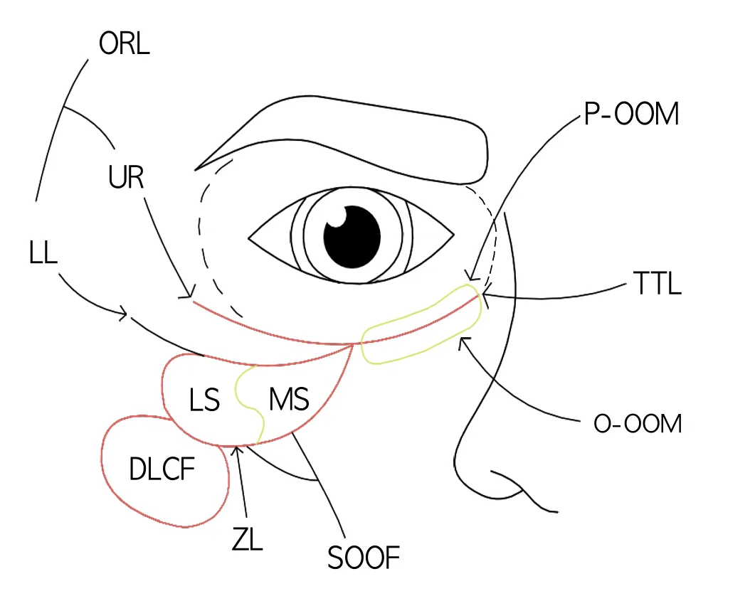 Fig 1. The tear-trough ligament (yellow) tethers skin to maxilla medially and continues laterally as the ORL over the SOOF; the SOOF is the safe deep pocket and the TTL is the structure that must be bridged, not overfilled - (Recognising the key tear trough ligaments, PMFA Journal, 2022, p.1).
> Sources: Recognising the key tear trough ligaments 2022 [18]; ligament scheme per Cotofana 2015 [3], Wong 2012 [5].
Fig 1. The tear-trough ligament (yellow) tethers skin to maxilla medially and continues laterally as the ORL over the SOOF; the SOOF is the safe deep pocket and the TTL is the structure that must be bridged, not overfilled - (Recognising the key tear trough ligaments, PMFA Journal, 2022, p.1).
> Sources: Recognising the key tear trough ligaments 2022 [18]; ligament scheme per Cotofana 2015 [3], Wong 2012 [5].
The SOOF and the tear trough as a landmark. The sub-orbicularis oculi fat (SOOF) sits deep to the orbicularis in layer 4, divided into a medial and a lateral segment. Cotofana 2019 [2] established that the tear trough is the superficial landmark for the medial end of the SOOF: the zygomaticocutaneous ligament fuses with the ORL where it crosses the vertical medial-pupillary line just before reaching the trough. Laterally, the multi-layered stack (SOOF over preperiosteal fat over periosteum, all under the ORL) is why the malar mound is difficult and why product placed too superficially here triggers premalar and malar edema [6][9].
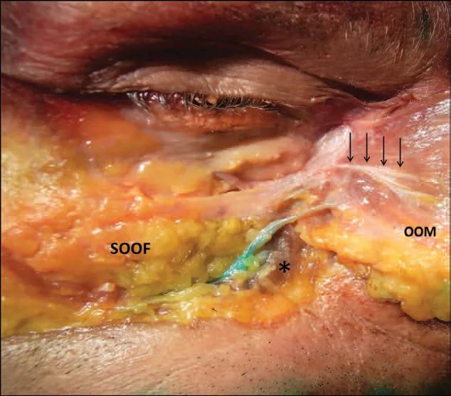 Fig 2. On dissection the SOOF is a real, forgiving pocket laterally, while the medial zone (toward the reflected muscle) is tethered to bone with almost no fat: the anatomical reason deep filler is placed laterally and milked medially - (Cotofana, 2015, p.6).
> Sources: Cotofana 2015 [3].
Fig 2. On dissection the SOOF is a real, forgiving pocket laterally, while the medial zone (toward the reflected muscle) is tethered to bone with almost no fat: the anatomical reason deep filler is placed laterally and milked medially - (Cotofana, 2015, p.6).
> Sources: Cotofana 2015 [3].
The muscle that dictates depth. The orbicularis oculi has an orbital ring (outer) and a palpebral/pretarsal part (inner, over the tarsus). Jones 2019 [6] frames it as "aviator glasses": most of the muscle glides freely over the SOOF, but the medial zone is firmly adherent to the infraorbital bony rim. Injecting on bone in that medial zone therefore deposits intramuscular product, which is squeezed and displaced by repeated blinking ("balls up" over time), the mechanism behind late medial nodules and the bluish medial swelling [6][11]. The lateral, non-adherent muscle over the SOOF is the region where deep product can sit and recreate contour with free muscle movement.
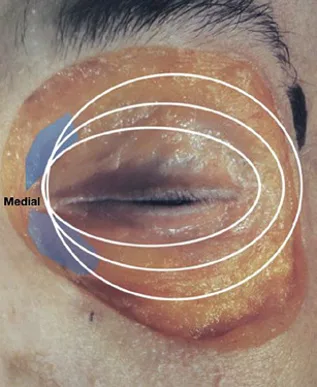 Fig 3. Fig 3 documents, region by region, why the medial trough is a bad place for a bolus: the muscle is glued to bone there, so "deep on bone" is intramuscular, whereas laterally the same muscle glides over the SOOF and tolerates a deep depot - (Jones, 2019, p.111).
> Sources: Jones 2019 [6].
Fig 3. Fig 3 documents, region by region, why the medial trough is a bad place for a bolus: the muscle is glued to bone there, so "deep on bone" is intramuscular, whereas laterally the same muscle glides over the SOOF and tolerates a deep depot - (Jones, 2019, p.111).
> Sources: Jones 2019 [6].
The prezygomatic space and the malar septum (why lateral overfill floods). Just beneath the ORL lies the prezygomatic space, bounded inferiorly by the zygomatic ligament (originating laterally at McGregor's patch) [18]. The malar septum encircles the orbit like a funnel and ends about 5 mm lateral to the lateral canthus; a superficial deposit in or above it traps lymph and produces premalar edema, an error the corpus authors report seeing frequently, triggered by even a tiny droplet left on withdrawal [6][9]. This is the anatomical basis for the rule that infraorbital enhancement lateral to the trough should come from treating the cheek, not from filling between the midline and the lateral canthus [18].
Why the skin colour is anatomy, not cosmetics. The eyelid skin is the thinnest of the body, under 1 mm, sebaceous- and adnexa-poor, with almost no hypodermis [25]. Melanin granules in the epidermis read blue or brown; papillary and subpapillary dermal capillaries read red or blue by oxy/deoxy haemoglobin; the near-absence of subcutaneous fat lets both show through [25][30]. The tear-trough shadow is a fourth mechanism, purely optical (light-and-shadow over the concavity). D5.5b turns this anatomy into a treatment-selecting classification; here the point is that the same shadow can be pigment, vessel, thin skin, or geometry, and the layer it lives in decides the tool.
The three confluent depressions, named separately (they are treated differently). The tear trough proper runs from the medial canthus along the ligament to the mid-pupil; the nasojugal fold continues inferolaterally as the medial cheek fat and skin thicken; the palpebromalar groove runs laterally along the ORL [7][24]. The medial tear trough is the ligamentous, three-layer, high-risk zone; the palpebromalar groove is a shallower, superficial-fat-atrophy problem corrected with a fine superficial pass; the nasojugal component often improves most from cheek support rather than direct fill. The PMFA anatomy review [18] notes the palpebromalar groove is driven mainly by superficial fat atrophy fixed by the ORL, not by the tear-trough ligament, which is why the two grooves respond to different planes.
The malar septum funnel, the reason lateral overfill floods. The malar septum runs from the orbital rim as a funnel and ends about 5 mm lateral to the lateral canthus on bone; a superficial deposit within or above it traps fluid and produces premalar edema from even a tiny droplet left on withdrawal [6]. Surek's midface analysis [48] shows the prezygomatic space in the deep sub-orbicularis plane and the zygomaticocutaneous ligament partitioning the infraorbital "malar" fat compartment above from the superficial cheek compartment below; a percutaneous injection aimed at lateral cheek projection but left too superficial fills the malar compartment and creates an iatrogenic malar mound, the anatomical counterpart of the festoon exclusion in D5.6.
Trampa clásica: treating "deep on bone" as safe everywhere in the trough. Medial to the facial vein the region is three layers and the orbicularis is glued to bone, so a periosteal deposit is intramuscular and next to the angular vessels; the forgiving deep pocket (SOOF) exists only laterally. The safe move is to enter laterally and milk product medially, never to bolus the medial rim [3][6].
D5.3 · Vessels, nerves and danger zone → J2 — Vascular Occlusion & Emergency Response
The vessels of the trough, with course, depth, plane and what fails if you hit them:
| Vessel | Origin | Where it is | Depth / behaviour | If occluded / injured |
|---|---|---|---|---|
| Angular artery | terminal facial a. | ascends from the nasolabial region toward the medial canthus, medial to the trough | variable subcutaneous-to-deep; anastomoses with the dorsal nasal (ophthalmic) at the canthus | retrograde embolus → ophthalmic → central retinal a. = blindness; skin necrosis [8][10] |
| Angular / facial vein | facial v. | runs ~4.2 ± 0.7 mm inferior to the inferior orbital rim, deep to orbicularis in the nasojugal groove | deep, oblique; it is the lateral/medial boundary of the trough (4-6 mm medial to mid-pupil) | venous route into the ophthalmic system; contributes the bluish medial hue [2][3] |
| Infraorbital a. + n. | maxillary a. / V2 | exit the infraorbital foramen, ~4-8 mm below the rim near the mid-pupillary vertical | on periosteum at the foramen, then superficial | a deposit too low at the mid-pupil can enter foramen vessels or bruise/anaesthetise the infraorbital nerve [11] |
| Dorsal nasal a. | terminal ophthalmic a. | medial canthal area | subcutaneous near canthus | the shortest retrograde route to the retina from a medial deposit [8] |
| Superficial vascular plexus | dermal | in the < 1 mm skin | intradermal | not a danger vessel but the substrate of the vascular dark circle [30] |
Consensus (the danger-zone rule everyone teaches). The infraorbital area is a high-risk zone for filler because it has "little buffer over bone and is highly vascular", with multiple communications between internal and external carotid circulations [8]. Rare but reported: blindness, stroke and skin necrosis from inadvertent intravascular injection, described with both needles and cannula [8][10]. The practical danger zone is the strip between a vertical line through the medial pupil and the lateral nasal wall; Haney 2020 [10], citing Hufschmidt 2019, teaches that safer practice keeps injections retromuscular / pre-orbital (supraperiosteal) and out of this medial triangle, and notes the region is not an FDA-approved filler indication in general and is left to advanced training. Fig 4 shows how close the medial injection point sits to the dye-marked angular and infraorbital vessels.
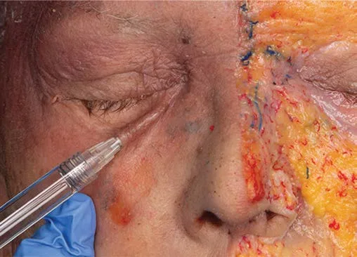 Fig 4. Fig 4 documents the spatial trap of the medial trough: the needle tip at the groove is millimetres from dye-filled angular and infraorbital branches that anastomose with the ophthalmic system, which is why low pressure, small aliquots and a moving cannula are non-negotiable here - (Pirayesh, 2020, p.141).
> Sources: Pirayesh 2020 [8]; carotid anastomosis concept per DeLorenzi via Carruthers [7].
Fig 4. Fig 4 documents the spatial trap of the medial trough: the needle tip at the groove is millimetres from dye-filled angular and infraorbital branches that anastomose with the ophthalmic system, which is why low pressure, small aliquots and a moving cannula are non-negotiable here - (Pirayesh, 2020, p.141).
> Sources: Pirayesh 2020 [8]; carotid anastomosis concept per DeLorenzi via Carruthers [7].
The mechanism, stated once (why this region blinds). A bolus injected under enough pressure into the angular, dorsal nasal or infraorbital branch can be pushed retrograde past the ophthalmic origin; when injection pressure drops, antegrade flow carries it into the central retinal artery, producing sudden, usually painful, monocular vision loss that hyaluronidase does not reliably reverse once the retina is embolised [8][7]. The theoretical rescue window is short (of the order of 90 minutes of retinal ischaemia), which is why the whole regional strategy is prevention over rescue [7]. Full algorithm and reversal pharmacology in J2 — Vascular Occlusion & Emergency Response and J3 — Hyaluronidase - Pharmacology & Clinical Protocols.
The safe-injection rule set for this region, each rule tied to its vessel. (1) Low injection pressure, so a bolus cannot be driven retrograde past the ophthalmic origin [8]. (2) Small aliquots (≤ 0.05-0.1 mL), so any intravascular volume is sub-embolic [6][17]. (3) Retrograde, moving needle or cannula, so the tip is never stationary in a vessel while pressure builds [8]. (4) Blunt cannula from a lateral port where feasible: fewer entries, less trauma, and it tends to push vessels aside, though it does not abolish intravascular entry [12][16]. (5) Ultrasound vascular mapping before deposit is increasingly treated as essential in this region because the angular vein and artery are variable [17][9]. (6) Reversible product only (HA) with hyaluronidase in the room [7][9]. (7) Stay on bone, 3-4 mm below the rim, no more medial than the inner limbus, and milk product medially from a lateral entry rather than bolus the medial trough [6].
Nerves (spare them; a numb cheek is a complication too). The infraorbital nerve (V2) supplies the lower lid, medial cheek, lateral nose and upper lip; a deposit driven into or beside the foramen (too low at the mid-pupil) can produce transient or lasting hypoesthesia and is intensely painful on injection [11]. The sentinel vein and superficial periorbital veins are visible landmarks to avoid on the skin surface [8].
Variants, and why frequency matters at the chair. The infraorbital foramen position varies (typically 4-8 mm below the rim, near the mid-pupillary vertical, occasionally a double foramen), so the "avoid the mid-pupil at the rim margin" rule points at a distribution, not a fixed spot [11]. The angular vein's depth and its exact distance below the rim vary around the mean of 4.2 mm [2], and the angular artery can run superficial or deep and medial or lateral to the trough; this variance is the argument for Doppler mapping over surface landmarks in any filler candidate here [17].
The aspiration debate, stated fairly. Aspirating before injection can detect an intravascular needle tip, but a negative aspiration does not guarantee an extravascular position: viscous gels and small-gauge needles give false negatives, and the tip can move between aspiration and injection [8][17]. It is one layer, not a licence. The stronger layers are plane choice (stay supraperiosteal or in the SOOF, off the vessels), low pressure, small aliquots, a moving tip, and direct ultrasound visualisation; aspiration supplements them and never replaces them.
Cannula versus needle, on the vascular question specifically. The blunt cannula tends to push vessels aside rather than pierce them and needs fewer entry points, which lowers (not abolishes) intravascular risk and bruising; the sharp needle gives a precise periosteal depot but passes through whatever is in its path [12][16]. Reports of vascular occlusion, including blindness, exist with both instruments [8], so neither is a substitute for the plane and pressure rules. In the trough the pragmatic split is a cannula from a lateral port for the broad deep-fat correction and a needle only for a precise, small, supraperiosteal medial depot under ultrasound.
Why hyaluronidase does not fully rescue the eye here. Once filler embolises the central retinal artery, the retina is ischaemic within minutes and enzymatic dissolution reaches the intraluminal gel unreliably; the retrobulbar and territory-flooding protocol is attempted because the downside of inaction is total, but established retinal embolism often does not recover even with correct, immediate treatment [7][8]. This is the anatomical reason the region's whole strategy is prevention over rescue, and why every HA here is a deliberate, reversible-by-design choice with the enzyme already drawn up.
Sensory and lymphatic collateral, quantified where the corpus gives it. The infraorbital nerve exits its foramen near the mid-pupillary vertical, typically 4-8 mm below the rim, so the "avoid the mid-pupil at the rim" rule protects both the nerve and the foramen vessels [11]. The lymphatic channels of the lower lid run superficially and are easily compressed; this is the anatomical basis for malar edema (D5.10) and the reason a superficial deposit is doubly penalised here, first for Tyndall and second for lymphatic obstruction [9].
Trampa clásica: placing a "safe deep bolus" on the medial rim at the mid-pupil to lift the deepest part of the trough. That is exactly where the orbicularis is glued to bone (intramuscular deposit), where the infraorbital foramen and its vessels sit, and where the angular vessels anastomose with the ophthalmic system. The safe medial correction is a small aliquot delivered from a lateral cannula entry and milked medially, or no filler at all [6][10].
D5.4 · Ageing of the region, in order
The order matters because it dictates the tool. Treating a bone/ligament problem with superficial filler, or a pigment problem with volume, is the commonest strategic error here. The sequence is layered and starts deep, but it is not one universal clock: a young patient can have a congenital tear-trough ligament groove with no volume loss at all, and a high-phototype patient can have deep pigment with a structurally intact orbit. Read the layer that actually failed.
| # | Structure | What it loses at the trough | Clinical sign | Correct lever |
|---|---|---|---|---|
| 1 | Bone (first) | orbital aperture widens; resorption greatest supero-medially and infero-laterally; maxilla and pyriform recede back and up; canthal tilt flattens | skeletonised, deeper rim shadow; senile enophthalmos (globe sinks, lid support lost) | supraperiosteal support or accept; bone is not reversed by trough filler [1][6] |
| 2 | Deep fat + retaining ligament | SOOF deflation and descent; ORL/tear-trough tether stays fixed while the fat below drops | lengthening lid-cheek junction, deep V-shaped hollow, mid-face "double convexity" | midface/SOOF support first, then residual trough fill [1][3][24] |
| 3 | Superficial fat | deflation of the thin premalar/subcutaneous fat below the lid-cheek line | flattening below the groove, loss of the smooth lid-cheek transition | soft superficial volume or biostimulation at the cheek, not the trough [3][4] |
| 4 | Skin / dermis | thinning (already < 1 mm), elastosis, laxity, fixed pigment | crepey lower-lid skin, static fine lines, festoons, worsening translucency | skin-quality tools (resurfacing, microneedling, boosters, PN); pigment ladder [14][19] |
| ↳ | Fat pseudoherniation | orbital septum weakens, lower-lid orbital fat prolapses over the ligament | "eye bags" above the groove that frame and deepen it | surgery (blepharoplasty), not filler, which would double the convexity [8][24] |
Consensus (the sequence, stated once). Cotofana 2016 [1] frames facial ageing as multi-tissue and deep-first. At the infraorbital region: the orbital aperture enlarges (bone resorbs supero-medially and infero-laterally), removing skeletal support and producing a relative senile enophthalmos; the maxilla and pyriform recede, dropping the platform under the medial trough; the SOOF deflates and descends while the tear-trough ligament and ORL hold the skin fixed, so the lid-cheek junction lengthens and the groove deepens; the thin superficial fat deflates; and the skin thins and loses elasticity last, on top of the already-changed frame [1][6][8]. Pirayesh 2020 [8] adds the surface reading: above the trough, thin preseptal skin with absent subcutaneous fat and hyperpigmentation; below it, thicker skin and more fat; and in advanced ageing, orbital fat herniation in the lower lid distracts from, and accentuates, the underlying trough and periorbital volume loss. Fig 5 shows the widened, resorbed aged orbital aperture that removes the skeletal platform.
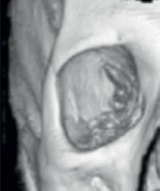 Fig 5. Bone first: the aged orbital aperture is larger and its rim resorbed supero-medially and infero-laterally, so the tear trough deepens from lost skeletal support before any soft-tissue change is visible; trough filler cannot restore this platform - (Jones, 2019, p.116).
> Sources: Jones 2019 [6]; skeletal-ageing pattern per Cotofana 2016 [1].
Fig 5. Bone first: the aged orbital aperture is larger and its rim resorbed supero-medially and infero-laterally, so the tear trough deepens from lost skeletal support before any soft-tissue change is visible; trough filler cannot restore this platform - (Jones, 2019, p.116).
> Sources: Jones 2019 [6]; skeletal-ageing pattern per Cotofana 2016 [1].
Deflation vs descent, and why it is not an idle debate here. One school reads infraorbital ageing as volume loss (SOOF and deep-fat deflation over a resorbing rim), another as ligamentous fixation with tissue descent (the tether stays while everything around it drops). In the trough the honest answer is both, in sequence: the deep fat deflates and the lid-cheek junction descends against a fixed ligament over a widening aperture [1][3]. The clinical consequence is that neither "just fill the line" nor "just support the cheek" is complete; the residual groove that remains after midface support is the true fillable defect, and the tether itself sometimes needs releasing rather than filling. Averaging the two models into "add HA to the line" reproduces the region's classic overfill and edema.
The congenital / young-patient variant, stated explicitly. The tear-trough ligament produces a groove regardless of age when the bone-to-skin tether is strong; Pirayesh 2020 [8] and the PMFA anatomy review [18] both note the deformity can be present in youth with no volume loss, because it is ligamentous, not deflationary. Adding volume to a young patient who has not lost any is a common error: the groove needs the tether bridged with a minimal deposit or a subcision-style release, not restoration of a volume that was never lost [18].
What ageing does NOT change (still state-of-the-art). The deep-first sequence and the SOOF / ORL / tear-trough-ligament mechanism come from 2015-2019 cadaveric and imaging work [2][3][5] and are reinforced, not revised, by later imaging. The three-versus-seven-layer regional split [3] and the SOOF-landmark relationship [2] remain the reference frame for where to place product.
The skeletal change, in detail. The orbital aperture enlarges with age, and the recession is regional, not uniform: greatest at the supero-medial and infero-lateral rim, so the medial trough loses its bony shelf while the inferolateral rim recedes under the lateral canthus [1][6]. The maxilla and pyriform region recede backward and upward, dropping the platform under the medial trough and deepening the nasojugal component; the canthal tilt flattens as the lateral canthal tendon descends with the remodelling bone [6]. The globe loses the lower-lid support it had when the orbit was smaller, producing a relative senile enophthalmos that a young orbit did not show. None of this is reversed by trough filler; where the deficit is skeletal, the honest options are supraperiosteal support at the rim/cheek or acceptance [1].
The soft-tissue sequence over the skeleton. Against this widening frame, the SOOF deflates and descends while the tear-trough ligament and ORL hold the overlying skin fixed, so the lid-cheek junction lengthens and a young single convexity becomes an aged double convexity (bag above, hollow below) [3][24]. The thin superficial fat deflates next, flattening the transition; the skin thins further (from an already sub-millimetre baseline), loses elasticity, and grabs static fine lines and festoons last [1][8]. Yang 2013 [35] confirmed on sectional anatomy that the tear trough and palpebromalar groove are anatomically distinct in young versus elderly adults, and Haddock 2009 [36] mapped the lid-cheek junction that lengthens; both underpin why the aged groove is a compound of skeletal, ligamentous and fat change rather than a single line.
Muscle ageing, as behaviour not dose. The orbicularis, like other facial muscles, shortens its amplitude and raises resting tone with age, so dynamic infraorbital lines become static and a pretarsal roll that once appeared only on smiling becomes visible at rest [1]. This is why a toxin decision made on a young lid does not transfer to an old one, and why the snap test (D5.5, D5.8) is repeated at every visit rather than assumed.
Ethnic and phototype variation in the sequence. The structural timeline above is broadly shared, but its surface expression differs: darker-skinned and South Asian, Middle Eastern and Mediterranean patients express the same hollow with a heavier constitutional pigment overlay, so the "ageing dark circle" reads as brown rather than the blue-grey of a fair-skinned vascular trough [15][30]. The bone and fat sequence is the same; the colour the patient complains about is not, which is why D5.5b classification is done before the aging story is used to justify a treatment.
Trampa clásica: reading a lengthening lid-cheek junction and a deep groove as "the trough needs more filler". The junction lengthened because the SOOF deflated and descended over a widened aperture; supporting the cheek/SOOF shortens it and often removes most of the shadow before a drop of trough filler is placed. Filling the line first, without midface support, chases a shadow that keeps returning and stacks volume in the worst plane [1][3][24].
D5.5 · Assessment: what is measured, photographed, tested dynamically, scanned
The assessment answers five questions in order: what phenotype is the darkness (D5.5b), how deep is the hollow, is the midface support adequate, is the lower lid competent (tone, fat, laxity), and is there a surgical exclusion (festoon, mound, pseudoherniation). Getting the first and last wrong produces the region's worst outcomes.
| What | How | Why it changes the plan |
|---|---|---|
| Phenotype of the darkness | daylight + Wood's lamp; stretch/traction test (see D5.5b) | pigment/vessel/shadow/mixed decide filler vs pigment vs laser vs referral [29][30] |
| Hollow depth / class | Hirmand nasojugal grade I (mild medial) · II (medial + central) · III (full trough + lid-cheek) | class I-II single session; class III staged, midface-first [17] |
| Lid-cheek vector | profile 45°/90°; is the cheek anterior or posterior to the cornea | a negative vector (posterior cheek) is a poor direct-fill candidate and a malar-edema risk [24] |
| Midface / SOOF support | palpate cheek projection; does lifting the cheek erase the trough? | if the trough vanishes on manual cheek lift, treat the cheek first (D5.9) [16][24] |
| Lower-lid tone | snap-back / distraction test: pull the lid down, time recoil | a sluggish snap = laxity: contraindicates lower-lid toxin and flags edema/ectropion risk [20][21] |
| Orbital fat / festoon | look up and squint; palpate for pseudoherniation and malar mounds | fat prolapse and festoons are surgical, not filler; volume worsens them [8][23] |
| Skin quality / pigment | pinch test, phototype, actinic change | thin, crepey or high-phototype skin shifts toward booster/PN and away from superficial HA [13][19] |
| Prior filler | high-frequency ultrasound: HA is anechoic (black), CaHA/PMMA hyperechoic (white) | undisclosed old filler changes the plan and explains refractory edema [9] |
| Vessel map | Doppler over the angular vessels and trough | locates the angular vein/artery before a deep pass; the region where this pays for itself [17][9] |
| Systemic contributors | sleep, salt, allergy/atopy, thyroid, renal, iron, fluid-retention history | periorbital edema and constitutional pigment are partly systemic; filler will not fix them [25][29] |
Consensus (what to document, always). Pinto 2009 [25] frames the periorbital examination as: skin characteristics and pigment degree, visible cutaneous vascularity, excess or deficit of periorbital/infraorbital fat, septal laxity and orbicularis atrophy/hypertrophy, and the degree of orbital and facial skeletonisation. Mosaheb 2022 [17] standardises the photograph: patient upright looking up, documented at 45°, 90° and 180°, before and at three-week review, with a thorough history to assess suitability. The two together are the minimum defensible record: a phenotype call, a class, a vector, a lid-competence test, and standard photographs.
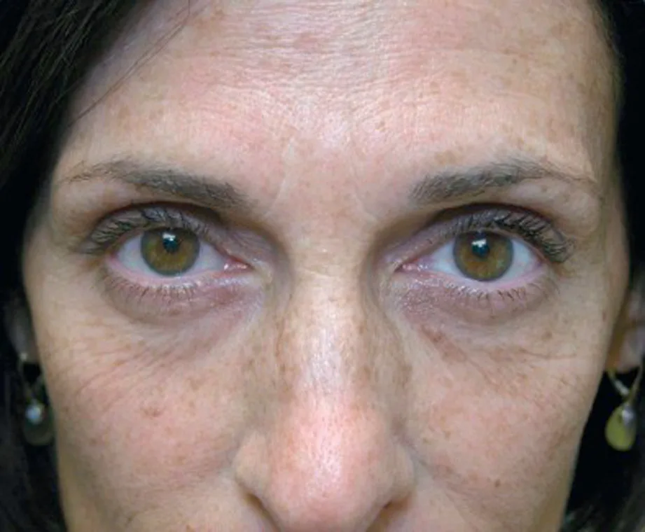 Fig 6. Fig 6 is the reason classification precedes treatment: the same face carries a fillable shadow, a non-fillable pigment, and a lid laxity that volume would worsen; the plan must name which component each treatment addresses - (Carruthers, 2018, p.117).
> Sources: Carruthers 2018 [7]; classification per Roh 2009 [29], Huang 2014 [30].
Fig 6. Fig 6 is the reason classification precedes treatment: the same face carries a fillable shadow, a non-fillable pigment, and a lid laxity that volume would worsen; the plan must name which component each treatment addresses - (Carruthers, 2018, p.117).
> Sources: Carruthers 2018 [7]; classification per Roh 2009 [29], Huang 2014 [30].
The dynamic tests that change the gesture. (1) Snap-back / distraction test: pull the lower lid inferiorly and release; slow recoil (a "sluggish snap") signals laxity and is a stop sign for lower-lid toxin and a predictor of edema and ectropion [20][21]. (2) Manual cheek lift: if elevating the cheek erases the trough, the primary deficit is midface support, and the trough should not be filled first [16][24]. (3) Look-up and squint: unmasks orbital fat pseudoherniation and malar mounds that worsen on animation, both surgical exclusions [8]. (4) Smile: a malar mound that balloons on smiling warns of compromised lymphatic drainage and edema risk [9].
Ultrasound, twice useful. High-frequency ultrasound serves two distinct purposes here: before treatment, Doppler locates the variable angular vein and artery in the intended plane, and it detects undisclosed prior filler, which van Loghem 2023 [9] shows appears anechoic (black) for hydrophilic HA and hyperechoic (white) for CaHA/PMMA. A refractory "edema" that is actually old hydrophilic gel is a common misdiagnosis that ultrasound resolves and hyaluronidase then treats.
Photography and consent specifics. Because the Tyndall/blue hue can appear only under flash photography, patients who are professionally photographed must be forewarned, and the pre-treatment photographs must be standardised for distance, head position and lighting so a follow-up is comparable [8][23]. Baseline asymmetry and any pre-existing bags, festoons or pigment are documented before, not after, or they become "your" complication.
Grading, so the plan and the follow-up are defensible. The Hirmand nasojugal grade sorts severity into three classes: I = mild, medial volume loss only; II = moderate, medial plus central infraorbital loss; III = severe, full trough with lid-cheek and lateral involvement [17]. Class I-II are single-session, small-volume, often direct; class III is staged and midface-first (D5.9). The manual cheek-lift and vector assessment convert this static grade into a treatment decision by asking whether the hollow is primary or a consequence of midface descent. Photographs at 45°/90°/180° upright, looking up, before and at the 3-4 week review, make any grade change objective rather than remembered [17].
Palpation, the step that catches the surgical exclusion. Beyond looking, the examiner palpates for orbital-fat pseudoherniation (a soft bulge above the groove that increases on upgaze), malar mounds (firm fullness lateral and below, worse on smile), festoons (redundant skin-muscle folds that persist at rest), and skin laxity (crepe on pinch, slow snap). Any of these reclassifies the patient from filler to surgery or energy (D5.6) before a plane or product is even considered.
Trampa clásica: skipping the snap-back test and the phenotype call, then injecting a lax, pigmented lower lid. The hollow is masked but the pigment remains, the lax lid retains fluid and festoons, and the record has no baseline to defend. Two tests (snap-back, traction) and one classification take a minute and reroute half of these patients away from filler [20][29].
D5.5b · Dark-circle etiologic classification (pigment / vascular / structural / mixed)
Added subchapter. The chapter title is "tear trough and dark circle"; the 10-block template covers the hollow but not the pigment/vascular half, and half of "dark circle" patients have no hollow to fill. This block classifies the darkness so the treatment (D5.6, D5.7, D5.7b) is chosen by phenotype, never by default HA.
The classification, answer first (Huang four-type + Roh cause map):
| Type | Colour / look | Mechanism | Bedside test | First-line lever |
|---|---|---|---|---|
| Pigmented | brown, matte | epidermal/dermal melanin: constitutional, post-inflammatory, atopic/rubbing, drug | stretch: colour persists / grows, no blanch; Wood's lamp accentuates epidermal melanin | topical depigment + photoprotection; peels; pigment laser (D5.7b); not filler [29][30][14] |
| Vascular | blue / purple / pink | visible orbicularis + engorged subdermal veins/capillaries through < 1 mm translucent skin; oxy/deoxy Hb | stretch: deepens to violaceous; diascopy blanches a vascular red | vascular laser/IPL; superficial masking HA sheet; skin thickening (PN/PRP); treat congestion [29][13][30] |
| Structural (shadow) | grey shadow, colour-neutral | optics: tear-trough concavity, orbital-fat bags, lid-cheek laxity cast a shadow | diminishes in bright overhead light / on cheek lift; worsens in downlight | HA on bone (the true filler indication); midface support; surgery for bags [29][32][6] |
| Mixed | combination | two or more of the above (the majority in practice) | mixed test response; decompose component by component | sequence: shadow → filler, pigment → topical/laser, vascular → laser/sheet [30][32] |
> The four-type scheme is Huang 2014 [30] (pigmented, vascular, structural, mixed, with a Wood's-lamp assessment score); Roh & Chung 2009 [29] give the same causes as three drivers (excess pigment, thin translucent skin over orbicularis, shadowing from laxity/trough). Friedmann & Goldman 2015 [32] add the treatment-by-etiology map. They agree on the essential clinical act: identify the cause before choosing the treatment, because a treatment matched to the wrong type does nothing or harms.
The stretch (traction) test, spelled out. Apply gentle downward traction to the infraorbital cheek [13][32]: (a) if the pigmented area grows proportionally with stretch and does not blanch or lighten, excess pigment is the driver; (b) if traction spreads the area and it turns a deeper violaceous, thin translucent skin over vessels (vascular) is the driver, because stretching thins the skin further and exposes the plexus; (c) if traction, especially in a brightly lit room, diminishes the darkening, shadow (structural) is the driver. This one test, plus a Wood's lamp, sorts most patients into a lever in under a minute.
Colour is a spectroscopy clue [25]. Melanin granules in the epidermis read blue or brown; papillary/subpapillary dermal capillaries read red or blue by oxygenated vs reduced haemoglobin; the thin cornified layer and any carotene read yellow. Because the skin is under 1 mm with almost no subcutaneous buffer, the colour of whatever sits deepest shows through, which is why the same "dark circle" is brown in one patient and blue in the next.
Ethnicity and phototype change the base rate and the risk [15][30]. A vascular origin predominates in fair-skinned patients; constitutional hyperpigmentation predominates in darker-skinned patients and in South Asian, Middle Eastern and Mediterranean populations. This matters twice: it shifts the likely diagnosis, and it raises the stakes of any energy or superficial procedure, because post-inflammatory hyperpigmentation is common in high phototypes and Q-switched lasers can worsen dermal melanin (D5.7b). A pigment-predominant high-phototype dark circle is the archetype of a patient who should not be lasered aggressively or filled.
Constitutional and acquired, and the systemic screen. Pinto 2009 [25] separates constitutional (hereditary/ethnic, present in old photographs) from acquired dark circles (recent onset), and lists the systemic contributors worth a history and, when indicated, bloods: sleep debt, tobacco, sun, iron deficiency, thyroid disease and vitamin deficiency. A recently darkened circle with fatigue and pallor is a haematology question before it is an aesthetics one.
How this routes the patient (bridge to D5.6/D5.7/D5.7b): structural/shadow with adequate support → HA on bone (D5.7); structural with poor support → midface first (D5.9); pigment → topicals/peel/pigment laser (D5.7b), never filler; vascular → vascular laser or superficial masking sheet plus skin thickening (D5.7b); fat bags / festoon / laxity → surgery/referral (D5.6). Mixed circles get a sequenced, multi-modal plan, addressed component by component rather than with one syringe. Fig 6 (D5.5) is the mixed archetype these rules decompose.
Tools that sharpen the classification. The Wood's lamp accentuates epidermal melanin (it becomes more contrasted) while dermal melanin changes little, separating a topical-responsive from a laser-resistant pigment [30]. Dermoscopy distinguishes the pigment network from telangiectatic vessels of the vascular type. Huang 2014 [30] formalised a DEC (dark eye circle) assessment score using Wood's lamp within the four-type scheme, giving a repeatable grade for follow-up. Photographs under standard and flash light document the shadow and the flash-revealed Tyndall separately.
The overlap that makes "mixed" the commonest call. In practice most patients carry more than one driver: a constitutional pigment on thin translucent skin over a ligamentous shadow, for example. Roh 2009 [29] and Friedmann 2015 [32] both stress that the treatment of a mixed circle is not a single modality but a sequenced, component-by-component plan, and that a patient who is told only "you need filler" for a mixed circle will be dissatisfied because the untreated pigment or vessel remains after the shadow is corrected.
Trampa clásica: calling every dark circle a "tear trough" and reaching for HA. Filler corrects only the structural/shadow component; on a pigmented or vascular circle it leaves the darkness unchanged, adds cost and risk, and in high phototypes the needle trauma itself can trigger post-inflammatory pigment that worsens the complaint [29][30].
D5.6 · Goal and patient selection: who benefits, who does not, what is referred
The goal is a smooth lid-cheek transition, not a filled line. Success is the disappearance of the shadow and the restoration of a gentle convexity from lid to cheek, achieved with the least product in the safest plane. Overcorrection is a worse outcome than undercorrection here, because the region punishes excess with edema and Tyndall that outlast the benefit.
Who benefits (the good direct-filler candidate):
| Feature | Why it predicts success |
|---|---|
| Structural/shadow phenotype (D5.5b) | filler corrects only the shadow; this is the one type it fixes [29][6] |
| Mild-to-moderate hollow (Hirmand I-II) | single session, small volume, low edema risk [17] |
| Thick, smooth skin, low phototype-independent quality | masks product; less Tyndall, less pigment risk [7] |
| Well-defined trough without protruding orbital fat or excess skin | no bag to accentuate, no laxity to worsen [7] |
| Adequate midface support / positive vector | trough is the only defect; no cascade to chase [24][16] |
| No history of periorbital edema / fluid retention | low malar-edema risk [9][23] |
| Realistic expectations, understands staging and reversibility | tolerates the "less is more", staged approach the region needs [7] |
Who does NOT benefit, and where they go instead (the tear-trough exclusions):
| Finding | Why filler is wrong | Correct route |
|---|---|---|
| Festoons (redundant malar skin/muscle folds) | volume worsens the fold and traps fluid | oculoplastic surgery; energy for mild skin laxity [23][24] |
| Malar mounds / malar bags | multi-layer lateral infraorbital structure; superficial or hydrophilic filler triggers refractory malar edema | surgery or cautious deep cheek support only; often referral [6][9] |
| Baseline malar / periorbital edema, fluid-retention history | compromised lymphatics; HA (especially hydrophilic) causes persistent edema | treat the cause; avoid HA or use minimal, less-hydrophilic gel [9][23] |
| Orbital fat pseudoherniation ("eye bags") | filling below a bag doubles the convexity, does not flatten it | blepharoplasty (fat repositioning/removal) [8][24] |
| Double convexity / negative vector (posterior cheek) | filling the trough over a receding maxilla deepens the two-bump look | midface/skeletal support first, or surgery [24] |
| Marked skin laxity / dermatochalasis | no injectable removes skin; volume adds weight | energy or surgery [23] |
| Pigment-only or vascular-only dark circle | filler leaves the colour unchanged | pigment/vascular ladder (D5.7b) [29][30] |
| Complex post-blepharoplasty anatomy (over-resected fat, scar, hollow lid) | distorted planes, adhesions, high complication risk | advanced/surgical hands; structural fat, not routine HA [12] |
| Body dysmorphia / unrealistic expectations | no result satisfies; medicolegal risk | decline, refer to psychology [7] |
Consensus (the selection rule everyone states). Carruthers 2018 [7] and Humphrey/Fagien: injections "work best in patients with thick and smooth skin and a well-defined tear trough, without excessively protruding eyelid fat or excess eyelid skin"; the IOH is "an unforgiving region". Haney 2020 [10]: the infraorbital area is not an FDA-approved indication for most fillers, requires advanced training and specific protocols, and carries a real danger of blindness/embolism/necrosis. Wong 2022 [16] and the surgical texts: when the deficit is a bag, festoon, mound or lax lid, the answer is surgery or energy, not filler. The double convexity (nasojugal + palpebromalar grooves framing a mid-face bulge) is a rejuvenation target for support and surgery, not a trough to be filled [24].
The single approved exception, and what it changes. Restylane Eyelight (NASHA, high G′) received FDA approval in 2023 specifically for infraorbital hollows, on a randomized controlled trial (n=333) [31]. This narrows, but does not remove, the "off-label, advanced-training" framing: it legitimises HA correction of the hollow in appropriate candidates, still excludes the pigment/vascular/bag/festoon phenotypes, and still demands the region's safety discipline. It also carries the school discrepancy of D5.7 (a firm gel where tradition taught soft only).
The consent conversation, region-specific. Beyond generic filler consent, the trough consultation must state: the darkness may be partly or wholly pigment/vessel that filler will not change; swelling and bruising are common and can last weeks; malar edema can be prolonged or, rarely, permanent; the Tyndall blue hue can appear even with correct depth; results usually need a staged approach and a review at 3-4 weeks; and the region carries a rare but real risk of vision loss [7][9][31].
Special populations and situational cautions. High phototypes carry a higher base rate of constitutional/dermal pigment and a higher PIH risk from needle trauma and energy, shifting the plan toward topicals and conservative devices and away from aggressive lasering [15][30]. Thyroid eye disease, myasthenia and prior periocular surgery must be identified before any injectable or toxin, because they change lid position and closure [24]. Fluid-retention states (renal, cardiac, salt/alcohol load, allergy) predispose to malar edema and are addressed before HA [9][25]. Dry-eye or a sluggish snap removes lower-lid toxin from the menu [20][21]. Anticoagulation and antiplatelets raise the already-high ecchymosis rate of this vascular region and warrant a bruising discussion and, where clinically appropriate, timing [12].
Undercorrect by design. Because the skin is sub-millimetre, the fat buffer minimal and HA hydrophilic, the region rewards deliberate undercorrection and a staged review at 3-4 weeks over a single full correction [6][17]. A patient who leaves slightly undercorrected and returns for a top-up has a better and safer trajectory than one corrected to the target in one visit and then edemas; this is a selection-and-consent point, not only a technique point, because the patient must accept the staged model at the outset.
Trampa clásica: accepting a festoon or malar-mound patient for "tear-trough filler" because they asked for it. The mound is multi-layered and lymphatically fragile; any volume, especially hydrophilic HA, converts a cosmetic complaint into months of refractory malar edema. The correct answer to a festoon is a surgeon, said at the consultation, not after the complication [9][23].
D5.7 · Technique, the full grid (product, instrument, plane, movement, school, volume)
This block is not closed with one technique; it is the full grid of options for the infraorbital region. Every row carries its school, plane, volume and evidence. What does not apply is marked "No apply" with the reason. Toxin is D5.8; the pigment/vascular non-injectable ladder is D5.7b.
Product axis (the complete list, with the region-specific verdict for each):
| Product | Region verdict | Plane / use | Note |
|---|---|---|---|
| Low-G′ / low-cohesivity HA (Volbella VYC-15: G′ ~271 Pa, cohesivity 19 gmf; Belotero Balance; Teoxane Redensity II / RHA 2; Restylane) | ✅ first line | deep supraperiosteal or subdermal sheet | soft, spreads evenly, low Tyndall; the traditional trough gel [6][7][8] |
| Firm NASHA HA (Restylane Eyelight, high G′, low water uptake) | ✅ evidence-backed alternative ⚠ discrepancy | deep on bone (needle or cannula) | FDA-approved 2023 for IOH; RCT n=333, 87.4% responders at 3 mo, no Tyndall reported [31] |
| Highly hydrophilic HA (Juvederm Ultra / Ultra Plus / Voluma) | ⛔ avoid in trough | (cheek support only) | Juvederm Ultra "should not be used in the tear trough": delayed sausage-like swelling months/years later [6][11] |
| CaHA (Radiesse) | ⛔ avoid in trough; cheek only | supraperiosteal cheek support | minimally hygroscopic but nodulogenic and non-reversible in thin skin; some use it, most avoid [11][17] |
| Hyperdilute CaHA | ○ adjunct, not the trough | subdermal cheek/periorbital biostimulation | skin-quality/collagen, not volume in the groove [19] |
| PLLA (Sculptra) | ⛔ avoid in/near trough | deep cheek volumising over sessions | nodule/granuloma risk in thin periorbital skin [11][22] |
| PCL (Ellanse) | ⛔ avoid in trough | (lower face/cheek) | semi-permanent, non-reversible, nodulogenic here |
| PMMA / permanent | ⛔ never | - | non-reversible, late granuloma; catastrophic in this skin [11] |
| Autologous fat (macro) | ○ surgical option | supraperiosteal/deep by surgeon | durable but lumpy/overcorrection-prone in the lid; specialist only [16][12] |
| Nanofat / microfat | ○ regenerative | subdermal | skin quality and pigment, not volume (D5.7b) [16] |
| Polynucleotides (PN) | ○ skin/vascular adjunct | intradermal | thickens skin, improves vascular/pigment component (D5.7b) [27] |
| Skin boosters (non-cross-linked HA) | ○ skin adjunct | intradermal | hydration/quality, not shadow correction [27] |
| Botulinum toxin | ○ narrow role | pretarsal (D5.8) | only selected dynamic pretarsal hypertrophy [20] |
Rheology grid (why one HA is right and another causes edema here). The trough selects a gel on three properties: G′ (firmness/lift), cohesivity (how the gel holds together and spreads), and water uptake / hydrophilicity (how much it swells after placement). In < 1 mm skin over compromised lymphatics, high water uptake is the enemy [34][9].
| Product | G′ | Cohesivity | Water uptake | Trough verdict |
|---|---|---|---|---|
| Juvederm Volbella (VYC-15) | low (~271 Pa) | low (~19 gmf) | low-moderate | ✅ soft, spreads evenly, superficial-safe [7] |
| Belotero Balance | low | high (integrates evenly) | moderate | ✅ even integration, low beading [8][11] |
| Teoxane Redensity II / RHA 2 | low-moderate | moderate | moderate | ✅ dedicated periorbital formulations [8] |
| Restylane / Restylane-L | moderate | moderate | moderate | ○ usable deep, blue haze if superficial [8] |
| Restylane Eyelight (NASHA) | high | moderate | low | ✅ RCT-backed on bone; low swell, no Tyndall reported ⚠ discrepancy [31] |
| Juvederm Ultra / Ultra Plus | moderate | high | high | ⛔ delayed sausage swelling; avoid in trough [6][11] |
| Juvederm Voluma | high | high | moderate | ⛔ trough; cheek support only [7] |
| Radiesse (CaHA) | very high | n/a | minimally hygroscopic | ⛔ trough (nodules, non-reversible); cheek only [11][17] |
The rheology discrepancy the region has not resolved: the classic school picks low G′ to avoid a visible firm ridge and Tyndall [8], while the Eyelight RCT shows a deliberately high-G′, low-water NASHA gel achieving high response with no reported Tyndall [31], arguing that low water uptake, not low G′, is the property that protects this location. Both are internally consistent; choose by skin thickness, injector experience and which evidence you weight, and never blend the two logics into a mid-G′ compromise gel chosen for neither reason.
Instrument axis:
| Instrument | Where / how | Trade-off |
|---|---|---|
| 30 G sharp needle | vertical serial puncture on the rim, deep supraperiosteal microbolus | focal periosteal precision; more entries, higher intravascular risk if mobile [6][17] |
| 32 G needle | very superficial subdermal "spot" to lift a focal area | for a tiny superficial lift only [12] |
| 25-27 G blunt cannula, 25-38 mm | from a lateral port, advance in deep fat under orbicularis, retrograde | fewer entries, less bruising, pushes vessels aside; not intravascular-proof [12][16][17] |
| 28 G cannula | superficial palpebromalar groove from below the lateral canthus | for the shallow lateral groove only [16] |
| Ultrasound guidance | Doppler map before, or real-time | sees the angular vessels and the plane; increasingly standard here [17][9] |
Plane axis (which plane for which job): supraperiosteal on the rim (deep, the main filler plane, medial and lateral) · SOOF / sub-orbicularis (deep, lateral only, the forgiving pocket) · subdermal microsheeting (superficial, to mask a vascular/Tyndall shadow, blended low-viscosity only) · intradermal (boosters/PN, skin quality) · NOT behind the orbital septum (prolonged edema) · NOT intramuscular in the medial adherent zone (balls up).
Movement axis: microbolus (≤ 0.05-0.1 mL, the deep workhorse [6]) · serial puncture 90° (0.05 mL supraperiosteal aliquots, the "sandwich" base [17]) · retrograde linear thread (cannula, milked medially) · fanning (broad lateral deep-fat distribution by cannula) · subdermal sheeting (thin even layer over muscle) · milking (digital moulding of a lateral deposit into the medial trough). Avoid: a single large bolus, cross-hatching in this thin skin, and any anterograde high-pressure push.
School axis (named techniques, kept side by side):
| School / author | The gesture | Volume | Where it fits |
|---|---|---|---|
| Deep supraperiosteal bolus/microbolus (Swift/Jones [6]; Carruthers [7]) | on bone below the rim, lateral entry, milk medially, no more medial than the inner limbus; bilayered for deep troughs | < 0.1 mL deep single; ≤ 0.05 mL if very deep | class I-II, structural shadow |
| Subdermal microsheeting (Swift/Jones [6]) | thin blended HA fanned over the orbicularis to mask the Tyndall/vascular plexus | minute microdroplets | vascular/dark component with mild hollow |
| Sandwich technique (Mosaheb [17]) | 0.05 mL supraperiosteal serial puncture at 90° along the nasojugal margin, then a superficial pass | 0.5 mL/side start, avoid > 1 mL | class II-III deep troughs |
| Deep-fat cannula (Wong [16]) | 25-28 G cannula 1.5 cm below the temporal orbital rim into deep fat under orbicularis, slow retrograde | 0.5-0.8 mL/side | broad infraorbital + lateral distribution |
| MD Codes support-first (de Maio, via [7][3]) | assess on animation and in oblique/profile/head-down; anchor the cheek support points first, correct the trough only as a residual | cheek volume + minimal trough | negative vector, support-deficient midface. ⚠ the trough itself is not a primary MD Code point; it is corrected via cheek anchors, not a named trough code |
 Fig 7. The target of the deep technique is the shadow-casting concavity of the medial trough and lid-cheek junction shown here; the goal is a smooth convexity restored with the least product, not a filled line - (Jones, 2019, p.113).
> Sources: Jones 2019 [6].
Fig 7. The target of the deep technique is the shadow-casting concavity of the medial trough and lid-cheek junction shown here; the goal is a smooth convexity restored with the least product, not a filled line - (Jones, 2019, p.113).
> Sources: Jones 2019 [6].
Volume ceilings, stated once (the number that prevents the region's edema): per point ≤ 0.05-0.1 mL; per side start 0.5 mL and treat ~1 mL/side as the ceiling before overfill (Wong's cannula range 0.5-0.8 mL/side; de Castro's needle range 0.2-0.5 mL/side) [6][17][16][12]; per session rarely more than 1-2 mL across both sides; and always undercorrect and stage a review at 3-4 weeks, because HA is hydrophilic and expands. Overfilling produces the festoon/malar-edema/Tyndall triad that defines a bad trough result [6][9].
Discrepancy 1 (needle vs cannula): kept, not averaged. > Consensus: low pressure, small aliquots, moving tip, hyaluronidase ready, whichever instrument [8][17]. > Discrepancy: Needle (Swift/Mosaheb [6][17]): focal periosteal precision, a controlled microdepot exactly on bone, best for the deep medial point. Cannula (Wong/de Castro [16][12]): blunt broad distribution through the deep fat, fewer entry points, less bruising, tends to displace vessels. Current randomized data do not make either universally superior. Decide: a precise medial supraperiosteal depot → needle; broad lateral deep-fat distribution and a vessel-dense map → cannula. Never average into a "medium" approach.
Discrepancy 2 (soft low-G′ vs firm NASHA): kept, not averaged. > Consensus: deep placement, small volume, avoid superficial hydrophilic gel [6][8]. > Discrepancy: Low-G′ soft-gel school [6][8]: only a soft, low-cohesivity gel is safe in < 1 mm skin; a firm gel will show and cast its own shadow. Firm-NASHA school [31]: the FDA-approved RCT product (Restylane Eyelight) is deliberately high G′ with low water uptake, achieved 87.4% responders at 3 months, and reported no Tyndall, arguing that low water absorption matters more than low G′ in this location. Decide: thin, crepey skin and injector unfamiliar with firm-gel technique → low-G′ soft gel; moderate hollow, injector experienced, evidence-first → firm NASHA on bone. The two are genuinely opposed schools; pick one per patient.
Non-injectable, when the correct answer is not a filler. If the phenotype is pigment or vascular (D5.7b), if the deficit is a bag/festoon/laxity (D5.6), or if the hollow is a receding maxilla, the correct tool is topical/laser/energy or surgery, not HA. The exhaustive grid explicitly includes "do not inject" as a valid row. The treatment target itself, the shadow-casting medial concavity, is shown in Fig 7.
Trampa clásica: delivering a single deep bolus on the medial rim with a needle "to save time". That is the intramuscular, angular-vessel, orbital-septum trap in one gesture: it balls up, it can embolise, and any excess floods behind the septum. The safe deep technique is a lateral cannula entry with retrograde milking, or 0.05 mL serial supraperiosteal punctures, never a bolus [6][10][17].
D5.7b · Pigment and vascular dark circle: the non-injectable and regenerative ladder
Added subchapter. D5.7 fills the shadow; this block treats the pigment, the vessel and the thin skin, which filler cannot touch. Most "dark circle" patients need something here, alone or combined.
Ladder by phenotype (answer first):
| Phenotype | First line | Second line | Caution |
|---|---|---|---|
| Pigmented (epidermal) | photoprotection + topical (hydroquinone 4-10%, retinoid, azelaic/kojic acid, vitamin C, arbutin); superficial peel | pigment laser (QS ruby/alexandrite) | high phototype: PIH risk, go slow [14][29] |
| Pigmented (dermal) | Q-switched ruby / alexandrite for dermal melanin | fractional resurfacing | QS lasers can worsen dermal melanin/PIH in some; test spot [14] |
| Vascular | 1064 nm Q-switched Nd:YAG (treats melanocytic + vascular), PDL 595 nm, KTP 532/1064 | superficial HA masking sheet (D5.7) | vascular lesions need vascular, not pigment, settings [13][14] |
| Thin translucent skin | skin thickening: polynucleotides, PRP/PRF, boosters, microneedling | fractional non-ablative laser | additive over months, not immediate [19][27] |
| Structural / shadow | HA on bone (D5.7) or midface support (D5.9) | surgery for bags | filler is the answer only here [6] |
| Post-inflammatory / atopic | treat the dermatitis, stop rubbing, topical | pigment ladder once quiet | rubbing perpetuates it [29][15] |
Pigment lane, detail. Photoprotection is the non-negotiable base of every pigment plan [14][29]. Topicals: hydroquinone (often 4%, up to 10% short-term), retinoids, azelaic and kojic acid, vitamin C, with peels for epidermal pigment [15][14]. Lasers: the Q-switched ruby (QSRL) and Q-switched alexandrite treat dermal melanin, with reports of 75-100% clearing when CO2 resurfacing is followed immediately by Q-switched alexandrite; the 1064 nm Q-switched Nd:YAG is valued because it addresses both melanocytic and vascular components [14][13]. The recurring caution: Q-switched lasers are largely ineffective and can worsen melasma-type and dermal pigment through melanophage induction and PIH, so a high-phototype, pigment-predominant dark circle is treated conservatively, with a test spot, not aggressively [14].
Vascular lane, detail. The vascular dark circle responds to vascular devices: the 595 nm PDL (also used to speed resolution of post-procedure purpura, e.g. 7.5 J/cm² at 10 mm, 6 ms) [13], long-pulsed 532/1064 nm KTP, and the 1064 nm Nd:YAG; a thin superficial HA sheet can additionally mask the deep plexus optically [13][6]. Vascular congestion from sleep, allergy or salt is addressed by history and lifestyle before devices [25].
Regenerative lane (skin quality, the thin-skin phenotype). These improve the substrate rather than the shadow, over months:
| Modality | Evidence in this region | Practical protocol |
|---|---|---|
| PRP | Mehryan 2014 [33]: single intradermal PRP to tear trough + crow's feet, significant improvement in infraorbital colour at 3 mo (no significant change in melanin content/hydration/wrinkle). Kang (via Hausauer [19]): split-face PRP vs PPP/saline, 3 monthly sessions, significant wrinkle + tone improvement. Nofal (via [19]): PRP vs carboxytherapy comparable for periorbital pigment | 3 sessions ~monthly; intradermal ± subdermal cannula [19][33] |
| PRF | longer growth-factor release than PRP | as PRP |
| Polynucleotides (PN) | salmon/trout DNA fragments, act via adenosine A2A receptor; fibroblast renewal and skin thickening; PN vs HA periocular split-face and real-world cohorts across Huang subtypes | 3 sessions ~2-4 weeks apart, intradermal [27] |
| Microneedling ± PRP | collagen induction; safe in skin of colour (no chromophore-selective heat), useful for periorbital melanosis | series, combine with PRP topically/intradermally [19] |
| Carboxytherapy | comparable to PRP for periorbital pigment in the cited comparison | series [19] |
| Hyperdilute CaHA / boosters | biostimulation, skin quality | subdermal/intradermal [19] |
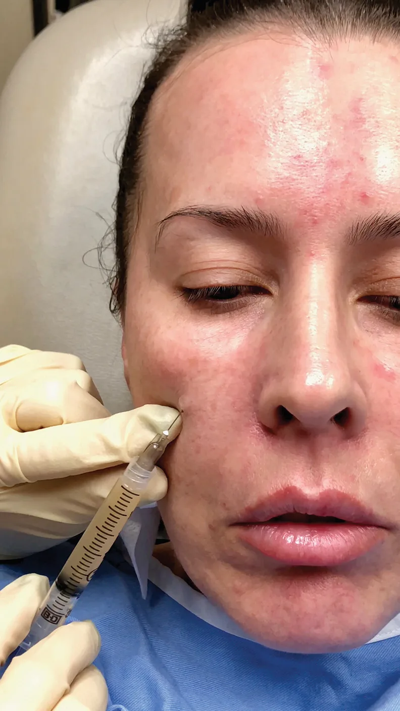 Fig 8. Fig 8 shows the regenerative target: the thin lower-lid skin itself, treated to improve colour homogeneity and thickness over months, the correct lever when the dark circle is pigment or translucency rather than a hollow - (Hausauer, 2019, p.51).
> Sources: Hausauer 2019 [19]; PRP dark-circle data per Mehryan 2014 [33].
Fig 8. Fig 8 shows the regenerative target: the thin lower-lid skin itself, treated to improve colour homogeneity and thickness over months, the correct lever when the dark circle is pigment or translucency rather than a hollow - (Hausauer, 2019, p.51).
> Sources: Hausauer 2019 [19]; PRP dark-circle data per Mehryan 2014 [33].
One hard interaction to remember. PRP and botulinum toxin are not delivered in the same product/session in the corpus protocol (Bulam, via Ordiz [27]); sequence them, do not mix. And PRP's evidence in this region is honest: it improves colour and tone more reliably than volume or wrinkle depth, and satisfaction is high with an excellent safety profile [33][19]. Fig 8 shows the regenerative deposit into the thin lower-lid skin that is its true target.
Surgery, the non-injectable endpoint. Festoons, malar mounds, orbital-fat pseudoherniation and marked laxity are corrected by lower-lid blepharoplasty (fat repositioning/removal, skin pinch, canthal support), sometimes combined with chemical blepharoplasty/peel for skin quality [24][43][44]. This is the "do not inject" branch of the exhaustive grid, and for the bag/festoon patient it is first line, not a fallback.
Topical actives, the daily base of any pigment plan. The workhorses are hydroquinone (typically 4%, higher short-term under supervision), retinoids (turnover and dispersion), azelaic and kojic acid, vitamin C and arbutin, layered on rigorous photoprotection which is itself the most important single measure [14][15]. Combination formulations and superficial peels (glycolic, mandelic, low-strength TCA, and the periorbital-specific weaker formulations to limit PIH) address the epidermal component; the periorbital skin is prepared with a weaker regimen than the face because it is thin and PIH-prone [44][15]. The realistic expectation is gradual lightening over months, not a single-session result.
Peels and resurfacing, the caveats specific to this skin. Erian 2012 [44] and the cosmetic-dermatology texts caution that periorbital and eyelid skin needs a weaker peel than facial skin and careful conditioning to reduce PIH, and that ablative resurfacing (CO2, Er:YAG) can help skin quality and shadow but carries prolonged erythema and, in high phototypes, a real PIH risk [14][44]. This is the "energy is not free in dark skin" rule that governs the whole pigment/vascular lane.
Sequencing the regenerative and device lanes. A pragmatic combined dark-circle plan runs topicals continuously, schedules a PN or PRP course (roughly three sessions weeks apart) for the thin-skin/vascular substrate, and adds vascular or pigment laser in its own sessions matched to phenotype and phototype, with filler reserved for the residual shadow. None of these is stacked on a freshly filled trough in the same visit, and PRP is not co-delivered with toxin [27][19].
Trampa clásica: lasering a pigment-predominant, high-phototype dark circle with a Q-switched device at full settings. Dermal melanin and melasma-type pigment can worsen with post-inflammatory hyperpigmentation, converting a cosmetic complaint into a harder problem. Photoprotection and topicals first, a test spot always, conservative energy in dark skin [14][29].
D5.8 · Toxin of the region: muscle, units/point, points, safety distances, antagonist spared
Toxin has a narrow, optional role here. It does not treat the hollow, the pigment or the vessel. It relaxes a hypertrophic pretarsal orbicularis that bulges as a "jelly roll" on smiling and softens fine infraorbital rhytids, and it slightly widens the palpebral aperture for a rounder, "wide-eyed" look. It is a small refinement, not a dark-circle treatment, and it is contraindicated in a lax lid.
| Parameter | Value | Source |
|---|---|---|
| Target muscle | superficial fibres of the pretarsal orbicularis oculi, lower lid | [20][21] |
| Dose per side | 1-2 U ona, no more than 3-4 U | [20] |
| Point (1-site) | mid-pupillary line, 2-3 mm below the lid margin, 1-2 mm lateral to mid-pupil | [20] |
| Points (2-site) | A1 at mid-pupil + A2 midway between mid-pupil and lateral canthus | [20] |
| Depth | intradermal / dermal-subcutaneous interface, a visible wheal, superficial | [20] |
| Technique | pull lid taut, needle ~45°, low-volume highly concentrated aliquot | [20] |
| Effect (2 U, 1 site) | palpebral aperture +~0.5 mm at rest, +1.3 mm at full smile | [20] |
| Effect (4 U, 2 sites) | +~1.75 mm rest, +2.5 mm smile | [20] |
| Onset / peak / duration | 3-5 days / 1-2 weeks / ~3 months | [21] |
| Mandatory pre-test | snap-back test; a sluggish snap is a stop sign | [20][21] |
Consensus (the seven treatment rules for the lower lid, Benedetto [20]). (1) Inject only 1-3 U, never more than 4 U, into the pretarsal midpupillary line. (2) A wheal confirms correct superficial depth. (3) Injecting at the intermediate point between lateral canthus and mid-pupil increases the risk of ectropion and a rounded lateral canthus. (4) Injecting medial to the mid-pupillary line can cause epiphora (blink-reflex weakening) or dry eye (persistent lagophthalmos, corneal exposure). (5) Injecting more than 3-4 mm below the margin can cause lip asymmetry and cheek ptosis by reaching the lip elevators. (6) Use low-volume, highly concentrated injections of the superficial pretarsal fibres. (7) A patient with prior periocular surgery and a sluggish snap test should not be treated.
The muscle deliberately spared (the "antagonist" here). The lower lid has no strong elevator antagonist; its position is held by the orbicularis tone, the retractors and the tarsoligamentous sling. The safety therefore is about not over-weakening or over-diffusing the one sphincter: keep the toxin in the superficial pretarsal fibres and spare the deeper preseptal/orbital orbicularis, which drives the lacrimal pump and lymphatic drainage. Weakening those produces dry eye, epiphora and infraorbital/malar edema. Diffusion inferomedially threatens the inferior oblique (diplopia) and inferolaterally the lip elevators (cheek ptosis), so the safe zone is a narrow superficial band at and just lateral to the mid-pupil [20][21].
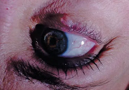 Fig 9. Fig 9 shows the exact gesture and its limits: a superficial pretarsal wheal at the mid-pupil, never lateral to the intermediate point (ectropion) nor medial to the mid-pupil (dry eye), and never deep (lip/cheek ptosis) - (Benedetto, 2011, p.106).
> Sources: Benedetto 2011 [20]; snap-test mandate per Berry 2021 [21].
Fig 9. Fig 9 shows the exact gesture and its limits: a superficial pretarsal wheal at the mid-pupil, never lateral to the intermediate point (ectropion) nor medial to the mid-pupil (dry eye), and never deep (lip/cheek ptosis) - (Benedetto, 2011, p.106).
> Sources: Benedetto 2011 [20]; snap-test mandate per Berry 2021 [21].
Why the snap test is mandatory. Berry 2021 [21] calls the snap test "particularly useful if not mandatory" before lower-lid toxin: the pretarsal injection weakens the sphincter, and in a lid that already recoils slowly this tips into ectropion, scleral show, dry eye and corneal exposure. Berry also stresses angling the needle away from the orbit to reduce ocular injury, and warns that many patients have creased, hyperdynamic preseptal lower lids that must be told they are outside the treatment zone and will not change. Fig 9 shows the exact superficial pretarsal gesture at the mid-pupil.
Discrepancy (widen the aperture vs leave it alone). > Consensus: a lax lid is never treated; the snap test governs [20][21]. > Discrepancy: the "wide-eyed" school treats the pretarsal orbicularis for aperture widening and jelly-roll softening in a competent young lid; the conservative school treats the lower lid only for a genuine hypertrophic pretarsal roll and otherwise leaves it, judging the millimetric aperture gain not worth the dry-eye/ectropion risk. Decide: young, competent lid, dynamic roll, good snap → treat 1-2 U; any laxity, dry-eye history, prior surgery, older lid → do not treat.
Trampa clásica: treating a "puffy lower lid" with pretarsal toxin when the puffiness is orbital-fat pseudoherniation or malar edema, not orbicularis hypertrophy. The toxin does nothing for fat or fluid, and if it diffuses to the preseptal/orbital fibres it weakens the lymphatic pump and worsens the edema. Diagnose the roll dynamically (it appears on smile) before treating [20][21].
D5.9 · Combination and sequence: what before, what after, at what interval → L2 — Combination & Sequencing
The sequence is support first, shadow second, colour and skin in parallel. The single most common strategic error is filling the trough before restoring the cheek that should support it.
| Step | What | When / interval | Why the order |
|---|---|---|---|
| 0 | classify phenotype (D5.5b), snap test, vector | consultation | routes to filler vs pigment vs surgery [29][20] |
| 1 | midface / SOOF / deep-cheek support if support-deficient | first, then reassess at 2-4 weeks | shortens the lid-cheek junction and often erases most of the shadow [16][24] |
| 2 | direct trough correction of the residual shadow | after reassessment (same visit if support adequate) | fill only what remains; less volume, less edema [6][17] |
| 3 | pigment / vascular treatment (topical ongoing; laser in separate sessions) | in parallel, own schedule | different mechanism, different device [14][29] |
| 4 | skin quality: PN / PRP / boosters / microneedling | series, own schedule | thickens skin, improves colour over months [19][27] |
| 5 | pretarsal toxin if indicated | separate, and not the same session as PRP | refinement last; toxin + PRP not co-delivered [20][27] |
Consensus (support before line). Wong 2022 [16] and the midface literature agree that when the cheek/SOOF is deficient, restoring it first shortens the lid-cheek junction and reduces or removes the trough shadow, so the residual defect fillable at the rim is smaller and safer. de Maio's MD Codes philosophy (via [7]) is the same idea formalised: anchor the cheek support points, treat the trough as a residual. The clinical test is the manual cheek lift (D5.5): if lifting the cheek erases the trough, do the cheek first.
Discrepancy (direct vs staged midface-first): kept, not averaged. > Consensus: both schools reassess before adding more, and both undercorrect and stage [6][16]. > Discrepancy: Direct correction treats a localized hollow at the rim in one step when midface support is adequate; it is faster and precise for the isolated class I-II trough [6][17]. Staged midface-first restores deficient cheek/SOOF support first and fills only the residual infraorbital defect, arguing that direct filling over a deficient midface chases a shadow that keeps returning and stacks volume in the worst plane [16][24]. Decide by the support deficit and vector: adequate cheek, positive vector, isolated groove → direct; deficient cheek, negative vector, long lid-cheek junction → midface first. Do not average into "a little of both in one plane".
Intervals and co-treatment rules, stated once. Review every trough correction at 3-4 weeks before adding product; HA is hydrophilic and expands, so early overcorrection reads as edema [12][6]. Pigment topicals run continuously as a baseline; pigment/vascular laser is scheduled in its own sessions and not stacked on fresh filler in the same visit (heat plus fresh HA is avoided). PN and PRP run as a course of ~3 sessions weeks apart and improve the substrate over months. PRP and toxin are not co-delivered (Bulam, via [27]); sequence them. Combination with energy and threads is coordinated in L2 — Combination & Sequencing.
What pairs well, and what does not. Well: midface support + minimal residual trough HA; trough HA + a skin-quality course (PN/booster) for the thin-skin component; pigment topicals + vascular laser for a mixed circle. Poorly: hydrophilic HA + a fluid-retaining lower lid (edema); aggressive laser + a freshly filled trough in the same visit; toxin + PRP in the same syringe pass; and any injectable stacked onto a festoon or bag that needs a surgeon.
Five archetypes, sequenced (the plan reads off the phenotype + support + lid state).
| Archetype | Phenotype / findings | Sequenced plan |
|---|---|---|
| Young ligamentous groove | isolated tear-trough shadow, no volume loss, good skin, positive vector | minimal deep supraperiosteal HA or a cannula subcision-style release; no cheek volume [18][6] |
| Aged deflated midface | long lid-cheek junction, hollow cheek, negative vector | cheek/SOOF support first, reassess at 2-4 weeks, then residual trough HA [16][24] |
| Pigment-dominant dark circle | brown, stretch-persistent, high phototype | photoprotection + topicals + cautious pigment ladder; no filler [29][14] |
| Vascular/thin-skin circle | blue-violet, stretch-deepens, fair skin | vascular laser and/or superficial masking sheet + skin thickening (PN/PRP); treat congestion [13][19] |
| Bag / festoon / laxity | orbital-fat prolapse, malar mound, slow snap | surgical referral (blepharoplasty); do not inject [24][43] |
Sequencing with adjacent regions. The trough is rarely treated alone. Its natural companions are the cheek (support platform, treated first), the midface and the lateral orbit / crow's feet (toxin, if combined, at least a week's mental separation from a fresh trough fill so edema is not misread), and skin-quality courses (PN/booster/microneedling) that run on their own calendar. The one firm ordering rule inside a single visit: support before line, and never stack an energy device on a freshly filled trough. Full cross-region logic in L2 — Combination & Sequencing.
Longevity and maintenance interval. HA in the periorbital region is unusually durable: it can persist years because of the low metabolic turnover and minimal movement of the medial trough [26]. This is a double message: touch-ups are infrequent, but a poorly placed deposit also lasts a long time, and a "dark circle" that reappears at 6-12 months is more often a re-shadowing from ongoing midface descent than resorption of the trough product, which again points to treating the cheek rather than re-filling the line [1][26].
Trampa clásica: treating the trough and the cheek as independent line items and filling the trough first "because that is what the patient pointed at". Over a deficient midface the trough re-shadows within weeks, the patient returns for "more", and the injector stacks volume that eventually festoons and edemas. Support the platform, reassess, then fill the residual [16][24].
D5.10 · Region-specific complications → J1 — Injectable Complications Overview · J2 — Vascular Occlusion & Emergency Response
These are the complications that happen here and almost nowhere else, or that behave differently here. The generic bruise is in J1; this block is the trough's own list.
| Complication | Mechanism (region-specific) | Signature / timing | Management |
|---|---|---|---|
| Malar edema | lymph trapped between the malar septum and the ORL; superficial, hydrophilic or over-volumed deposit | boggy malar swelling, worse mornings; can persist months to years, refractory | head elevation, massage, lymphatic drainage; hyaluronidase for HA; wait/dissolve [9][26][23] |
| Prolonged periorbital edema | product behind the orbital septum along the rim | pathognomonic unresolving lid swelling | hyaluronidase (needed for resolution) [6] |
| Tyndall effect | superficial HA in < 1 mm skin scatters blue light; can occur even after deep placement or by anterior migration | blue-grey hue, worse on flash photography | hyaluronidase; prevent with deep placement + low-viscosity thin sheet [6][7][23] |
| Product visibility / beading / contour irregularity | no subcutaneous buffer; too-superficial deposit | visible/palpable ridge or lump | massage early; hyaluronidase if HA [8][23] |
| Medial "balling up" / nodule | intramuscular deposit in the bone-adherent medial zone, squeezed by blinking | late medial lump | hyaluronidase; avoid by not injecting the adherent medial zone [6] |
| Migration | anterior/gravitational displacement over time (worse with permanent/semi-permanent) | delayed bluish swelling months-years later | hyaluronidase for HA; excision for permanent [8][26] |
| Post-inflammatory hyperpigmentation | needle trauma / prolonged bruise in a pigment-prone lid | a "bruise" lasting > 2-3 weeks | hydroquinone, photoprotection, time [11] |
| Ecchymosis | dense vascular plexus, thin skin | very common (up to ~50% for some techniques), up to ~10 days | pressure, cold, PDL to speed purpura [12][13] |
| Delayed inflammatory nodule / biofilm | HA persists years periorbitally; late immune/infective trigger | late erythema, swelling, tenderness | distinguish infection vs inflammation; antibiotics/steroid; cautious hyaluronidase [23][26] |
| Vision loss / retinal artery occlusion | retrograde embolus via angular/dorsal nasal/infraorbital → ophthalmic → central retinal a. | sudden painful monocular vision loss, ophthalmoplegia, skin mottling | EMERGENCY: retrobulbar hyaluronidase 150-200 U/mL, ~90-min window; ophthalmology now → J2 — Vascular Occlusion & Emergency Response [7][8] |
The signature complication (malar edema), in detail. van Loghem 2023 [9]: malar edema is "serious and long-lasting", reported with all fillers injected into the infraorbital hollow and tear troughs; superficial injection is the usual cause, and the risk rises with high-elasticity, high-viscosity or hydrophilic gels, because the region's lymphatics are easily compromised. The fluid is trapped between the malar septum and the orbicularis retaining ligament. Management is lymphatic drainage, massage and night head-elevation; persistent HA-induced edema needs hyaluronidase; CaHA-induced edema resolves less readily [9][26]. Tejero 2024 [26] adds that a superficial deposit, a high-G′ product or excess volume in the ojera area is the typical technical cause and that the edema can become chronic and permanent, refractory to treatment, as Fig 10 documents years after injection. The blue-grey Tyndall artefact that governs product choice and depth is shown in Fig 11.
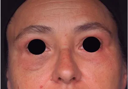 Fig 10. Fig 10 documents why "less is more" is a safety rule here: superficial, hydrophilic or over-volumed infraorbital filler traps lymph between the malar septum and the ORL, producing malar edema that persisted for years in this patient - (UPO Sorted, Tejero, 2024, p.154).
> Sources: UPO Sorted, Tejero 2024 [26] (
Fig 10. Fig 10 documents why "less is more" is a safety rule here: superficial, hydrophilic or over-volumed infraorbital filler traps lymph between the malar septum and the ORL, producing malar edema that persisted for years in this patient - (UPO Sorted, Tejero, 2024, p.154).
> Sources: UPO Sorted, Tejero 2024 [26] ([D], corroborated by van Loghem 2023 [9]); mechanism per van Loghem [9].
The optical complication (Tyndall), in detail. Because the eyelid skin is under 1 mm, superficially placed HA preferentially scatters blue light, producing a blue-grey hue that is worse under flash photography [6][7][23]. It can appear even after deep placement and can develop over time as product migrates anteriorly; a highly hydrophilic gel (Juvederm Ultra) is the classic offender, which is why it is contraindicated in the trough [6][11]. Treatment is hyaluronidase, with the caveat that dissolving the filler also removes the correction, so counsel accordingly [23].
 Fig 11. Fig 11 is the Tyndall trap that governs product choice and depth: HA in < 1 mm skin scatters blue light into a grey-blue shadow that reads as a persistent dark circle and needs hyaluronidase to clear - (Advances in Cosmetic Surgery, 2018, p.152).
> Sources: Hwang & Perry, Advances in Cosmetic Surgery 2018 [23].
Fig 11. Fig 11 is the Tyndall trap that governs product choice and depth: HA in < 1 mm skin scatters blue light into a grey-blue shadow that reads as a persistent dark circle and needs hyaluronidase to clear - (Advances in Cosmetic Surgery, 2018, p.152).
> Sources: Hwang & Perry, Advances in Cosmetic Surgery 2018 [23].
The catastrophe (vision loss), and the region's discipline. Vascular occlusion of the ophthalmic system is rare but reported for the infraorbital area; the incidence of vascular compromise across the face has been estimated around 3 in 1000 injections, and no technique is 100% protective [23][8]. Any visual symptom, severe ocular pain, ophthalmoplegia or spreading skin mottling after a trough injection is a time-critical emergency: stop, do not inject more, flood the territory and consider retrobulbar hyaluronidase 150-200 U/mL within the ~90-minute window, and get ophthalmology immediately. Full algorithm in J2 — Vascular Occlusion & Emergency Response and reversal pharmacology in J3 — Hyaluronidase - Pharmacology & Clinical Protocols [7][8].
The management principle that ties them together. Most trough complications are HA in the wrong plane and are reversed by hyaluronidase; the injector must distinguish these from infection/biofilm (delayed erythema, tenderness, sometimes fluctuance: culture, antibiotics, cautious enzyme) and from non-HA material (CaHA/PLLA/PMMA nodules, which hyaluronidase will not touch and which may need steroid, 5-FU or excision) [23][26]. Ultrasound (D5.5) resolves most of these questions at the bedside.
Hyaluronidase in this region, the practical notes. For a cosmetic HA complication (Tyndall, lump, malar edema, prolonged periseptal swelling), hyaluronidase is titrated to the deposit and can be repeated; the corpus documents dramatic reversals, including a periorbital HA that had caused pitting eyelid edema for years resolving within 2 days of enzyme, and a nasolacrimal-duct obstruction after tear-trough HA relieved by duct irrigation and enzyme [26]. For the vascular emergency the dose is entirely different and urgent: high-volume flooding of the ischaemic territory and retrobulbar hyaluronidase 150-200 U/mL within the ~90-minute window, per J3 — Hyaluronidase - Pharmacology & Clinical Protocols [7]. The enzyme does not touch CaHA, PLLA or PMMA, which is a second reason those products are wrong in the trough: a complication from them is not reversible [9][26].
Timelines that tell you which complication you have. Ecchymosis appears immediately and clears within ~10 days [7][12]; transient edema/erythema clears within 1-2 weeks; malar edema is boggy, worse in the morning, and persists weeks to years [9][26]; Tyndall is a stable blue-grey from the first days and does not resolve without enzyme [23]; a delayed inflammatory nodule or biofilm appears weeks to months later as tender erythema and warrants an infection-versus-inflammation work-up [23][26]; migration produces new bluish swelling months to years out [8]. Reading the timeline plus the ultrasound (D5.5) usually names the complication without guesswork.
The FOS caveat (overfilling as its own diagnosis). Facial overfilling syndrome (Tingson Lim, 2017), with the ojera among the affected zones, is the endpoint of repeated superficial infraorbital and periorbital filling: an unnatural, expanded, sometimes fibrotic appearance that no single injection caused and that hyaluronidase only partly reverses [26]. It is the cumulative form of this chapter's core warning, and the reason "undercorrect and stage" is a safety rule, not a stylistic one.
Trampa clásica: treating a persistent post-filler "dark circle" that is actually Tyndall or malar edema with more filler. The blue-grey is superficial gel and the boggy fullness is trapped lymph; adding product deepens both. The correct move is hyaluronidase and time, not another syringe [6][9].
Coverage vs UPO
Three columns: what the UPO master teaches, its status in this chapter, and what the atlas adds. UPO material is valuable and ages fastest; a dose resting on a single UPO slide is never_sufficient_alone.
| UPO teaches (source) | Status here | What the atlas adds |
|---|---|---|
| Tear trough = low/medium-consistency HA, below orbicularis, inferior to the bony rim, palpate the rim with the non-injecting index finger (Arenas [47]) | ✅ D5.7 | the 3-vs-7-layer split, medial-intramuscular caveat, lateral-entry milking, needle-vs-cannula controversy, firm-NASHA RCT evidence, explicit volume ceilings, ultrasound guidance |
| Malar area supraperiosteal support for midface projection (Arenas [47]) | ✅ D5.9 | support-first sequencing, the manual cheek-lift test, negative-vector selection, MD Codes anchoring |
| Angular artery and vein anatomy; vein ~4 mm below the infraorbital rim, between orbicularis fibres (Tejero [26]) | ✅ D5.3 | quantified depth (4.2 ± 0.7 mm), the medial-pupil-to-nasal-wall danger zone, the ophthalmic retrograde mechanism, the retrobulbar dose |
| Complications: malar edema, Tyndall, migration, overfill (FOS), nasolacrimal obstruction; HA persists years periorbitally (Tejero [26]) | ✅ D5.10 | the malar-septum/ORL mechanism, hydrophilic/high-G′ causation, ultrasound differentiation, the HA-vs-infection-vs-non-HA management fork |
| Toxin + filler combination for periorbital rhytids (Arenas [47]) | ✅ D5.8 / D5.9 | pretarsal dosing map, mandatory snap test, sequencing rules, the PRP-toxin non-co-delivery rule |
| Mesotherapy / polynucleotides / PRP for skin quality and dark circles (Ordiz [27]) | ✅ D5.7b | phenotype-matched ladder, the PRP dark-circle RCT evidence, PN mechanism |
| Iatrogenic malar mound from superficial midface injection (Surek [48], UPO article) | ✅ D5.6 / D5.10 | the prezygomatic-space / zygomaticocutaneous-ligament mechanism, made a selection exclusion |
What UPO does NOT cover (atlas-only, and why it matters): - Dark-circle etiologic classification (pigment / vascular / structural / mixed): UPO treats the trough as a hollow; the classification (Huang [30], Roh [29]) is what routes half these patients away from filler. New ground. - Pigment and vascular laser ladder (QS ruby/alexandrite, 1064 Nd:YAG, PDL) matched to phenotype and phototype (Nouri [14], Fabbrocini [13]). New ground. - The traction/stretch test to separate pigment, vessel and shadow at the bedside [13][32]. New ground. - Restylane Eyelight FDA approval and RCT, and the low-G′-vs-firm-NASHA discrepancy [31]. Post-dates the master material. - Ultrasound-guided injection and prior-filler detection (anechoic HA vs hyperechoic CaHA/PMMA) [9]. New ground. - Lower-eyelid surgical exclusions (festoons, malar mounds, bags, double convexity, post-blepharoplasty) as referral criteria, not difficult cases [24]. New ground.
The one place the atlas nuances UPO. UPO teaches placement "below the orbicularis" generically [47]. The atlas keeps that as the lateral rule but flags that medially the orbicularis is fused to bone (three-layer zone), so "below the muscle" there is intramuscular or intraorbital, which is why the safe medial deposit comes from a lateral entry milked medially, not a medial sub-muscular bolus [3][6]. Same teaching, a regional exception the slide does not state.
Self-assessment
Ten active-recall questions built only from facts already in this chapter. Answers fold open.
- Before treating any "dark circle", what is the single first question, and what four answers does it have?
answer
Is the darkness a hollow (shadow), pigment, vessel, or skin/edema? Only the structural/shadow phenotype is a filler target (D5.1, D5.5b). - How many tissue layers does the tear trough have medially versus laterally, and what marks the boundary?
answer
Two (skin + bone-adherent orbicularis) medially, seven laterally; the boundary is the facial vein, ~4-6 mm medial to the mid-pupillary line (D5.2). - What are the length and course of the tear-trough ligament, and what does it become laterally?
answer
A single-lamina osteocutaneous ligament ~5-7 mm from the medial canthus to the mid-pupil, becoming the bilaminar orbicularis retaining ligament laterally (~15 mm) (D5.2). - How far below the infraorbital rim does the angular vein run, and why does it matter?
answer
~4.2 ± 0.7 mm below the rim, deep to orbicularis in the nasojugal groove; it is a venous route to the ophthalmic system and the medial/lateral boundary of the trough (D5.3). - Why is a deep bolus on the medial rim at the mid-pupil dangerous on three counts?
answer
The orbicularis is glued to bone there (intramuscular, balls up), the infraorbital foramen and vessels sit there, and the angular/dorsal-nasal vessels anastomose with the ophthalmic system (blindness) (D5.3). - In what order does the infraorbital region age, and which is first?
answer
Bone first (orbital aperture widens supero-medially and infero-laterally, senile enophthalmos), then deep fat/SOOF deflation and descent against a fixed ligament, then superficial fat, then skin; fat pseudoherniation can add "bags" (D5.4). - Which three lower-eyelid findings are surgical exclusions that volume worsens?
answer
Festoons, malar mounds/bags, and orbital-fat pseudoherniation (also marked laxity/double convexity) (D5.6). - What is the deep single-technique volume limit in the medial trough, and the per-side start and ceiling?
answer
≤ 0.05-0.1 mL microbolus deep; start 0.5 mL/side, ceiling ~1 mL/side (D5.7). - State the lower-lid toxin dose, the two "never" zones, and the mandatory pre-test.
answer
1-2 U (max 3-4) pretarsal at the mid-pupil; never lateral to the intermediate point (ectropion) or medial to the mid-pupil (dry eye/epiphora); mandatory snap-back test (D5.8). - A persistent post-filler blue-grey hue and a boggy malar fullness: what are they, and what is the wrong treatment?
answer
Tyndall (superficial HA) and malar edema (trapped lymph); the wrong treatment is more filler; the right one is hyaluronidase and time (D5.10).
What's new and trends
What changed in roughly the last two to three years:
| Year | Change | Maturity | Consequence |
|---|---|---|---|
| 2023 | Restylane Eyelight FDA-approved for infraorbital hollows (NASHA, high G′, low water uptake) [31] | clinically actionable now | on-label HA for the hollow; challenges the low-G′-only dogma |
| 2024 | Pivotal RCT: n=333, 87.4% responders at 3 months, no Tyndall reported [31] | clinically actionable now | low water uptake may matter more than low G′ for Tyndall/edema here |
| 2023-2026 | Ultrasound (Doppler) before periorbital filler + prior-filler detection [9][17] | clinically actionable now | vessel mapping and anechoic/hyperechoic filler ID at the bedside |
| 2023-2026 | Polynucleotides (salmon/trout DNA, adenosine A2A) for periorbital skin/dark circles; PN-vs-HA split-face and Huang-subtype cohorts [27] | promising but not validated | regenerative option for the thin-skin/vascular phenotype, mostly observational |
| 2023-2026 | Nanofat / microfat + PRP for skin quality and colour [16][19][46] | promising but not validated | autologous skin-quality lever; growth factors may aid fat-graft survival |
| 2017-2026 | Facial overfilling syndrome (FOS) named (Tingson Lim, 2017), ojera among affected zones [26] | promising but not validated | named endpoint for the "keep filling" trap; no objective diagnostic tool yet |
| ongoing | Polynucleotide pigment-specific benefit claimed beyond skin quality [27] | preclinical/speculative | dermal A2A action shown; a pigment-lightening effect is not established |
| ongoing | Under-eye filler marketed as a permanent dark-circle cure / single-session fix [29][31] | unsupported commercial claim | no product removes pigment or vessels; only the structural shadow is fillable |
What did NOT change, and why the older references are still state of the art. The anatomy that grounds every technique here is stable: the tear-trough ligament (Wong 2012 [5]), the SOOF / ORL frame and the three-vs-seven-layer split (Cotofana 2015/2019 [2][3]), the young-vs-elderly sectional anatomy (Yang 2013 [35]) and the lid-cheek junction anatomy (Haddock 2009 [36]) are reinforced, not revised, by newer imaging. The deep-first ageing sequence (Cotofana 2016 [1]) is unchanged. The etiologic classification of dark circles (Roh 2009 [29], Huang 2014 [30]) still governs treatment selection; no newer scheme has replaced it. The earliest HA tear-trough technique (Kane 2005 [37]) and the rheology science that explains product behaviour (Sundaram 2013 [34]) remain the reference frame, as does the regenerative rationale for autologous therapies (Zenker [45], Khan [28]). Hyaluronidase remains the single most important reversal agent, and the region's core discipline (classify first, support before line, small aliquots, low pressure, undercorrect and stage) is unchanged [7][9]. The lane that ages fastest is the UPO master material on specific products and doses; its anatomy and classification content is durable, its brand/dose content is not, which is exactly why a single UPO slide is never sufficient alone.
Unexplored directions (AI speculation)
> Disclaimer. The following are AI-generated research directions, not clinical recommendations. Each is tagged [IA-ESPEC], is not evidence, and must never be acted on clinically. Each states an anchor (a cited fact already in this chapter), a proposal, and what would settle it. None carries a dose, a product choice, or a protocol.
[IA-ESPEC]Tyndall may track water uptake, not gel firmness. Anchor: a high-G′ NASHA gel reported no Tyndall in its infraorbital RCT [31], contradicting the low-G′-only teaching [8]. Proposal: the optical blue-scatter may be driven by a gel's water uptake and refractive behaviour more than by its G′. Expected effect: at matched firmness, lower-water gels produce fewer blue-grey outcomes. Confounder: injection depth and skin thickness vary between arms and drive Tyndall independently. What would settle it: a head-to-head trial of gels matched on G′ but differing in water uptake, with Tyndall scored by objective spectrophotometry rather than by eye.[IA-ESPEC]Regenerative agents may help only the vascular/thin-skin subtype. Anchor: PRP improved infraorbital colour more reliably than volume or wrinkle depth [33], and polynucleotides act on the dermis via adenosine signalling [27]. Proposal: the benefit of regenerative injectables in dark circles may be concentrated in the vascular and thin-skin phenotypes and near-absent in pure pigment. Expected effect: a measurable colour and erythema gain in the vascular/thin-skin subtype, minimal in pure pigment. Confounder: mixed phenotypes and concurrent photoprotection blur subtype-specific effects. What would settle it: an RCT stratified by Huang subtype with objective melanin and erythema indices as endpoints.[IA-ESPEC]Routine Doppler may lower the region's two commonest harms. Anchor: ultrasound already locates the variable angular vein and identifies prior filler at the bedside [9][17]. Proposal: mandatory pre-injection Doppler mapping could reduce both vascular occlusion and malar edema by keeping the deposit off vessels and out of the wrong plane. Expected effect: lower occlusion and malar-edema rates in the guided arm. Confounder: operator experience and case mix differ between ultrasound-adopting and landmark practices. What would settle it: a prospective registry comparing ultrasound-guided with landmark-only trough injection for occlusion and edema rates.[IA-ESPEC]The congenital ligamentous groove may be a release problem, not a volume problem. Anchor: the medial orbicularis is fused to bone by the tear-trough ligament, producing a groove even without volume loss in young patients [3][18]. Proposal: a blunt subcision-style release of the tether, adding no volume, may correct the young ligamentous groove more physiologically than filling it. Expected effect: durable groove improvement in young ligamentous troughs without added volume. Confounder: unrecognised subclinical volume loss and variable ligament strength between patients. What would settle it: a controlled comparison of cannula release versus minimal filler in young troughs with documented normal volume.[IA-ESPEC]A bedside lymphatic-competence test might predict malar edema. Anchor: malar edema is lymph trapped between the malar septum and the ORL, and a history of fluid retention predicts it [9][23]. Proposal: a provocation test of infraorbital lymphatic clearance before treatment could flag the patient who will edema, converting a post-hoc complication into a selection criterion. Expected effect: the test identifies the subset that later develops malar edema. Confounder: systemic fluid status and product hydrophilicity also drive edema independently of lymphatic competence. What would settle it: a cohort correlating a standardised bedside lymphatic/edema-provocation test with subsequent malar edema after filler.
Safety
The region's non-negotiable safety rules, consolidated. The infraorbital area is a high-risk, largely off-label filler zone with thin tissue over bone and dense vascular anastomoses to the ophthalmic system [10][8]. Every treatment here obeys the same discipline.
Vascular occlusion and vision loss (the catastrophe). Prevent with low injection pressure, aliquots ≤ 0.05-0.1 mL, retrograde moving needle/cannula, lateral entry milked medially, and ultrasound vessel mapping; keep HA-only and hyaluronidase in the room [7][8][9]. Any visual symptom, ocular pain, ophthalmoplegia or spreading skin mottling is a time-critical emergency: stop, flood the territory, consider retrobulbar hyaluronidase 150-200 U/mL within the ~90-minute window, and refer to ophthalmology immediately, per J2 — Vascular Occlusion & Emergency Response and J3 — Hyaluronidase - Pharmacology & Clinical Protocols [7].
Malar and periorbital edema (the signature). Avoid superficial, hydrophilic and over-volumed deposits; stay on bone 3-4 mm below the rim, never behind the orbital septum; screen for a fluid-retention history and festoon/mound phenotype and do not inject them [9][23][26]. Persistent HA edema is treated with hyaluronidase, head elevation and lymphatic drainage.
Product safety. Use reversible HA in the trough; avoid highly hydrophilic HA (Juvederm Ultra), and avoid CaHA, PLLA, PCL and PMMA in the trough proper (nodules, non-reversible, granuloma) [6][11][22]. Semipermanent and permanent fillers in this skin are a durable-complication risk out of proportion to any benefit.
Toxin safety. Lower-lid pretarsal toxin is optional and small: 1-2 U, max 3-4, superficial pretarsal at the mid-pupil, only with a good snap test [20][38][39]. Never inject lateral to the intermediate point (ectropion, rounded canthus), never medial to the mid-pupil (epiphora, dry eye, corneal exposure), never deeper than 3-4 mm below the margin (lip/cheek ptosis), and angle the needle away from the orbit [20][21][40]. Spare the preseptal/orbital orbicularis (lacrimal pump, lymphatic drainage).
Phenotype and expectation safety. Classify before treating: pigment and vascular circles are not filler targets, and filling them wastes money, adds risk, and in high phototypes the trauma can trigger post-inflammatory hyperpigmentation [29][30][14]. Festoons, malar mounds, orbital-fat pseudoherniation and marked laxity are surgical and worsen with volume; refer them [24][43][44][41]. Document baseline asymmetry, pigment and bags before treatment, warn about flash-photography Tyndall, and stage a review at 3-4 weeks [8][42].
Systemic and consent safety. Screen sleep, salt, atopy, thyroid, renal and iron status for the systemic contributors to periorbital darkness and edema [25]. Consent must state explicitly: the darkness may be pigment/vessel that filler will not change; swelling and bruising are common and can last weeks; malar edema can be prolonged or permanent; Tyndall can occur even with correct depth; and vision loss, though rare, is possible [7][9][31].
References
- Cotofana S, et al. Anatomy of the Aging Face: A Review. Facial Plast Surg. 2016.
[MEDLIB][B]DOI 10.1055/s-0036-1582234 - Cotofana S, Lachman N. Anatomy of the Facial Fat Compartments and their Relevance in Aesthetic Surgery. J Dtsch Dermatol Ges. 2019.
[MEDLIB][B]DOI 10.1111/ddg.13737 - Cotofana S, et al. Midface: Clinical Anatomy and Regional Approaches with Injectable Fillers. Plast Reconstr Surg. 2015.
[MEDLIB][B]DOI 10.1097/PRS.0000000000001837 - Schenck TL, et al. Functional Anatomy of the Facial Superficial Fat Compartments. Plast Reconstr Surg. 2018.
[MEDLIB][B]DOI 10.1097/PRS.0000000000004364 - Wong CH, Hsieh MK, Mendelson B. The tear trough ligament: anatomical basis for the tear trough deformity. Plast Reconstr Surg. 2012;129(6):1392-1402.
[B] - Swift A, Raspaldo H. Periorbital Rejuvenation. In: Jones D, ed. Injectable Fillers: Principles and Practice. Wiley; 2019. ISBN 9781119046967.
[MEDLIB][C] - Carruthers J, Carruthers A, eds. Soft Tissue Augmentation. 4th ed. Elsevier; 2018. ISBN 9780323476584.
[MEDLIB][C] - Pirayesh A, et al. Aesthetic Facial Anatomy Essentials for Injections. CRC Press; 2020.
[MEDLIB][C] - van Loghem J, ed. Soft Tissue Filler Complications: Prevention and Management. CRC Press; 2023. ISBN 9781032440460.
[MEDLIB][C] - Haney B. Aesthetic Procedures. Springer; 2020. ISBN 9783030199470.
[MEDLIB][C] - Kontis TC, Lacombe V, eds. Cosmetic Injection Techniques. 2nd ed. Thieme; 2019. ISBN 9781626234574.
[MEDLIB][C] - de Castro CC. Correction of the tear trough deformity with hyaluronic acid. In: Midface Surgery. Elsevier; 2009. ISBN 9780702030871.
[MEDLIB][C] - Neckman JP, Brauer J, Geronemus RG. Laser for Periorbital Rejuvenation. In: Fabbrocini G, et al. Nonsurgical Lip and Eye Rejuvenation Techniques. Springer; 2016. ISBN 9783319232690.
[MEDLIB][C] - Nouri K, ed. Lasers in Dermatology and Medicine. 2nd ed. Springer; 2018. ISBN 9783319761169.
[MEDLIB][C] - Baran R, Maibach HI, eds. Textbook of Cosmetic Dermatology. 5th ed. CRC Press; 2017. ISBN 9781482257342.
[MEDLIB][C] - Wong V. Decision Making in Aesthetic Practice. CRC Press; 2022. ISBN 9781032046037.
[MEDLIB][C] - Mosaheb R. Tear trough rejuvenation. PMFA Journal. 2022;10(5).
[MEDLIB][C] - Recognising the key tear trough ligaments. PMFA Journal. 2022;10(1).
[MEDLIB][C] - Hausauer AK, Humphrey S, eds. PRP and Microneedling in Aesthetic Medicine. Thieme; 2019. ISBN 9781626239050.
[MEDLIB][C] - Benedetto AV. Botulinum Toxins in Clinical Aesthetic Practice. 2nd ed. Informa Healthcare; 2011. ISBN 9781841845098.
[MEDLIB][C] - Berry M. Botulinum Toxin in Clinical Practice. Springer; 2021. ISBN 9783030806705.
[MEDLIB][C] - Connell B, et al. Aesthetic Rejuvenation of the Face and Neck. Thieme; 2016. ISBN 9781626230897.
[MEDLIB][C] - Hwang CJ, Perry JD. Filler Complications. Advances in Cosmetic Surgery. 2018.
[MEDLIB][C] - Massry GG, Murphy MR, Azizzadeh B, eds. Master Techniques in Blepharoplasty and Periorbital Rejuvenation. Springer; 2011. ISBN 9781461400660.
[MEDLIB][C] - Pinto R. Manual Práctico de Medicina Estética. 4th ed. 2009.
[MEDLIB][C] - Tejero P, Mota S. Efectos Adversos de los Materiales de Relleno Inyectables. UPO Sorted, Máster de Medicina Estética. 2024.
[MEDLIB][D](never_sufficient_alone) - Ordiz I. Mesoterapia: técnicas de administración transcutánea (polinucleótidos, PRP). UPO Sorted, Máster de Medicina Estética.
[MEDLIB][D](never_sufficient_alone) - Khan U, ed. Regenerative Medicine in Aesthetic Treatments. CRC Press; 2022. ISBN 9780367431471.
[MEDLIB][C] - Roh MR, Chung KY. Infraorbital dark circles: definition, causes, and treatment options. Dermatol Surg. 2009;35(8):1163-1171.
[B]PMID 19469797 · DOI 10.1111/j.1524-4725.2009.01213.x - Huang YL, Chang SL, Ma L, Lee MC, Hu S. Clinical analysis and classification of dark eye circle. Int J Dermatol. 2014;53(2):164-170.
[B]PMID 23879616 · DOI 10.1111/j.1365-4632.2012.05701.x - Biesman BS, et al. A Multicenter, Randomized, Evaluator-Blinded Study to Examine the Safety and Effectiveness of Hyaluronic Acid Filler in the Correction of Infraorbital Hollows (Restylane Eyelight). Aesthet Surg J. 2024.
[B]PMID 38573527 · DOI 10.1093/asj/sjae073 - Friedmann DP, Goldman MP. Dark circles: etiology and management options. Clin Plast Surg. 2015;42(1):33-50.
[B]DOI 10.1016/j.cps.2014.08.007 - Mehryan P, Zartab H, Rajabi A, et al. Assessment of efficacy of platelet-rich plasma (PRP) on infraorbital dark circles and crow's feet wrinkles. J Cosmet Dermatol. 2014;13(1):72-78.
[B] - Sundaram H, Cassuto D. Biophysical characteristics of hyaluronic acid soft-tissue fillers and their relevance to aesthetic applications. Plast Reconstr Surg. 2013;132(4 Suppl 2):5S-21S.
[B] - Yang C, Zhang P, Xing X. Tear trough and palpebromalar groove in young versus elderly adults: a sectional anatomy study. Plast Reconstr Surg. 2013;132(4):796-808.
[B] - Haddock NT, Saadeh PB, Boutros S, et al. The tear trough and lid/cheek junction: anatomy and implications for surgical correction. Plast Reconstr Surg. 2009;123(4):1332-1340.
[B] - Kane MA. Treatment of tear trough deformity and lower lid bowing with injectable hyaluronic acid. Aesthetic Plast Surg. 2005;29(5):363-367.
[B] - Benedetto AV. Botulinum Toxin in Clinical Dermatology. Taylor & Francis; 2006. ISBN 9780203495056.
[MEDLIB][C] - Truong D, Dressler D, Hallett M, eds. Manual of Botulinum Toxin Therapy. Cambridge University Press; 2009. ISBN 9780521694421.
[MEDLIB][C] - Blitzer A, et al. Botulinum Neurotoxin for Head and Neck Disorders. Thieme; 2020. ISBN 9781684200955.
[MEDLIB][C] - Master Techniques in Facial Rejuvenation. 2nd ed. Elsevier; 2018. ISBN 9780323358767.
[MEDLIB][C] - Towne WS, Bentsianov B, Waldman A. Neurotoxins and Fillers in Facial Esthetic Surgery. Wiley; 2019. ISBN 9781119294283.
[MEDLIB][C] - Sadick NS, Lawrence N, Moy R. Concise Manual of Cosmetic Dermatologic Surgery. McGraw-Hill; 2008. ISBN 9780071593281.
[MEDLIB][C] - Erian A, Shiffman MA, eds. Advanced Surgical Facial Rejuvenation. Springer; 2012. ISBN 9783642178375.
[MEDLIB][C] - Zenker S. Platelet-rich plasma from science to clinical results. In: Cosmetic Medicine & Surgery. CRC Press; 2016.
[MEDLIB][C] - Jacono A. Park Avenue Face. 2019. ISBN 9781948836234.
[MEDLIB][C] - Arenas D. Revisión de Materiales de Relleno; Técnicas Avanzadas de Rellenos. UPO Sorted, Máster de Medicina Estética.
[MEDLIB][D](never_sufficient_alone) - Surek CC, et al. Pertinent anatomy for midface volumizing (iatrogenic malar mound mechanism: prezygomatic space, orbitomalar and zygomaticocutaneous ligaments). 2015.
[MEDLIB][B](UPO corpus article)
Verification: All 10 template blocks written, plus two added subchapters, D5.5b (dark-circle etiologic classification: pigment/vascular/structural/mixed) and D5.7b (pigment/vascular non-injectable + regenerative ladder), because the chapter title covers "dark circle" and the 10-block template only covers the hollow, so half of these patients would otherwise have no home; declared here per the coverage>counts rule. Region absorbs the nasojugal groove and the lower-eyelid referral exclusions (festoons, malar mounds, double convexity, post-blepharoplasty) salvaged from D2.4. Corpus pass: evaluation/runs/D5.2-D5.10.jsonl (fresh, exit 0; D5.1 run failed exit 1 and retains the 2026-08-10 file, logged to AA.log). Evidence: MEDLIB corpus (RAG) + external verified (Roh 2009, Huang 2014, Biesman 2024, Friedmann-Goldman 2015). 11 figures, each opened with Read before captioning; figure-pick receipt at _images/D5/figure-pick-receipt.json. Salvage: closed --no-prior because no genuine prior tear-trough chapter exists. The old D5 code was a reused-code collision (the registry, chapter_brief.py l.691/901, flags it): D5 — Perioral Region & Lips.es.md is a legacy perioral file whose content belongs to D10 "Labio y perioral", not to tear-trough D5 (0 tear-trough hits on grep). It was preserved to docs/salvage/perioral-lips-legacy.md and retired from wiki/Themes/ with send2trash (reversible; git history intact) so the correctly-coded tear-trough D5 can publish; its perioral content must be re-homed to D10 by a future D10 pass. D2 is the current, separate brow chapter, not a prior version of D5. Due diligence: salvage_diff --cross-lang was still run against D2.1/D2.2/D2.4 (docs/salvage/D5.salvage.json); its 8 residual LOST facts are all brow/temple/forehead-specific (glabellar 20 U, temple 1.0 mL, supratrochlear 2.2 cm, Charpy 1909, forehead compartments 2007) that belong to D2 and correctly do not enter a tear-trough chapter. The lower-eyelid referral exclusions the brief routes from D2.4 to D5 (festoons, malar mounds, double convexity, post-blepharoplasty) were authored fresh in D5.6 from corpus evidence. Every DOI/PMID written as a resolvable link for RM refverify (Crossref + Europe PMC).