D14 · Escote (Décolletage)
> Currency and provenance — 43 references · median 2018, range 2000-2026, 30 % from 2022 on · provenance: verified external 44 % (19) · MEDLIB corpus 56 % (24).
Domain: D, Region-by-Region - Face & Head · Region unit: anterior chest / V of the neckline, from the clavicles and suprasternal notch to the upper breast, laterally to the anterior axillary lines. Neck = D13; earlobe = M5; torn/keloid lobe = M7/M2. IPL/pigment-vascular device physics = G4; skin-quality modality mechanics = E2, those chapters LINK here, they do not house the region.
> Evidence legend: [A] datasheet, guideline or dated consensus · [B] primary literature with PMID/DOI · [C] monograph/textbook · [D] slide or opinion, never sufficient alone · [MEDLIB] own corpus · [MODELO] structure only, never a figure · [IA-ESPEC] AI speculation, never actionable · [MATERIAL GAP] corpus/literature silence, declared not invented · ⚠ disputed or stale figure.
> ⚠ Governing red line of the whole chapter, the décolletage is NOT the face and does not accept facial parameters. The skin is thin, the dermis is of low pilosebaceous-unit density, and re-epithelialisation depends on those adnexal reservoirs, so repair is worse. It is the region of aesthetic medicine with the highest iatrogenic hypertrophic-scarring and permanent-dyschromia rate. Treated softer, in more sessions, always with a test spot. Every energy fluence and every ablative depth that is safe on the face is potentially scarring here.
Subchapters
- [x] D14.1 · In 30 seconds: region doses, volumes, planes, red lines
- [x] D14.2 · Layered anatomy skin to bone (6-layer diagram), this region only
- [x] D14.3 · Vessels, nerves and the danger zone → J2
- [x] D14.4 · Ageing of the region, and in what order
- [x] D14.5 · Assessment: measured, photographed, tested dynamically, scanned
- [x] D14.6 · Goal and patient selection
- [x] D14.7 · Technique, full regional grid (7a injectables · 7b resurfacing/EBD)
- [x] D14.8 · Toxin of the region: microtox dose, points, safety distances
- [x] D14.9 · Combination and sequence → L2
- [x] D14.10 · Region-specific complications → J1-J8
D14.1 · In 30 seconds
The décolletage is a skin-quality region, not a volume region. The dominant problems are photodamage (dyschromia, telangiectasia, poikiloderma), crepey atrophy and mechanical/sleep rhytides, not deficient projection. Direct filler of a wrinkle is the classic error. The correct toolbox is, in order of yield: photoprotection + topical prep → resurfacing/EBD (IPL first for poikiloderma) → biostimulator or skin booster for dermal quality → intradermal microtoxin for the dynamic/platysmal component → HA only for a discrete static rhytid.
One-glance region parameter grid, every figure below is region-specific, softer than the face:
| Modality | Product / device | Plane | Instrument | Dose / setting (décolleté) | Sessions | Red line |
|---|---|---|---|---|---|---|
| IPL (1st line poikiloderma) [5,15] | broadband 515–755 nm cut-off | epidermal-dermal | crystal/handpiece | fluence ~25–30% below facial, longer pulse, double-pass OFF | 3, q3–4 wk | ⚠ test spot; never on tanned skin; hypopigmented "footprints" are the region's signature disfigurement |
| Non-ablative fractional (thulium 1927 nm; 1550 nm) [32,33] | fiber laser | dermis to ~200 µm | fractional handpiece | low energy, low density (≤10–15%) | 3–5, q4 wk | ⚠ pilosebaceous-poor skin = raise density = scar |
| Ablative fractional CO₂/Er:YAG [1,39,40] | fractional | mid-reticular | scanner | low density, 1–2 passes, reduced fluence | 1–3 | ⚠ full-field ablation off-face = HTS; neck/chest scars documented |
| Chemical peel | Jessner / TCA 20–25% (superficial-medium) | epidermis–papillary | n/a | build up over sessions; prime 2–4 wk | 3–4 | ⚠ never deep TCA on chest |
| Hyperdilute CaHA [4,25] | CaHA + saline/lidocaine | deep subdermal, linear | 25G cannula pref. | 1:2 normal / 1:4 thin / 1:6 atrophic skin; ≤1 syringe/session | 2–3, q3 mo | ⚠ irreversible in a poor-healing zone; nodules |
| PLLA [24] | poly-L-lactic acid | deep subdermal | cannula | body dilution ≥16–18 mL, hydrate 24 h, massage | 2–3, q4–6 wk | ⚠ delayed papules/nodules; overcorrection is late |
| HA skin booster [27] | non/low-crosslinked HA | mid-dermis | 30G needle / cannula | microdroplet grid, ~0.01–0.02 mL/point | 3, q3 wk + maintenance | palpable beading if too superficial |
| HA filler (discrete rhytid only) [22] | low-G′ HA | deep dermal/subdermal, retrograde | 30G / cannula | microaliquots, linear threading, not bolus | 1 + touch-up | ⚠ visible nodule in thin skin; Tyndall |
| Microtoxin (chest/platysmal) [3] | onabotulinumtoxinA, intradermal | intradermal/immediate subdermal | 32–33G | 1–2 U per 0.05 mL microdroplet, 1 cm grid; ~ 20–30 U total chest field | q3–4 mo | ⚠ never if laxity dominant; no deep IM injection over ribs |
| Microneedling ± RF [29,30] | needle depth 1–2 mm | epidermis–dermis | pen / RF microneedle | conservative depth/energy | 3–4, q4 wk | infection, tracking, PIH in high phototype |
PN / PDRN [28] [MATERIAL GAP] |
polynucleotide booster | mid-dermis | 30G | facial protocol extrapolated | 3, q2–3 wk | region-specific RCT absent, see D14.7 |
Governing numbers (memorise): - ⚠ Fluence/energy reduced 25–30% vs the face, and more, softer sessions replace one aggressive pass [1,17]. - ⚠ Test spot mandatory before any IPL/laser/peel; read it at 2–4 weeks, not the same day [5,15]. - ⚠ Dermatologic pre-screen first: actinic keratoses and atypical pigmented lesions are referred and biopsied BEFORE any aesthetic treatment, the sun-damaged chest is AK and melanoma territory [20,23]. - ⚠ CaHA/PLLA are irreversible biostimulators in a bad-healing zone, go diffuse, deep, diluted, ≤1 syringe/session [4,24]. - ⚠ Poikiloderma of Civatte = reticulate pigment + telangiectasia + atrophy with submental sparing (the shaded island under the chin) as the diagnostic key; IPL first line [5,17]. - ⚠ Fitzpatrick IV–VI and any keloid/HTS history = the sternal chest is a top keloid site (up to ~16% in skin of color) → contraindicate energy/ablation or maximal caution [7,40]. - ⚠ IPL for poikiloderma clears ~75–80% of vascular + pigment components over ~3 sessions with ~5% (mostly transient) side-effects, the region's highest-satisfaction indication [5,15]. - ⚠ Chemical peel ceiling off-face = Jessner + TCA 20–25%; deep peels are contraindicated on the chest [34,35]. - ⚠ "Deep" on the chest = the chest wall. There is no safe facial-style supraperiosteal bony plane; deep between ribs is the pleura [8]. - ⚠ HA is the only enzymatically reversible product here (hyaluronidase); CaHA, PLLA, PCL, PMMA are not, irreversibility weighs more in a poor-healing zone [4,22].
Red lines (the six that prevent the harm): 1. No facial parameters on chest skin, reduce every energy and every depth. 2. No treatment over an undiagnosed pigmented or keratotic lesion, dermatology first. 3. No IPL/laser on tanned skin, and no treatment without a healed test spot. 4. No wrinkle-bolus filler in thin chest skin, quality, not volume; threads, not boluses. 5. No deep intramuscular toxin over the chest wall, intradermal microdroplets only; pneumothorax is the anatomical hazard of going deep between ribs. 6. No promise of one-session clearance, the chest rewards patience and punishes aggression.
Face vs chest, the deltas that change every decision (memorise the column, not the sentence):
| Parameter | Face | Décolletage |
|---|---|---|
| Pilosebaceous density | high | low → worse healing, higher scar/dyschromia |
| Musculo-aponeurotic layer | SMAS + fat compartments | no SMAS, no compartments; caudal platysma only |
| "Deep" plane | supraperiosteal on facial bones | chest wall / intercostal space / pleura |
| Danger vessels | facial/angular/supratrochlear (necrosis + blindness) | internal-thoracic perforators (necrosis) + pneumothorax |
| Energy fluence | reference | −25–30%, longer pulse, lower density |
| Filler goal | volume/projection | skin quality; HA only for a discrete rhytid |
| Toxin | IM, muscle paralysis | intradermal microtox only |
| Sessions | fewer, stronger | more, softer, test spot first |
| Reversal margin | facial skin heals well | poor healing amplifies any irreversible error |
Classic trap: transferring the face's IPL/laser recipe and fluence to the chest "because it is the same patient." The chest re-epithelialises worse, so identical energy that is safe on the cheek leaves a bordered hypopigmented footprint or a hypertrophic scar on the sternum [1,40].
D14.2 · Layered anatomy, skin to bone, in THIS region
Answer first, the facial 6-layer model does NOT hold on the chest, and treating as if it did is the anatomical root of the region's complications. The canonical face has skin · dermis · superficial fat · SMAS · deep fat · periosteum with discrete fat compartments and a continuous musculo-aponeurotic sheet [9,10]. The décolletage has no SMAS, no septated facial fat compartments, and a discontinuous, variable muscular layer (the caudal fibres of platysma over the upper chest). What it does have is thin skin over a thin, adnexa-poor dermis directly over subcutaneous fat and the pectoral fascia [8,11].
The fixed 6-layer diagram, annotated for what each layer actually IS here:
  FACIAL TEMPLATE  →  DÉCOLLETAGE REALITY (why it matters)
  ─────────────────────────────────────────────────────────────
  1 SKIN          →  THIN epidermis; LOW pilosebaceous-unit density.
                     Adnexa = the re-epithelialisation reservoir → poor
                     healing, high scar/dyschromia risk. Injection/energy
                     target for quality work.                        [1,13]
  2 DERMIS        →  THINNED, photo-elastotic (solar elastosis), sparse
                     appendages. Crepey texture and rhytids live here.
                     Skin boosters / microtox / fractional work this layer. [13,17]
  3 SUPERFICIAL   →  Real but scant subcutaneous fat; NO discrete septated
    FAT              compartments as on the face. Diffuse. Subdermal biostim
                     plane sits at its top.                          [9,10]
  4 "SMAS"        →  ABSENT as a face-type sheet. Instead: the caudal/
                     decussating fibres of PLATYSMA extend over the upper
                     chest (origin ~2nd rib / pectoralis & deltoid fascia).
                     This is the anatomical substrate of chest microtox and
                     of vertical bands continuing off the neck.       [3,8]
  5 DEEP FAT      →  Minimal; effectively the plane on the pectoral fascia.
                     NOT a filler target, deep here = chest wall.    [8,11]
  6 PERIOSTEUM    →  Periosteum of STERNUM and RIBS, with intercostal
                     spaces between. "Supraperiosteal" here = over bone or,
                     dangerously, over an intercostal space → pleura.  [8,12]
The platysma's thoracic extension, the one muscle that matters here. The platysma is a thin, broad subcutaneous muscle; its lower fibres arise from the fascia over the upper pectoralis major and deltoid, roughly at the level of the 2nd rib, and decussate variably across the midline as they ascend [3,8]. Clinically this means: (a) the vertical "necklace/tech-neck" bands and the fine horizontal chest crinkle have a dynamic muscular component that intradermal microtoxin can soften; (b) the depth of this muscle is immediately subdermal, so toxin is placed intradermally, never in a deep plane; (c) laxity of this thin sheet is the reason toxin fails once true skin redundancy dominates.
Why there are no facial fat compartments to respect (and why that is a trap of omission, not of commission). The Cotofana and Schenck maps of superficial/deep facial fat compartments, the framework that governs cheek, temple and periorbital injection, stops at the mandible and does not extend onto the chest [9,10]. There is no deep medial cheek-fat equivalent to fill, no ligamentous boundary to respect, no compartment to overfill. The injector who "thinks in compartments" on the chest fills a diffuse, unforgiving subcutaneous space and produces palpable product in thin skin.
Skin thickness gradient across the region (governs depth and dose):
| Sub-site | Relative skin thickness | Practical consequence |
|---|---|---|
| Over the manubrium/sternum | Thinnest, tightest, over bone | Highest scar/keloid risk; least forgiving of any bolus [7,40] |
| Parasternal / medial V | Thin | Telangiectasia and ITA-perforant danger converge (D14.3) [8,12] |
| Lateral chest toward axilla | Slightly thicker, more mobile | Better tolerates cannula threading |
| Upper breast slope | Thicker, glandular below | Filler migration and asymmetry risk |
Surface landmarks that bound the region and guide injection: superiorly the clavicles and suprasternal (jugular) notch; medially the sternum/manubrium; the 2nd rib / sternal angle (of Louis) marks the platysma-origin level and the dominant internal-thoracic perforator space; laterally the anterior axillary lines; inferiorly the upper breast slope. Microtox starts 2–3 cm below the clavicles; the parasternal danger strip runs 1–2 cm either side of the sternal edge; the sternal-angle level is where perforator caution peaks [8,12].
Ultrasound anatomy (when you scan, D14.5): high-frequency US shows a thin, hyperechoic epidermis–dermis, a variable hypoechoic subcutis, and the hyperechoic pectoral fascia; the intercostal spaces and pleural line are identifiable and are the structure to avoid on any deep manoeuvre [8]. Doppler locates parasternal internal-thoracic perforators before a medial injection.
Histology that explains the clinical behaviour: chest skin is thin with a reduced dermal thickness and, critically, a low density of pilosebaceous units relative to the face, and because re-epithelialisation after an ablative/energy wound proceeds largely from adnexal (follicular/glandular) reservoirs, fewer adnexa means slower, more scar-prone healing [1,13]. Photoageing adds solar elastosis (accumulated abnormal elastotic material), collagen loss and a disorganised dermal matrix, which is why the chest looks "crepey" and why biostimulation (new collagen) is the rational quality intervention [13]. Melanocyte and vascular changes give the mottled pigment and telangiectasia (D14.4).
Platysma decussation, variant patterns (predict the band you will see): the paired platysma muscles meet at the midline in variable patterns, some individuals have extensive midline decussation, others have a wide gap with unopposed medial borders. This variation, described for the neck, continues onto the upper chest and determines whether vertical bands are central and paired or lateralised; read the pattern on dynamic contraction before planning microtox (D14.8) [3,8].
Consensus: across schools the chest is treated as a skin-quality plane, intradermal to deep-subdermal, and the deep IM/periosteal planes of the face are avoided because "deep" over the chest wall means the intercostal space and pleura [8,11,12]. Discrepancy: none that changes the gesture at the layer level, every source agrees the face's SMAS/compartment logic does not transfer. Where schools differ (product choice, D14.7) it is a technique dispute, not an anatomy one.
Classic trap: injecting a "supraperiosteal" bolus on the chest as one would on the zygoma. On the sternum that is a bolus under tight thin skin over bone (nodule, visible lump); one intercostal space lateral, "deep" is the route to the pleura. There is no safe facial-style deep bony plane on the décolletage [8,12].
D14.3 · Vessels, nerves and the danger zone → J2
Answer first, the danger zone is the parasternal strip. The internal thoracic (internal mammary) artery perforators emerge through the intercostal spaces within ~1–2 cm of the lateral sternal border, are largest in the 2nd (and 1st–3rd) intercostal spaces, and run superficially to supply the medial chest and the breast [8,12]. An intra-arterial injection here can occlude a perforator (skin necrosis of the medial V) and, retrograde, is the theoretical route to a devastating central embolic event; the deep needle between ribs threatens the pleura (pneumothorax) [8,12]. This is the one place on the décolletage where a filler complication is not merely cosmetic.
Regional vascular map, course, depth, what occludes if you fail:
| Vessel | Course / where | Depth | Variant frequency | What is lost if injected/occluded |
|---|---|---|---|---|
| Internal thoracic (mammary) perforators | parasternal, through ICS 1–3, 2nd ICS dominant, within 1–2 cm of sternal edge | pierce deep→superficial, become subcutaneous | present in essentially all; dominant space varies (usually 2nd) [8,12] | medial chest/upper-breast skin necrosis; feeds IMAP flap territory |
| Anterior intercostal perforators | segmental, each ICS laterally | between ribs, deep | consistent, small | localised ischaemia; deep route = intercostal vessel/pleura |
| Thoracoacromial (deltopectoral) branches | infraclavicular, deltopectoral groove | deep to pectoral fascia | consistent | upper-lateral chest supply |
| Lateral thoracic artery | lateral chest wall toward axilla | deep, along pec major border | consistent | lateral chest/breast |
| Subdermal plexus (diffuse) | throughout thin dermis | intradermal/subdermal | universal | the plane where microtox/boosters sit, bruising, not necrosis |
Nerves of the region (sensory only, no motor nerve to injure, unlike the face): - Supraclavicular nerves (C3–C4) cross the clavicle to innervate the skin of the upper chest and shoulder cap, the reason a "chest" sensation can refer to the neck; block/topical for comfort, not a danger structure [8,11]. - Anterior and lateral cutaneous branches of the intercostal nerves (T2–T6) supply the chest skin segmentally; the anterior branches emerge parasternally beside the ITA perforators, the parasternal strip is neurovascular, not just vascular [8]. - There is no facial-motor analogue here: the platysma is supplied by the cervical branch of the facial nerve proximally, but chest microtox targets muscle fibres directly and does not risk a named motor nerve of the region.
Depth discipline that keeps the region safe: - Quality work (boosters, microtox, non-ablative energy) lives intradermal to immediate-subdermal, above every named vessel. - Biostimulator threading lives in the deep subcutaneous plane on the pectoral fascia, placed by cannula, aspiration or not being the safeguard against the perforators; stay ≥1–2 cm off the sternal edge and inject in the sub-site, not the parasternal gutter [4,8]. - Never aim a needle deep into an intercostal space. "Deep" on the chest is the chest wall.
Perforator variant frequency (why "the 2nd space" is a rule of thumb, not a guarantee): the internal-thoracic perforators are present in essentially every patient, but the dominant perforator is usually in the 2nd intercostal space and may instead be the 1st or 3rd; the medial-row breast blood supply (the basis of the internal-mammary-artery perforator flap) confirms these are large, named, reliable vessels, not incidental capillaries [8,12]. Because the dominant space varies, the safe rule is spatial (stay ≥1–2 cm off the sternal edge) rather than "avoid one space" [8].
Venous and lymphatic notes: superficial chest veins can be prominent in thin/fair skin and are a target for vascular lasers when cosmetically bothersome, distinct from the fine telangiectasia of poikiloderma [31]. Lymphatic drainage of the medial chest runs to the parasternal (internal-mammary) nodes and laterally to the axillary nodes; aggressive or repeated energy can produce transient oedema of the dependent lower chest that resolves but should be anticipated in counselling [8].
Consensus: cannula over needle for any subdermal filler/biostimulator on the medial chest specifically to reduce perforator cannulation; know the parasternal danger strip; Doppler it if injecting medially [4,8,12]. Discrepancy (changes the gesture): needle vs cannula for skin boosters, needle users accept the intradermal plane is above the perforators and prize precision (microdroplet grid), cannula users prioritise perforator safety on the deeper threads. Decide by plane and sub-site: intradermal/central → 30–33G needle acceptable; deep-subdermal/parasternal → cannula [4,27].
Occlusion recognition timeline (medial chest), read it in order, act at the first sign:
  seconds     : disproportionate PAIN + immediate BLANCHING at/around the injection
  minutes     : dusky, reticulate (livedo) discolouration of the medial V
  hours       : mottled violaceous patch, sluggish capillary refill, cool skin
  1–3 days    : dark eschar demarcation if untreated (necrosis)
  ACT at "seconds–minutes": stop → HA = flood hyaluronidase → warmth → J2
Blanching that does not refill and pain out of proportion are the two signs that must never be watched, they are treated. For a non-HA product there is no antidote, which is another reason to prefer HA/booster on the medial chest [8,12], J2.
Link: intravascular event management (hyaluronidase cascade for HA, warm compress/observation, imaging, necrosis care) is the J2 protocol; this block defines where the vessels are so J2 is never needed.
Classic trap: a medial-V bolus "to fill a crease" placed in the parasternal gutter, the exact location of the internal-thoracic perforator, producing blanching, then a dusky reticulate patch of impending necrosis. The signature is immediate pain and blanching over the medial chest; the failure is treating the parasternal strip as if it were inert facial skin [8,12].
D14.4 · Ageing of the region, what is lost, and in what order
Answer first, the chest inverts the facial ageing cascade. The face ages top-down through the deep structures (bone resorption → deep-fat deflation → ligament laxity → superficial-fat descent → skin) [13]. The décolletage has little bone remodelling, negligible fat compartments and no facial ligaments, so its ageing is skin-first and photodriven, with a mechanical folding overlay. Ranking the drivers by contribution is what selects the treatment.
Order of loss on the chest (dominant → minor):
| Rank | What is lost | Mechanism | Clinical sign | First-line answer |
|---|---|---|---|---|
| 1 (dominant) | Dermal/epidermal photo-integrity | chronic UV → solar elastosis, collagen loss, dyschromia, telangiectasia | dyspigmentation, redness, poikiloderma, crepey atrophy | photoprotection + IPL/laser + dermal biostim [5,13,17] |
| 2 | Skin elasticity / dermal collagen | intrinsic + photoageing, ↓ type I/III collagen | laxity, fine crinkle, "creping" | biostimulator, non-ablative fractional, RF [24,32] |
| 3 (mechanical) | Skin folding resistance | side-sleeping compression + loss of recoil | vertical/oblique sleep or "necklace" lines | posture change first; then microtox/booster/resurfacing [3,6] |
| 4 (minor) | Superficial subcutaneous fat | modest volume/redistribution | mild upper-chest hollowing between breasts | rarely treated; diffuse biostim if at all [4] |
| 5 (negligible) | Bone / deep fat / ligament | minimal on the chest wall | not a driver of chest ageing | (do not treat a non-problem) [8] |
Poikiloderma of Civatte, the named diagnosis of chest ageing you must recognise before you treat. It is the chronic-UV triad of (a) reticulate hyper- and hypo-pigmentation, (b) telangiectasia, and (c) superficial atrophy, in a symmetric distribution over the lateral neck and the V of the chest [5,17]. - ⚠ The diagnostic key is submental sparing: the strip of skin shaded under the chin is spared, an island of normal skin that betrays the UV aetiology and separates poikiloderma from other pigmentary dermatoses [5]. If the submental region is involved, reconsider the diagnosis. - Fragrance/cosmetic photosensitisation is a proposed co-factor (the "berloque"/perfume pattern overlaps the same distribution) [42]. - Histology after IPL shows a more homogeneous melanin distribution and increased non-fragmented elastic fibres, collagen density and intensity, correlating with clinical improvement, i.e. the response is structural, not just cosmetic [16]. - First line = IPL, because it addresses vascular and pigmentary components simultaneously (D14.7) [5,15].
The photoageing signs, catalogued (each maps to a different tool): - Dyschromia / lentigines, solar lentigines and mottled pigment; IPL, Q-switched/pico pigment laser, non-ablative 1927 nm, topical lighteners [14,31,32]. - Telangiectasia / erythrosis interfollicularis colli, dilated vessels of the lateral neck/chest; IPL or pulsed-dye laser (PDL) [5,31]. - Solar elastosis, yellowed, thickened, "plucked-chicken" dermis; resurfacing + biostimulation [13]. - Horizontal chest rhytids, creases parallel to the clavicles, partly gravitational/postural. - Vertical/oblique "sleep" or "necklace" lines, from lateral-decubitus compression of the medial breasts against each other; mechanical, so they recur unless sleep posture changes [3,6]. - Crepey atrophy, fine wrinkling of thinned skin; booster/biostim/fractional, not filler.
Grading anchors (so you measure ageing, not eyeball it): the Fabi–Bolton 5-point Chest Wrinkle Scale (1 absent → 5 very deep with redundant folds) [6], the validated décolleté wrinkle-at-rest, wrinkle-dynamic and pigmentation scales [17], and Glogau photoageing class frame the baseline and the endpoint (D14.5).
Intrinsic vs extrinsic ageing, the split that sets the ceiling: intrinsic (chronologic) ageing thins the dermis and reduces collagen/elastin turnover uniformly; extrinsic (photo)ageing dominates the chest because the V is chronically UV-exposed and rarely photoprotected, adding solar elastosis, dyspigmentation and telangiectasia on top [13]. The clinical consequence: the reversible fraction of chest ageing is mostly the extrinsic part (pigment, vessels, some texture), which is why photoprotection and pigment/vascular treatment give the highest yield, and why the intrinsic laxity component has a low non-surgical ceiling [13,38].
Telangiectasia and elastosis, the two dermal signatures, catalogued: - Erythrosis interfollicularis colli, the reticulate telangiectatic erythema of the lateral neck/chest that forms the vascular arm of poikiloderma; IPL/PDL target [5,31]. - Poikilodermatous atrophy, thinned, shiny skin that tolerates less energy, a reason to de-rate further [5]. - Solar (actinic) elastosis, yellowed, coarsely wrinkled, thickened dermis ("cutis rhomboidalis"-type change on the chest); resurfacing + biostimulation, not filler [13]. - Comedonal/nodular elastosis (Favre–Racouchot spectrum), periorbital classically, but chest photodamage can show open comedones in elastotic skin; a marker of severe cumulative UV, and a prompt to intensify screening [13,20].
Consensus: chest ageing is photodamage plus mechanical folding on a thin dermis; the deep-structure filling logic of the face does not apply, and the highest-yield interventions act on skin quality and pigment/vessel, not on volume; the reversible fraction is mostly extrinsic [5,13,17]. Discrepancy (changes the gesture): how much of the vertical rhytid is muscular vs mechanical, proponents of chest microtox attribute a real dynamic platysmal component and treat it [3]; skeptics attribute it to sleep compression and treat posture + resurfacing first [6]. Decide by dynamic exam (D14.5): if the line deepens on active platysmal contraction, microtox helps; if it is a static crease reproduced by lateral decubitus, it will not.
Classic trap: reading poikiloderma as "just sun spots" and firing a pigment device at it, missing that the vascular component needs simultaneous treatment (IPL, not a pure pigment laser) and, worse, missing an actinic keratosis or atypical lentigo hiding in the mottle. The submental-sparing check is the 5-second habit that prevents both errors [5,20].
Fig 1 documents the facial layered-ageing cascade, bone → deep fat → muscle → dermis, with the cross-sections showing how loss propagates upward through the layers, and is included here as the contrast: this top-down deep-structure cascade is what the chest does not do, because it inverts to skin-first photoageing (the reason facial deep-plane filling logic fails on the décolletage) [8,13].
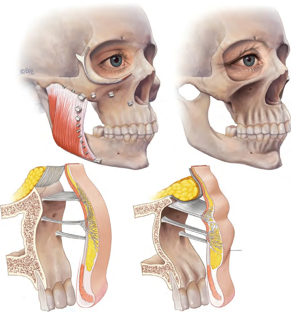 Fig 1. Linking of ageing changes through the tissue layers of the face, the top-down cascade the décolletage inverts. (Standring, Gray's Anatomy, 2016, p.960).
> Fuentes: Standring, Gray's Anatomy 41e [2016] [MEDLIB]
Fig 1. Linking of ageing changes through the tissue layers of the face, the top-down cascade the décolletage inverts. (Standring, Gray's Anatomy, 2016, p.960).
> Fuentes: Standring, Gray's Anatomy 41e [2016] [MEDLIB] [C], p.960 [8].
D14.5 · Assessment, measured, photographed, tested dynamically, scanned
Answer first, the décolletage consult has one step the face does not: a dermatologic pre-screen that gates everything else. Before a single aesthetic decision, the chest is examined as skin at risk: actinic keratoses, atypical or changing pigmented lesions, and any non-healing/bleeding lesion are referred and biopsied first, an aesthetic treatment over an undiagnosed lesion delays a cancer diagnosis and may destroy the lesion before it is read [20,23]. Only a cleared chest proceeds.
The assessment checklist, what is measured / photographed / tested / scanned:
| Domain | What you capture | Tool | Why it changes the plan |
|---|---|---|---|
| Dermatologic safety | AK, atypical nevi, lentigo maligna, non-healing lesions | naked-eye + dermoscopy; biopsy/refer | gate to any treatment; melanoma/AK territory [20,23] |
| Phototype | Fitzpatrick I–VI + current tan | history + exam | IV–VI ⇒ PIH/hypopigmentation risk ⇒ lower fluence, longer wavelength, test spot [7,20] |
| Scar diathesis | keloid/HTS history, prior chest scars | history + inspect old scars | positive ⇒ contraindicate/defer energy & ablation (D14.6, D14.10) [7,40] |
| Wrinkles at rest | horizontal + vertical/oblique creases | Fabi–Bolton 5-point Chest Wrinkle Scale; Merz décolleté-wrinkle-at-rest | baseline + endpoint; grades severity objectively [6,17] |
| Wrinkles dynamic | change on platysmal contraction; sleep-line reproduction in lateral decubitus | Merz décolleté-wrinkle-dynamic scale; posture manoeuvre | separates muscular (microtox-responsive) from mechanical (posture) rhytid [3,17] |
| Pigment / vascular | mottled pigment, telangiectasia, poikiloderma, submental sparing | Merz décolleté-pigmentation scale; cross-polarised photography | selects IPL vs pigment vs vascular device [5,17] |
| Photoageing class | global sun damage | Glogau I–IV | frames aggressiveness and prognosis [13] |
| Patient-reported | appearance-related distress, expectations | BODY-Q Décolletage scale | aligns goals; documents PRO endpoint [19] |
| Skin quality biometrics (optional) | elasticity, hydration, dermal density, epidermal thickness | cutometer, US, corneometer | objective response tracking in trials/high-end practice [27] |
| Ultrasound | skin/subcutis thickness, pectoral fascia, intercostal space/pleural line, Doppler ITA perforators | high-freq US + Doppler | maps depth, confirms the danger strip before medial injection [8] |
Standardised photography (non-negotiable for a region judged on subtle change): - Same position, distance, angle and lighting every visit; the chest looks entirely different lying vs standing and under raking vs flat light. - Cross-polarised for pigment/vascular (subsurface), parallel-polarised/raking for texture and rhytid depth. - A dynamic pair (rest + platysmal contraction) and a lateral-decubitus frame to document sleep-line reproduction. - Bra/clothing line marked consistently; jewellery removed. - Fix the technical variables: same camera/distance, fixed focal length, neutral background, consistent exposure/white balance; a colour/scale reference in frame aids pigment comparison across visits [17].
What goes in the record (medicolegal, and it protects the patient): the documented dermatologic clearance (or referral), phototype and scar-history, the validated baseline scores (Fabi–Bolton / Merz / BODY-Q), the test-spot result read at 2–4 weeks, standardised baseline photos, the specific device/settings or product/dilution used, and the consent covering irreversibility, permanent-dyschromia/scar risk, session number and lifelong photoprotection [17,20,24]. A décolletage record without a lesion-screen entry and a test-spot read is incomplete.
Dynamic and provocative tests (the part clinicians skip and then mis-treat): - Platysmal contraction ("grimace"/forced lower-lip depression): bands and crinkle that deepen have a muscular component → microtox candidate [3]. - Lateral-decubitus reproduction: press the medial breasts together as in side-sleeping; a vertical crease that appears is a mechanical sleep line → posture + resurfacing, not toxin [6]. - Pinch/recoil: slow recoil = elastosis/laxity → biostimulator/energy; the pinch also estimates skin thickness for depth. - Ask sleep position and photosensitiser exposure (retinoid, doxycycline, St John's wort, fragrances on the chest), both change timing and risk [5].
Ultrasound scanning protocol (when a deep or medial injection is planned):
  high-frequency linear probe (≈15–22 MHz), transverse over the medial chest
  ── epidermis+dermis  : thin hyperechoic band  → target for quality work
  ── subcutis          : hypoechoic, variable    → deep-subdermal thread plane
  ── pectoral fascia    : hyperechoic line        → the floor of safe injection
  ── ribs (hypoechoic w/ shadow) + INTERCOSTAL SPACE
  ── PLEURAL LINE (bright, sliding)  ← the structure to keep the needle AWAY from
  + colour Doppler parasternally → mark internal-thoracic PERFORATORS before a medial thread
Scan first, mark the perforators and the pleural line, then inject; if the medial field cannot be cleared of a perforator, move laterally or switch to a pure intradermal plane [8].
Assessment sequence (the order that gates the treatment): screen lesions → phototype + scar history → validated scales (rest/dynamic/pigment) → dynamic + lateral-decubitus manoeuvres → tan/photosensitiser check → (if deep/medial injection) ultrasound → standardised photos → plan. A step skipped upstream (e.g. lesion screen) invalidates everything downstream [17,20].
Differential diagnosis of chest dyschromia/erythema, the can't-miss list before you call it "photodamage": the V of the chest is a classic site for systemic-disease signs, and an aesthetic device fired at one delays a diagnosis.
| Look-alike | Distinguishing feature | Why it must not be treated as cosmetic |
|---|---|---|
| Poikiloderma of Civatte | reticulate pigment + telangiectasia + atrophy, submental sparing | benign, IPL-treatable, but confirm the sparing sign first [5] |
| Dermatomyositis (V-sign / shawl sign) | violaceous erythema over the V and shoulders, ± Gottron papules, proximal weakness, photosensitivity | ⚠ autoimmune red flag, refer for myositis/malignancy workup, never laser [23] |
| Melasma extending to chest / poikilodermatous | hormonally-driven, symmetric, Wood's-light pattern | different treatment (lighteners), high PIH risk with energy [14] |
| Post-inflammatory hyperpigmentation | follows prior inflammation/lesion | treating the pigment without the cause recurs [14] |
| Drug-induced pigmentation (amiodarone, minocycline, chemo) | slate-grey/blue, drug history | stop/adjust drug; laser response unpredictable [14] |
| Riehl's melanosis / pigmented contact dermatitis | reticulate grey-brown, cosmetic/fragrance contact | patch test; stop allergen [42] |
| Poikilodermatous mycosis fungoides | atrophic patches, may mimic poikiloderma | ⚠ biopsy, a lymphoma, not sun damage [23] |
| Actinic keratosis / field cancerisation | rough, scaly, tender macules on the mottle | ⚠ dermatology/biopsy; premalignant [20,23] |
| Amyloidosis / other deposition | rippled pigmentation, pruritus | biopsy if atypical [23] |
| Berloque dermatitis | streaky pigment in perfume-drip pattern | photosensitiser + UV; remove trigger [42] |
Consensus: validated photonumeric scales + standardised, polarised photography + a documented dermatologic clearance are the minimum record; the dynamic exam decides muscular vs mechanical rhytid; the differential above is screened before any device [6,17,19,23]. Discrepancy: routine ultrasound, high-end/medial-injection practice scans perforators and pleura before deep threads [8]; many treat the chest safely with surface anatomy alone. Decide by manoeuvre: any deep or parasternal injection ⇒ scan; pure intradermal quality work ⇒ optional.
Classic trap: photographing the chest inconsistently (standing this visit, reclined next) so that a real improvement is invisible and a stable poikiloderma looks "worse", driving over-treatment. Standardise the frame or do not photograph at all [17].
D14.6 · Goal and patient selection, who benefits, who does not, who is referred
Answer first, the ideal chest patient has skin-quality ageing, a low phototype, no scar diathesis, and realistic expectations; the patient who wants "the crease filled" or "one session" is the one who gets harmed. The realistic goal is improved skin quality, pigment and vessel clearance, and softened fine rhytids, not the erasure of deep folds or the correction of true skin redundancy, which is a surgical problem [6,38].
Selection grid:
| Good candidate | Caution / modify | Contraindicate / refer | |
|---|---|---|---|
| Phototype | I–III | IV (longer wavelength, lower fluence, test spot) | V–VI for aggressive IPL/ablation → PIH/hypopigmentation risk [7,20] |
| Problem type | dyschromia, telangiectasia, poikiloderma, crepey, fine rhytid | moderate laxity (energy has a low ceiling) | true skin redundancy → plastic surgery (excision/mastopexy) [38] |
| Scar history | none | isolated fine scar | keloid/HTS history → the sternal chest is a top keloid site (≤~16% in skin of color) [7,40] |
| Lesions | screened clear | benign, documented | AK / atypical pigmented / non-healing → dermatology + biopsy [20,23] |
| Skin status | intact, untanned | mild dermatitis (treat first) | tanned / recent sun, active infection/eczema → defer |
| Systemic | healthy | photosensitiser use | recent isotretinoin (ablative timing debated), pregnancy (defer elective) |
| Expectations | quality, gradual, multi-session | wants faster result | wants volume / one-session clearance / a "new" chest → re-counsel or decline |
Who genuinely benefits (match the tool to the driver, D14.4): - Poikiloderma of Civatte / dyschromia / telangiectasia → IPL first line; the single highest-satisfaction chest indication [5,15]. - Crepey atrophy / early laxity + fine rhytid → biostimulator (CaHA/PLLA) and/or non-ablative fractional + boosters; gradual, realistic [24,32]. - Dynamic crinkle / platysmal band continuing off the neck → intradermal microtox (D14.8) [3]. - A single discrete static crease in reasonable-thickness skin → a threaded microaliquot of low-G′ HA, counselled on nodule risk (D14.7) [22].
Who does not benefit (and what they need instead): - True skin redundancy / significant laxity, non-surgical energy tightens modestly at best; the honest referral is to surgery. Selling five RF sessions for a mastopexy problem is the region's commonest mis-sell [38]. - Deep static folds from posture, will recur without a sleep-posture change; treat the cause first [6]. - The volume-seeker, there is little to volumise on the chest wall, and attempting it risks perforator injury and nodules (D14.3).
Who is referred, and to where: - Dermatology / dermatopathology, any AK, atypical or changing pigmented lesion, non-healing lesion (gate step) [20,23]. - Plastic surgery, genuine skin excess, ptotic skin folds, post-massive-weight-loss chest [38]. - M2/M7, an existing keloid or a lesion needing excision. - Oncology, a suspicious lesion confirmed malignant.
Fitzpatrick-specific modifications (the phototype dial, applied):
| Phototype | Energy posture | Preferred tools | Avoid |
|---|---|---|---|
| I–II | standard de-rated chest settings; still test-spot | IPL, fractional, peels | complacency (fair skin still scars) |
| III | lower fluence, prime with lightener, test spot | IPL (higher cut-off), 1927 nm | high-density ablation |
| IV | markedly conservative, longer wavelength, longer pulse | 1927 nm, microneedling ± RF, boosters, topicals | aggressive IPL/ablation |
| V–VI | prefer non-light modalities; decline aggressive energy | microneedling-RF (bulk heat below epidermis), boosters, topicals, PN | IPL/pigment laser/ablation without extreme caution [7,20] |
The rule scales monotonically: as phototype rises, shift from light-based to mechanical/regenerative modalities and lower every energy, RF-microneedling is comparatively phototype-agnostic because the epidermis is spared [20,30].
Consent points specific to the region: irreversibility of biostimulators in a poor-healing zone; the real risk of permanent hypopigmentation/footprints and hypertrophic scarring; the need for a test spot and multiple sessions; the mandatory, lifelong photoprotection without which any pigment/vascular result recurs [5,24].
Structured counselling script (say these things, in these words, before consent): 1. "The chest is not the face, it heals worse, so I treat it more gently, in more sessions, and I test a small area first." (sets the de-rating expectation) [1]. 2. "First I check this skin for spots that a dermatologist should see; anything suspicious goes there before we do anything cosmetic." (the pre-screen gate) [20]. 3. "The biggest gains here are colour and vessels and texture, not filling wrinkles; some products I use are permanent and cannot be dissolved, which matters more here because healing is worse." (goal + irreversibility) [4,24]. 4. "There is a real risk of a lasting pale mark or a raised scar if we are too aggressive, especially with your skin type / your history of keloids, that is why we go slow." (phototype/scar honesty) [7,40]. 5. "Sun protection every day is not advice, it is the treatment that keeps the result; without it, this comes back." (maintenance as treatment) [5]. 6. "If your main problem is loose, hanging skin, no injection or laser fixes that well, that is a surgical question, and I will tell you honestly." (the surgical referral) [38].
Decision shortcut, what does THIS chest need first? - Mottled colour / redness / poikiloderma → IPL (D14.7b). - Crepey/atrophic texture, early laxity → biostimulator + boosters ± non-ablative fractional (D14.7). - Dynamic crinkle / bands off the neck → microtox (D14.8). - A single deep static crease in decent skin → threaded HA (D14.7a). - Loose redundant skin → surgery (refer) [38]. - Any undiagnosed lesion → dermatology first (D14.5) [20].
Consensus: treat skin quality, screen aggressively, and decline or refer the laxity-dominant and the volume-seeking patient; phototype and scar history are hard gates; the counselling itself is part of the safety system [7,20,38]. Discrepancy: high phototype and energy, some operators treat IV–V with conservative long-pulse/longer-wavelength settings and rigorous test spots [20]; others decline IPL/ablation in V–VI entirely and use topicals/boosters. Decide by the test spot read at 2–4 weeks, not by the same-day response.
Classic trap: accepting the laxity-dominant patient for "tightening" because they refuse surgery, delivering repeated energy that neither tightens meaningfully nor is free of scar risk, spending the patient's money and the region's safety margin on a surgical problem [38].
D14.7 · Technique, the full regional grid
Per the exhaustiveness contract, the region is not closed with one technique but with the complete grid of options, each row carrying school, dose, plane and evidence. It is split into 7a Injectables/biostimulators and 7b Resurfacing & energy. What does not apply is written No aplica with the reason.
Master lane-selection (read before either grid): the décolletage's dominant problem decides the lane, and most chests need both in sequence, not one. Pigment, vessels and poikiloderma go to the energy/light lane first (7b), because no injectable clears dyschromia or telangiectasia [5,15]. Crepey atrophy, laxity and fine rhytid go to the biostimulator/booster lane (7a), because that is a collagen/hydration problem, not a resurfacing one [4,24,27]. A single discrete static crease is the only true filler indication, and even then it is threaded, never bolused [22]. A dynamic crinkle or platysmal band goes to intradermal toxin (D14.8) [3]. The through-rule for both lanes is identical to the region's governing law: de-rate every facial parameter, prefer the reversible and the diffuse, and stage rather than stack [1]. The two grids below enumerate every product, instrument, plane, movement, school, volume and non-injectable alternative so that the choice within a lane is explicit, not defaulted.
| Dominant problem | Lane | First move | Detail |
|---|---|---|---|
| Pigment / vessel / poikiloderma | energy (7b) | IPL | [5,15] |
| Crepey / atrophy / laxity | injectable (7a) | biostimulator + booster | [4,24] |
| Discrete static crease | injectable (7a) | threaded low-G′ HA | [22] |
| Dynamic crinkle / band | toxin (D14.8) | intradermal microtox | [3] |
| Texture + photodamage | energy (7b) | non-ablative fractional | [32] |
| True redundancy | neither | surgical referral | [38] |
D14.7a · Injectable and biostimulator grid
Answer first, on the chest, biostimulators and boosters (dermal quality) beat fillers (volume); HA is reserved for a discrete static rhytid, threaded not bolused. The unifying rule: diffuse, deep-to-mid, diluted, cannula-preferred, ≤1 syringe of any biostimulator per session, because every product here is placed in thin, poorly-healing skin over a neurovascular parasternal strip [4,8,22].
Product axis, enumerated in full:
| Product | Rheology / form | Role on chest | Plane | Instrument | Dose (region-specific) | Reversible? | Evidence |
|---|---|---|---|---|---|---|---|
| HA, low G′, low crosslink | soft, spreadable | a single discrete static rhytid; NOT field volumising | deep dermal/subdermal, retrograde threads | 30G needle or 25G cannula | microaliquots along the crease; no bolus | yes (hyaluronidase) | [22] |
| HA, high G′ structural | firm | ⚠ No aplica, no bony/deep compartment to project on the chest; high nodule risk in thin skin | n/a | , | n/a | yes | [22] |
| HA skin booster (non/low-crosslink) | fluid, hydrating | field skin quality, crepey texture | mid-dermis | 30G needle grid or cannula | microdroplets ~0.01–0.02 mL/point, 1 cm grid; 3 sessions q3 wk + maintenance | yes | [27] |
| CaHA, normal (1:1 "diluted") | biostimulator | ⚠ too concentrated for thin chest skin → nodules; avoid | deep subdermal | cannula | not recommended undiluted | no | [4] |
| CaHA, hyperdilute (≥1:2) | biostimulator, spread | field collagen stimulation, laxity/quality | deep subdermal, linear/fan | 25G cannula preferred | 1:2 normal / 1:4 thin / 1:6 atrophic; ≤1 syringe/session; massage; q3 mo ×2–3 | no | [4,25] |
| PLLA (poly-L-lactic acid) | biostimulator, delayed | photodamage + laxity, gradual collagen | deep subdermal | 25G cannula | body reconstitution ≥16–18 mL, hydrate ≥24 h; 2–3 sessions q4–6 wk; massage 5×5×5 | no | [24] |
| PCL (polycaprolactone) | biostimulator, longer | analogous to CaHA/PLLA; less region-specific data | deep subdermal | cannula | facial protocol extrapolated [MATERIAL GAP] |
no | [24] |
| PMMA (permanent) | permanent microspheres | ⚠ No aplica, permanent product in a poor-healing, high-complication zone; do not use | n/a | , | n/a | no (permanent) | [36] |
| Autologous fat | graft | possible for the inter-mammary hollow; operator-dependent, off-topic for skin quality | deep subcutaneous | blunt cannula | small aliquots, not for skin quality | partial | [38] |
| Polynucleotides / PDRN (Rejuran, Plinest) | regenerative booster | dermal quality; region-specific RCT absent | mid-dermis | 30G | facial/neck protocol extrapolated; 3 sessions q2–3 wk [MATERIAL GAP] |
yes (biodegradable) | [28] |
| Exosomes | regenerative topical/injectable | investigational; corpus = one commercial-magazine article, no region RCT | topical post-microneedling / mid-dermis | n/a | undefined [MATERIAL GAP] |
n/a | [MATERIAL GAP] |
| PRP | autologous | adjunct with microneedling; modest | intradermal / topical | 30G / microneedle | 2–3 sessions | yes (autologous) | [29] |
| Toxin (intradermal microtox) | neuromodulator | dynamic crinkle / platysmal bands → D14.8 | intradermal | 32–33G | see D14.8 | wears off | [3] |
HA by rheology (the discrete-rhytid choice, per the exhaustiveness grid):
| Rheology class | G′ / cohesivity | Chest use | Why |
|---|---|---|---|
| Fluid / booster (non–low crosslink) | very low G′, low cohesivity | field skin quality | spreads, hydrates, no projection, the safe default [27] |
| Soft, low G′ | low G′, moderate cohesivity | a single discrete static rhytid, threaded | enough support without a palpable lump in thin skin [22] |
| Medium G′ | medium | rarely, only a deep, defined crease, cautiously | nodule risk rises with G′ in thin skin [22] |
| High G′, high cohesivity (structural) | high | ⚠ No aplica, no deep/bony chest target; high visible-lump risk | designed for facial deep-plane projection [22] |
The chest inverts the facial rheology logic: on the face high-G′ product goes deep for projection; on the chest there is no projection target, so the useful HA is the low-G′/fluid end, placed to support or hydrate, never to volumise [22,27].
Instrument axis, enumerated: - Needle 30G ½", intradermal boosters, microdroplet grids, discrete-rhytid threading; precise, more bruising. - Needle 32–33G, microtox microdroplets (D14.8). - Cannula 25G 50 mm, the workhorse for deep-subdermal biostimulator threads on the medial chest; blunt tip reduces perforator cannulation [4,8]. - Cannula 27G, finer threads laterally. - With ultrasound guidance, for medial/parasternal deep threads to keep off the ITA perforators and the intercostal space; without, acceptable for pure intradermal central quality work [8].
Cannula entry-point map and safeguards (deep biostimulator threads): - Entry ports: 2–3 lateral ports per hemi-chest, so threads run lateral→medial and the cannula tip approaches (never enters) the parasternal strip from the side, decelerating before the danger zone [4,8]. - Stay ≥1–2 cm off the sternal edge with the tip; if the intended target is parasternal, switch to a superficial intradermal plane instead of a deep thread [8]. - Blunt 25G cannula for the medial field (perforator safety), finer 27G laterally; move while injecting (retrograde), never deposit into a stationary deep point [4]. - Aspiration is unreliable with fine cannulas/low-G′ product, so it is a weak safeguard, plane, cannula, entry geometry and staying off the sternal edge are the real safety layers [8,12]. - Immediate physician massage disperses product and is part of nodule prevention, not an optional finish [4].
Plane axis: supraperiosteal → ⚠ No aplica (chest wall/pleura); deep fat → effectively pectoral fascia, biostimulator threads sit just above it; SMAS → absent; superficial fat → diffuse subdermal biostim plane; subdermal → biostimulator/quality; intradermal → boosters, microtox, PN.
Movement axis, enumerated: retrograde linear threading (the default for biostimulators and the discrete rhytid) · fan (from a single lateral entry to cover a sub-site) · cross-hatch (two directions for field coverage, deep subdermal) · serial microdroplet/puncture (boosters, microtox, the 1 cm grid) · microbolus (tiny deposits for hyperdilute CaHA, immediately massaged) · bolus → ⚠ No aplica (visible nodule in thin skin, perforator risk) · tower → ⚠ No aplica (a facial midline-projection technique with no chest analogue).
School axis, named techniques and the one that does the opposite: - Hyperdilute CaHA field biostimulation (Casabona; de Almeida-type consensus), dilute to skin thickness (1:2→1:6), thread deep-subdermal, massage; skin-quality goal, not volume [4,25]. - PLLA collagen-stimulation (Christen body consensus; Goldberg décolletage work), high-dilution, delayed collagen, staged sessions [24]. - MD Codes (de Maio), ⚠ No aplica: MD Codes is a facial alphanumeric injection-point system; there is no décolletage MD Code. Declaring the absence is the honest answer, not inventing one. - The opposite school, "fill the wrinkle" HA bolus, historically used, rejected here: bolus HA into a chest crease gives short benefit and high visible-nodule/Tyndall risk in thin skin [22]. The discrepancy below is real and changes the gesture.
Point-by-point technique (the discrete static rhytid, HA, the only true "filler" gesture on the chest): 1. Plane: deep dermal to immediate subdermal, under the crease. 2. Entry: lateral to the crease, single point; retrograde. 3. Direction: along the length of the crease, parallel, never perpendicular/bolus. 4. Instrument: 30G needle for a short crease, 25G cannula for a longer one. 5. Volume per point/pass: microaliquots (sub-0.05 mL), linear; total rarely more than a fraction of a syringe over the field; standard injection-technique atlases describe the retrograde linear thread used here [37]. 6. Touch-up: at 2–4 weeks, never same-session stacking; assess for beading first.
Point-by-point technique (hyperdilute CaHA field, the main quality gesture): 1. Reconstitute to skin thickness (1:2/1:4/1:6 with saline ± lidocaine) [4]. 2. Entry: 2–3 lateral cannula ports; fan/cross-hatch the sub-site. 3. Plane: deep subdermal on the pectoral fascia plane, staying ≥1–2 cm off the sternal edge. 4. Deposit: retrograde linear threads / microboluses; ≤1 syringe of reconstituted product per session. 5. Immediate physician massage to disperse, then patient self-massage twice daily 3–7 days [4]. 6. Interval: q3 months ×2–3; effect is gradual (collagen), reviewed at 3–6 months [4,25].
Point-by-point technique (skin booster / mesotherapy grid, the reversible quality gesture): 1. Plane: mid-dermis, a superficial bleb risks beading in thin skin [27]. 2. Entry: serial 30G microdroplet grid ~1 cm apart, or a cannula for a mannitol-stabilised fluid HA. 3. Deposit: ~0.01–0.02 mL/point, symmetric across the V. 4. Course: 3 sessions q3 weeks, then maintenance every few months; the mesotherapy tradition (vitamins/amino acids/oligoelements) uses the same serial superficial grid [26,27]. 5. Endpoint: skin hydration/elasticity/texture (biometrics if tracked), not volume [27].
Point-by-point (PN/PDRN, extrapolated, the gap declared): the same mid-dermal microdroplet grid, ~3 sessions q2–3 weeks, is used by extrapolation from facial/neck protocols; there is no décolletage-specific RCT, so this is [MATERIAL GAP] and is offered as a regenerative adjunct, not an evidence-backed regional protocol [28].
Biostimulator mechanism, why CaHA, PLLA and PCL are not interchangeable (and must not be blended): - CaHA, calcium-hydroxylapatite microspheres in a gel carrier; a mechanotransductive stimulus with a partly immediate scaffold effect, faster onset, dissolvable-resistant [4,25]. - PLLA, poly-L-lactic acid microparticles; a macrophage-mediated inflammatory stimulus producing delayed type I/III collagen over 6–24 months; slowest onset, most delayed complications [24]. - PCL, polycaprolactone in a CMC gel; longer-lasting collagen stimulation, positioned between CaHA and PLLA; least region-specific data [24]. - The morphologic difference (particle shape/mechanism) means the same crepey chest can be approached by two valid but different routes; choose one, dilute it to skin thickness, and do not mix protocols in one session [24].
Non-injectable, when the correct answer is NOT an injectable: if the dominant problem is pigment/vascular/poikiloderma → IPL/laser (D14.7b), not filler; if texture/crepey with photodamage → fractional resurfacing + boosters; if true redundancy → surgery. An injectable is correct only for dermal-quality biostimulation or a single discrete rhytid.
Volume ceilings and overcorrection (the number that prevents the nodule): - Hyperdilute CaHA: ≤1 syringe of reconstituted product per session over the treated field; gradual, staged q3 months; overcorrection is judged at 3 months, not on the table, because collagen is delayed [4,25]. - PLLA: the ceiling is dilution and time, not immediate volume, high dilution (≥16–18 mL), hydrate ≥24 h, and do not re-treat sooner than 4–6 weeks; early "correction" chasing an immediate result is the route to late papules [24]. - HA booster/filler: microaliquots only; the visible-beading threshold in thin chest skin is low, so total volume over the field is a fraction of a syringe, reassessed at 2–4 weeks before any top-up [22,27]. - The chest has no "fill to correction" endpoint, unlike a facial fold, there is no volume deficit to fill; the endpoint is skin quality, so more product ≠ better result, it means more risk [8].
Reversal / salvage by product (know before you inject, irreversibility is the region's amplifier):
| Product | Reversible? | Salvage if it goes wrong |
|---|---|---|
| HA (booster/filler) | Yes | hyaluronidase, the reason HA is the safest "filler" choice here [22] |
| Hyperdilute CaHA | No (not enzymatically dissolvable) | massage, time, intralesional saline/steroid (limited); nodule may persist [4] |
| PLLA | No | intralesional steroid ± 5-FU for papules; time [24] |
| PCL | No | as CaHA/PLLA; limited options [24] |
| PMMA | No, permanent | surgical; do not use on the chest [36] |
| Autologous fat | Partial | not a skin-quality tool; overgrafting is hard to reverse [38] |
Consensus: biostimulator + booster for quality, cannula-preferred, diluted, diffuse, ≤1 syringe/session; HA reserved and threaded; the reversible product is preferred in a poor-healing zone [4,22,24,27]. Discrepancy (changes the gesture): HA vs CaHA vs PLLA for the same crepey chest, there is no consensus on which patient goes to which; Alam's evidence review explicitly finds insufficient comparative data [23]. CaHA proponents cite faster, mechanotransductive collagen [4,25]; PLLA proponents cite longer, macrophage-mediated remodelling [24]; booster/HA proponents cite reversibility in a risky zone [27]. Decide by: reversibility need (favours HA/booster), atrophy severity (favours PLLA/CaHA hyperdilute), and phototype/scar risk (favours the reversible, most-diluted option). Never average the dilutions or blend the protocols.
Classic trap: using the facial CaHA dilution (1:1) or a bolus on thin chest skin, producing palpable, sometimes visible, biostimulator nodules that are irreversible and sit in a poor-healing field, the single most regretted décolletage injectable error [4].
D14.7b · Resurfacing and energy grid
Answer first, IPL is the first-line device for the commonest chest indication (pigment + vascular + poikiloderma); everything ablative is de-rated relative to the face, and fully-ablative resurfacing off-face is the region's classic scarring error. The governing dial is reduced fluence/energy and reduced density, more sessions [1,17].
Device grid, enumerated in full, all settings region-de-rated:
| Device / peel | Target | Chest setting (vs face) | Sessions | Best chest indication | Caveat | Evidence |
|---|---|---|---|---|---|---|
| IPL (broadband, 515–755 nm cut-offs) | melanin + oxyhaemoglobin | fluence ~25–30% lower, longer pulse, single pass, larger spot | 3, q3–4 wk | poikiloderma of Civatte, dyschromia, telangiectasia, 1st line (75–80% clearance, ~5% AE) | ⚠ never tanned; hypopigmented footprints; test spot | [5,15,17] |
| Pulsed-dye laser (PDL, 585–595 nm) | oxyhaemoglobin | purpuric or sub-purpuric | 2–4 | isolated telangiectasia/erythema | purpura, mottling, scarring if stacked | [31] |
| Q-switched / picosecond (pigment) | melanin | conservative | 2–4 | discrete lentigines (not the vascular part) | misses vessels → wrong for poikiloderma alone | [14,31] |
| Non-ablative fractional 1927 nm (thulium) | epidermal/dermal water, pigment | low energy, low density | 3–5, q4 wk | diffuse dyschromia + texture; all phototypes | ⚠ raise density = scar in adnexa-poor skin | [32,33] |
| Non-ablative fractional 1550/1565 nm (erbium-glass) | dermal water | low density, mid-energy | 3–5 | rhytid/texture, laxity-lite | multiple sessions for modest gain | [31,33] |
| Ablative fractional Er:YAG (2940 nm) | water, superficial | shallow, low density, 1 pass | 1–3 | fine rhytid/texture, faster healing | ⚠ still off-face caution | [1,31] |
| Ablative fractional CO₂ (10 600 nm) | water, deeper | low density, 1–2 passes, reduced pulse energy | 1–3 | deeper rhytid/photoaging when justified | ⚠ HTS of neck/chest documented; conservative only | [1,39,40] |
| Fully-ablative CO₂/Er:YAG (full field) | full resurfacing | ⚠ No aplica / contraindicated off-face | n/a | none | scarring, dyschromia, the classic error | [40] |
| Chemical peel, superficial (Jessner, GA, salicylic) | epidermis | series, primed | 3–6 | dyschromia, texture, priming | modest depth | [34] |
| Chemical peel, medium (Jessner + TCA 20–25%) | epidermis–papillary dermis | cap at 20–25% TCA, build over sessions | 2–4 | photodamage, dyschromia | ⚠ deeper TCA = scar off-face | [34,35] |
| Chemical peel, deep (phenol/high TCA) | reticular dermis | ⚠ No aplica on the chest | n/a | none | scarring | [35] |
| Microneedling (± PRP) | mechanical collagen induction | depth 1–2 mm, conservative | 3–4, q4 wk | crepey texture, early laxity, phototype-flexible | infection, tracking, PIH | [29] |
| RF microneedling / fractional RF | dermal heating | conservative energy/depth | 3–4 | laxity-lite + texture, safer in higher phototype (bulk heating below epidermis) | modest; nodularity if over-treated | [29,30] |
| Monopolar/bipolar RF (bulk) | dermal/subdermal heat | device protocol | 1–4 | mild laxity, quality | low ceiling for true redundancy | [30] |
| HIFU/microfocused US | SMAS-depth on face; here fascia/dermis | device protocol | 1–2 | mild laxity (adjunct) | low chest evidence; combine with CaHA [4] | [4] |
| Carboxytherapy | CO₂-induced microcirculation | series | 4–6 | adjunct for quality/laxity | weak evidence, adjunct only | [30] |
Poikiloderma protocol (the flagship chest treatment), point by point: 1. Confirm diagnosis (triad + submental sparing), screen lesions, confirm untanned skin [5,20]. 2. Test spot on a lateral chest area; read at 2–4 weeks. 3. IPL cut-off filter to phototype (higher cut-off/longer wavelength as phototype rises), fluence 25–30% below facial, longer pulse duration, single non-overlapping pass with contact cooling [5,15]. 4. 3 sessions at 3–4-week intervals; expect 75–80% clearance of vascular + pigment components [5,15]. 5. If a residual pure-vascular component: add PDL; residual pure-pigment: add pigment laser or 1927 nm [31]. 6. Lifelong photoprotection as maintenance, without it, recurrence is guaranteed and the sessions are wasted [5].
Device selection by dominant sign (pick by what you see, not by what you own):
| Dominant sign on the chest | First device | Second line / add-on |
|---|---|---|
| Poikiloderma (pigment + vessel) | IPL | PDL (residual vessel) / pigment laser (residual spot) |
| Mottled pigment / lentigines only | IPL or 1927 nm | Q-switched/pico pigment laser |
| Telangiectasia / erythema only | PDL or IPL | vascular laser |
| Diffuse dyschromia + texture | 1927 nm non-ablative fractional | superficial peel series |
| Crepey texture / fine rhytid | non-ablative fractional (1550/1927) | microneedling ± RF, boosters |
| Deeper photoageing rhytid (selected) | low-density ablative fractional | with test spot + selection |
| Mild laxity | RF / microneedling-RF | + hyperdilute CaHA |
| Field priming / mild dyschromia | superficial/medium peel | topicals |
⚠ Never pick the deepest device you have "to save sessions", on the chest, matching the device to the sign at conservative settings beats one aggressive pass every time [1,40].
Parameter discipline that keeps energy safe on the chest (the dials, not just the device): - IPL: raise the cut-off filter / wavelength as phototype rises (more melanin-sparing); lengthen pulse duration; single, non-overlapping pass; contact/air cooling; larger spot; and the fluence set 25–30% below the facial value for the same complaint [2,5,15]. - Non-ablative fractional: keep density low (single digits to low teens %); energy modest; multiple gentle sessions rather than one deep pass, the adnexa-poor dermis punishes density [32,33]. - Ablative fractional: reduced pulse energy, low density, 1–2 passes; the neck/chest scars in the literature followed near-facial settings [40]. - PDL: sub-purpuric or lightly purpuric settings for isolated vessels; ⚠ avoid stacking (mottling/scarring) [31]. - Cooling and test spot are not optional on the chest, the test spot is read at 2–4 weeks because delayed hypopigmentation/scar is the failure mode, not an immediate one [5].
Chemical-peel technique for the chest (why "cap at 20–25% TCA"): - Prime 2–4 weeks with retinoid/AHA/lightener to even the field and reduce PIH [34]. - Degrease; apply a superficial-to-medium agent (Jessner then TCA 20–25%) to a light-to-medium frost, built over sessions, never a single deep application [34,35]. - ⚠ Deep peels (phenol/high TCA) are contraindicated off-face, the chest scars [35].
Peel agents by depth (chest ceiling in bold):
| Depth | Agent | Chest use |
|---|---|---|
| Very superficial | glycolic 20–50%, salicylic 20–30%, lactic, mandelic | priming, mild dyschromia, series [34] |
| Superficial | Jessner's solution, TCA 10–15% | dyschromia/texture, build over sessions [34] |
| Superficial–medium (ceiling) | Jessner + TCA 20–25% | photodamage/dyschromia, do not exceed off-face [34,35] |
| Medium | TCA 35% / TCA+CO₂ / Coleman | ⚠ face-oriented; too deep for routine chest |
| Deep | phenol–croton oil, high TCA | ⚠ contraindicated on the chest [35] |
Priming (retinoid/AHA/lightener) 2–4 weeks first lowers PIH and evens the frost; a chest peel is always built up across sessions, never taken deep in one pass [34]. - Neutralise/soothe; strict photoprotection; expect desquamation ~5–7 days [34].
Consensus: IPL first for poikiloderma/dyschromia/vascular; everything ablative de-rated by density and passes; test spot and photoprotection mandatory; more, softer sessions [1,5,15,17]. Discrepancy (the region's defining controversy, changes the gesture): - School A, off-face ablative is contraindicated / minimal only (Tannous): hypertrophic scarring of the neck and chest is documented after ablative fractional CO₂; non-facial skin's low pilosebaceous density makes full-field ablation unsafe, and even fractional needs extreme caution [40]. - School B, low-density fractional ablative is safe on the chest (Truswell): with reduced density, reduced fluence and fewer passes, fractional ablative and non-ablative devices treat the chest with shorter healing and acceptable safety [1]. - Both are right within their limits and must not be averaged: fully-ablative off-face is contraindicated (A); low-density fractional is acceptable with de-rated parameters and patient selection (B). Decide by: density and passes (low), phototype (lower is safer), scar history (none), and a test spot. The wrong reading is to take B as licence for facial-strength settings.
Classic trap: transferring facial fractional-CO₂ density/energy to the chest for "better results in one pass", the documented route to vertical hyperpigmented striations and hypertrophic scars of the sternal skin. The chest is treated at a fraction of facial density, or not ablatively at all [40].
Fig 2 documents an IPL chest photorejuvenation before/after: panel (A) the pre-treatment décolletage with mottled dyschromia, telangiectasia and lentigines in the V distribution; panel (B) the same chest after IPL, with the pigment and vascular components cleared, the flagship décolletage result and the visual argument for IPL as first line [5,15].
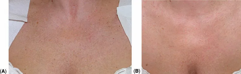 Fig 2. Use of intense pulsed light (IPL) for photorejuvenation of the chest, before (A) and after (B). (Lim, Photodermatology, 2007, p.427).
> Fuentes: Lim, Photodermatology [2007] [MEDLIB]
Fig 2. Use of intense pulsed light (IPL) for photorejuvenation of the chest, before (A) and after (B). (Lim, Photodermatology, 2007, p.427).
> Fuentes: Lim, Photodermatology [2007] [MEDLIB] [C], p.427 [42]; mechanism/efficacy corroborated by Weiss 2000 [5] and Rusciani 2008 [15].
D14.8 · Toxin of the region, muscle, dose, points, safety distances, antagonist untouched
Answer first, the only correct chest toxin is intradermal microtoxin (microbotox/mesobotox), never a deep intramuscular injection. The target is the caudal/decussating platysma fibres and the subdermal muscular tone producing the fine horizontal crinkle and the vertical bands that continue off the neck. It is placed as 1–2 U per ~0.05 mL microdroplet on a ~1 cm grid, intradermal to immediate-subdermal, with a 32–33G needle, for a chest field total of roughly 20–30 U onabotulinumtoxinA-equivalent [3]. It works only when a dynamic component exists; it does nothing for static laxity or a mechanical sleep crease.
Muscle / target map:
| Structure | Role in chest ageing | Treat? | Note |
|---|---|---|---|
| Platysma, thoracic/decussating fibres (from ~2nd rib, pectoral & deltoid fascia) | vertical bands + horizontal crinkle (dynamic) | Yes, intradermal microtox | immediately subdermal; the substrate of chest microtox [3,8] |
| Subdermal dermal-muscular interface | fine crinkle, skin tone | Yes, microdroplets | microbotox mechanism: partial, superficial chemodenervation preserving movement [3] |
| Pectoralis major | functional adductor/flexor of the arm | ⚠ No, antagonist/functional muscle, never injected | deep IM here = weakness + chest-wall/pleura risk |
| Intercostal muscles | respiration | ⚠ No, never | deep between ribs = pneumothorax |
Dose grid (region-specific, onabotulinumtoxinA units; convert 1:1 inco-, ~2.5:1 abobotulinumtoxinA) [3,36]:
| Parameter | Value | Source |
|---|---|---|
| Concentration of microtox solution | 20–28 U ONA per mL of prepared solution | [3] |
| Base reconstitution | 100 U vial in ~2.5 mL saline, then further diluted in-syringe | [3] |
| Microdroplet volume | ~0.05 mL per point | [3] |
| Units per microdroplet | 1–2 U (dilution-dependent) | [3] |
| Grid spacing | ~1 cm apart, serial puncture | [3] |
| Plane | intradermal / immediate subdermal, a raised bleb, never deep | [3] |
| Chest field total | ~20–30 U (≈100–150 microdroplets over the treated V) | [3] |
| Needle | 32–33G, bevel only into dermis | [3] |
| Onset / duration | days to ~2 weeks / ~3–4 months | [3,36] |
Safety distances and rules: - Start 2–3 cm below the platysma origin / clavicles, working down the V; do not chase fibres onto the pectoralis bulk [3]. - Bevel intradermal only, a visible bleb confirms plane; if the needle "drops in", you are too deep, withdraw. - Never cross into a deep plane over an intercostal space, the anatomical hazard of the chest is the pleura, not a facial-nerve branch [8]. - Symmetric grid, marked; photograph rest + contraction first (D14.5). - Antagonist untouched: the pectoralis major is a functional muscle and is never part of the field; unlike the face, the goal is not to balance an elevator against a depressor but to soften the subdermal/platysmal tone while preserving chest movement [3].
Point-by-point technique: 1. Mark a grid over the dynamic crinkle/bands, starting 2–3 cm below the clavicles/origin, 1 cm spacing [3]. 2. Prepare 20–28 U/mL solution; load a 32–33G syringe. 3. Raise intradermal blebs, 1–2 U each, serial puncture, symmetric L/R. 4. Total ~20–30 U over the chest V; stop if the field is covered, do not stack. 5. No massage into deep planes; upright ~4 h; review at 2 weeks for a touch-up of undertreated squares.
Safety-distance map (where the microdroplets go, and where they never do):
        clavicle ─────────────────────────
             (start 2–3 cm BELOW this line)
        ····· intradermal microdroplet grid, 1 cm spacing ·····
        ····· 1–2 U each, symmetric L/R over the V ·············
   ┌───────────────┐
   │  STERNUM       │  ← thin skin over bone: keep grid superficial (bleb)
   │  parasternal   │  ← perforator strip: intradermal ok, NEVER deep here
   └───────────────┘
   PECTORALIS MAJOR  ← functional muscle: NOT injected (no deep IM)
   INTERCOSTAL SPACE ← deep = pleura = pneumothorax: forbidden depth
The plane is the safety device: an intradermal bleb means you are above every hazard; if the needle "drops in", withdraw [3,8].
What microtox does NOT do (set the expectation): it does not tighten lax skin, does not erase a static/mechanical sleep crease, does not restore volume, and does not improve pigment or telangiectasia, it softens a dynamic crinkle/band. Selling it for laxity or dyschromia is a mismatch that disappoints [3,6].
Adjunct sequencing: microtox pairs well after the pigment/vascular and quality work is under way (it addresses the residual dynamic line), and it can share a visit with a different-plane booster; keep it away from same-site aggressive heat/massage that would diffuse the toxin (D14.9) [3].
Non-toxin alternative (when the correct answer is not toxin): if the exam shows a static crease (sleep line reproduced in lateral decubitus) or laxity dominates, the answer is posture change + resurfacing/biostimulator, not toxin, injecting toxin into a static or lax chest wastes product and disappoints [3,6].
Brand conversion for the microtox field (approximate, region unit ~20–30 U ONA-equivalent) [3,36]:
| Toxin | Approx. equivalence to onabotulinumtoxinA | Chest-field note |
|---|---|---|
| onabotulinumtoxinA (Botox) | 1:1 (reference) | 20–28 U/mL solution, 1–2 U/microdroplet [3] |
| incobotulinumtoxinA (Xeomin/Bocouture) | ~1:1 | comparable dilution/dose [36] |
| abobotulinumtoxinA (Dysport) | ~2.5:1 (≈2.5 speywood U ≈ 1 ONA U) | reconstitute the 500 U vial in ~2.5 mL, further dilute in-syringe [3] |
| prabotulinumtoxinA / others | ~1:1 (ONA-like) | limited region data; extrapolate [MATERIAL GAP] [36] |
⚠ Never average or interconvert loosely, a Dysport-for-Botox 1:1 error triples the microtox dose. Fix the brand and its dilution for the session [3,36].
Necklace ("Venus") lines vs microtox target, a distinction that changes the plan: horizontal cervicomental "necklace lines" are largely dermal tethering/ligamentous creases of the neck, and they respond poorly to toxin; the chest crinkle and the vertical bands that continue off the platysma are the dynamic targets microtox actually helps. If the exam shows fixed horizontal necklace grooves, the answer is a booster/energy or a threaded HA, not more toxin (D13, D14.4) [3,6].
Microtox vs conventional toxin, why the chest never gets conventional dosing: conventional (deeper, higher-per-point) botulinum injection aims to paralyse a muscle; on the chest that would mean the pectoralis/intercostal plane, functional muscles with a pleural hazard behind them. Microtox delivers a partial, superficial chemodenervation of the subdermal/dermal-muscular interface that softens tone while preserving movement, which is the only rationale that fits the region's anatomy [3,8].
Consensus: intradermal microdroplet technique only, low total dose, dynamic-component patients only, pectoralis and deep planes off-limits; brand and dilution fixed per session [3,36]. Discrepancy (changes the gesture): how much of the vertical band is platysmal vs mechanical, treat with microtox only the component that deepens on active platysmal contraction; do not treat the static crease with toxin [3,6]. Dilution ratio also varies by school (higher dilution = more superficial, more diffuse effect); do not average, pick a dilution and keep the plane intradermal [3].
Classic trap: injecting the chest toxin too deep (into or toward the pectoralis/intercostal plane) chasing a stronger effect, producing no skin benefit, possible chest-wall weakness, and, at worst, the intercostal/pleural hazard. Microtox is a dermal technique; the raised bleb is the safety signal [3,8].
D14.9 · Combination and sequence, before, after, interval → L2
Answer first, on the chest, sequence conservatively: prep the skin, clear pigment/vessels first, resurface texture next, biostimulate for collagen, and maintain with boosters + photoprotection; keep aggressive modalities apart in time because the skin heals slowly. The governing safety move is a test spot before anything device-based and de-rated parameters throughout [1,17].
Skin preparation (before), 2–4 weeks: - Topical retinoid + antioxidant (vitamin C) + a lightener (e.g. hydroquinone/azelaic/kojic where indicated) to prime the dermis and pre-empt PIH, especially phototype III+ [13,34]. - ⚠ Stop the retinoid ~5–7 days before any ablative/fractional pass to avoid delayed re-epithelialisation, then resume after healing. - ⚠ No sun / no tan for weeks before energy; a tanned chest is not treated [5]. - Antiviral prophylaxis if a herpes-prone area is in field for ablative work.
Session-level sequencing (what goes with what):
| First in a session | Can follow same day | Keep to a separate visit | Rationale |
|---|---|---|---|
| IPL / pigment-vascular laser | light superficial peel (careful) | ablative fractional | do not stack two epidermal insults on thin skin |
| Microneedling | topical PRP/exosome, boosters | IPL/laser (epidermal heat + open channels) | avoid pigment device over freshly needled skin |
| Biostimulator (deep subdermal, cannula) | n/a | same-day surface ablation | keep the injectable plane and the wound separate |
| Microtox (intradermal) | boosters (different plane) | heavy energy same site | avoid diffusing toxin with aggressive massage/heat |
Programme-level sequence (a realistic chest plan over months): 1. Screen + prep (dermatology clearance, 2–4 wk topical prep) [20,34]. 2. Test spot, read at 2–4 weeks [5]. 3. Pigment/vascular clearance, IPL series (poikiloderma/dyschromia/telangiectasia), 3 sessions q3–4 wk [5,15]. 4. Texture/resurfacing, non-ablative (or low-density ablative) fractional, once pigment is controlled, 3–5 sessions q4 wk [1,32]. 5. Collagen/quality, hyperdilute CaHA or PLLA, 2–3 sessions q3 mo, and/or HA boosters q3 wk ×3; CaHA may be combined with microfocused ultrasound/RF for laxity [4,25]. 6. Dynamic component, intradermal microtox where a platysmal/crinkle component persists (D14.8) [3]. 7. Maintenance, boosters + lifelong photoprotection, the step that preserves every prior result [5].
Example calendar (illustrative, not prescriptive, the region is staged):
| Week | Action | Gate |
|---|---|---|
| −4 to −2 | topical prep (retinoid/C/lightener), photoprotection | dermatology clearance done |
| −2 | test spot; book read at +2–4 wk | untanned skin confirmed |
| 0 | IPL #1 (poikiloderma/dyschromia) | test spot healed clean |
| 3–4 | IPL #2 | no PIH/footprint |
| 6–8 | IPL #3 ± add resurfacing once pigment controlled | photos show clearing |
| 12 | biostimulator #1 (hyperdilute CaHA / PLLA) | quality still the goal |
| 12+ | microtox if a dynamic component persists | dynamic exam positive |
| ~24 | biostimulator #2, review at 3–6 mo | collagen effect judged |
| ongoing | boosters + photoprotection | recurrence prevention |
Intervals (the numbers): - IPL/laser sessions: q3–4 weeks; do not compress [5,15]. - Biostimulator sessions: q3 months (CaHA) / q4–6 weeks (PLLA build), effect judged at 3–6 months [4,24]. - Boosters/PN: q2–3 weeks ×3, then maintenance [27,28]. - Microtox: q3–4 months as it wears off [3]. - ⚠ Leave ≥2–4 weeks between an ablative pass and a same-site injectable, longer than facial habit, because the chest re-epithelialises slowly [1].
Combination that is evidence-supported specifically for the region: hyperdilute/diluted CaHA + microfocused ultrasound for neck/décolletage laxity and lines (Casabona), improving skin quality beyond either alone; microfocused ultrasound alone also lifts/tightens the décolletage [4,18]. IPL + topical prep for poikiloderma durability. Microneedling + PRP for texture in phototypes where light-based devices are riskier [29].
Combination evidence grade (what is region-supported vs extrapolated):
| Combination | Rationale | Region evidence |
|---|---|---|
| IPL → topical prep/maintenance | durability of poikiloderma clearance | region-supported [5,15] |
| Diluted CaHA + microfocused US / RF | laxity + quality synergy on neck/décolletage | region-supported (Casabona) [4] |
| Non-ablative fractional + boosters | pigment/texture + hydration | supported, sequence-dependent [27,32] |
| Microneedling + PRP | texture, phototype-flexible | supported [29] |
| PLLA/CaHA + energy tightening | collagen + heat | plausible, less region-specific data [24] |
| PN/PDRN + microneedling/booster | regeneration adjunct | ⚠ extrapolated [MATERIAL GAP] [28] |
| Two ablative devices same session | n/a | ⚠ avoid on the chest (stacking) [1,40] |
Downtime by modality (set expectations, and space the calendar around it):
| Modality | Typical downtime | Return of erythema/pigment risk if rushed |
|---|---|---|
| IPL | erythema/darkening of spots hours–days; crusting of lentigines ~1 wk | ⚠ re-treat only q3–4 wk; stacking → PIH [5,15] |
| Non-ablative fractional (1927/1550 nm) | erythema/bronzing 2–5 days | ⚠ low density; higher density on chest → prolonged erythema/scar [32] |
| Ablative fractional (low density) | oozing/crusting ~5–10 days (slower than face) | ⚠ the extra healing days are the whole reason to de-rate [1,40] |
| Medium peel (Jessner+TCA 20–25%) | frosting → desquamation ~5–7 days | ⚠ deeper = scar; prime first [34] |
| Microneedling ± RF | erythema 1–3 days | infection/tracking if aftercare poor [29] |
| Biostimulator (CaHA/PLLA) | bruising/swelling days; massage required | nodule if massage skipped [4,24] |
| Microtox | pinpoint bruising, minimal | avoid heat/massage that diffuses toxin [3] |
Peri-procedure skincare regimen (the "before/after" that de-risks the region): - 2–4 weeks before device work: topical retinoid + vitamin C + a lightener (phototype III+), strict photoprotection [13,34]. - ~5–7 days before ablative/fractional: pause the retinoid; continue photoprotection [34]. - Day of: confirm untanned skin, test-spot healed, antiviral prophylaxis if ablative over a herpes-prone area. - After: bland emollient + rigorous SPF; resume actives only once re-epithelialised; no sun, no picking during healing to prevent PIH/scar [5,34]. - Maintenance: lifelong daily photoprotection + periodic boosters, the step that preserves every prior result [5].
Consensus: prep → clear → resurface → biostimulate → maintain, with wider intervals than the face and photoprotection as the through-line; combine CaHA with energy for laxity; space the calendar to the region's slower healing [4,17]. Discrepancy (changes the gesture): same-day multimodal vs staged, high-volume practices combine a light epidermal treatment with a deep injectable in one visit to save patient time [4]; conservative practice on this poor-healing region stages them to avoid stacking insults. Decide by phototype and healing history: phototype III+ or any prior dyschromia ⇒ stage; low phototype, robust healer ⇒ limited same-day combination acceptable.
Classic trap: stacking an ablative fractional pass and a biostimulator (or two energy devices) in the same session on thin chest skin "to be efficient", overwhelming a slow-healing field and precipitating PIH or a hypertrophic response, the chest is the region where staging beats combining [1,40].
D14.10 · Region-specific complications → J1–J8
Answer first, the décolletage owns two complication classes that are far rarer on the face: permanent disfiguring dyschromia/scarring from over-energetic treatment, and internal-thoracic-perforator vascular events with a pneumothorax hazard from deep injection. These are consequences of the region's biology (thin, adnexa-poor, poor-healing skin) and its anatomy (parasternal perforators over the pleura). The generic complications (bruising, oedema, transient erythema) are handled in J1; this block is the ones that only happen here.
Region-specific complication grid:
| Complication | Mechanism (why HERE) | Signature | Prevention | Management | Link |
|---|---|---|---|---|---|
| Hypertrophic scar / keloid | sternal chest = top-tier keloid site (high tension + mobility); adnexa-poor skin heals poorly; ↑ in Fitzpatrick IV–VI (≤~16% in skin of color) | raised, sometimes spreading scar after energy/ablation/injection track | phototype+scar-history gate; de-rate energy; avoid full-field ablation; test spot | intralesional steroid/5-FU, silicone, PDL, pressure; early | [7,40], M2 |
| Permanent hypopigmentation / "shot footprints" | IPL/laser over-fluence in adnexa-poor skin; bordered device geometry visible | sharp-edged pale rectangles/circles, reticulate leukoderma | never tanned; fluence 25–30% down; single pass; test spot; feather edges | difficult, camouflage, excimer/topicals partial; largely irreversible | [5,15], J8 |
| Post-inflammatory hyperpigmentation / permanent dyschromia | any inflammatory insult in a photodamaged, higher-phototype chest | diffuse or patchy darkening persisting months–permanently | prep (retinoid/lightener), lower energy, sun avoidance | lighteners, gentle 1927 nm, time; may be permanent | [14,20,34], J8 |
| Irreversible biostimulator nodule | CaHA/PLLA too concentrated/superficial/bolus in thin skin; poor clearance | palpable ± visible firm nodule weeks–months later | hyperdilute to skin thickness, deep-subdermal, cannula, ≤1 syringe, massage | massage, saline/steroid (limited for CaHA); PLLA papule → intralesional steroid; not dissolvable | [4,24], J3 |
| HA nodule / Tyndall / beading | bolus or too-superficial HA in thin chest skin | bluish or lumpy visible deposit | thread deep, low-G′, microaliquots, no bolus | hyaluronidase (the one reversible filler) | [22], J2 |
| Internal-thoracic perforator occlusion / skin necrosis | intra-arterial filler in the parasternal strip (ITA perforator, 2nd ICS dominant) | immediate pain + blanching → dusky reticulate medial V | stay ≥1–2 cm off sternal edge; cannula; know the strip; Doppler | J2 occlusion cascade: hyaluronidase (HA), warmth, observation, imaging; necrosis care | [8,12], J2 |
| Pneumothorax | needle driven deep into an intercostal space | dyspnoea, pleuritic pain, ↓ breath sounds | never inject deep between ribs; intradermal/subdermal only | urgent chest imaging/decompression | [8], J6 |
| Telangiectatic matting | vascular device stimulating fine neovessels | fine new red network after laser/IPL | conservative vascular settings | further gentle vascular laser; often self-limited | [31], J8 |
| Contact / photosensitised dermatitis | fragrances/cosmetics on chest + UV (berloque/poikiloderma co-factor) | eczematous or pigmented reaction in applied pattern | stop the agent; patch history | topical steroid; avoid trigger | [5,42] |
| Delayed PLLA papules | subclinical clumping, biofilm, or superficial placement | small papules months later | high dilution, hydrate, deep plane, massage | intralesional steroid ± 5-FU; time | [24], J3 |
| Herpes reactivation | ablative resurfacing over a dermatome | grouped vesicles/erosions post-laser | antiviral prophylaxis for ablative work | antivirals | [1], J1 |
Keloid / hypertrophic-scar ladder (the sternal chest is a top site, prevent first, then step up):
| Step | Intervention | Note |
|---|---|---|
| Prevent | patient selection (phototype/history gate), de-rated energy, no full-field ablation, tension-aware technique | the sternal V is a high-tension keloid zone (Fig 3) [7,40] |
| 1st line | intralesional triamcinolone (± 5-FU), silicone sheeting/gel, pressure | mainstay; combine steroid + 5-FU to reduce atrophy [7,21] |
| 2nd line | pulsed-dye laser for vascularity/erythema; cryotherapy (selected) | PDL flattens and de-reddens active scar [31] |
| Refractory | surgical excision WITH adjuvant (intralesional steroid and/or radiation) | ⚠ excision alone recurs at high rates, never do it alone [7] |
| Skin of color | escalate caution; PIH-aware settings | higher incidence + worse QoL outcomes [7,20] |
Why the chest heals worse (the one-line pathophysiology behind every complication): low pilosebaceous-unit density → fewer adnexal reservoirs for re-epithelialisation → slower closure → longer inflammation → higher hypertrophic/keloid and dyschromia risk; this single chain is why every parameter in this chapter is de-rated [1,13].
The two that define the region, expanded: - Permanent dyschromia/scarring is the characteristic chest complication. It is why every parameter is de-rated and every energy treatment carries a test spot. The literature that anchors it is the documented hypertrophic scarring of the neck after ablative fractional CO₂ and the off-face scarring caution [40], plus the low-pilosebaceous-density biology that makes re-epithelialisation slow [1,13]. A hypopigmented "footprint" or a sternal hypertrophic scar is often irreversible, prevention is the whole game. - Vascular occlusion here is not just cosmetic. The parasternal ITA perforators (D14.3) mean a medial-chest filler necrosis is a real event, and the deep needle threatens the pleura. This is the one décolletage complication that can be an emergency; management routes through J2 (occlusion) and, for pneumothorax, J6 [8,12].
What is NOT in this block (handled generically in J1): bruising, transient oedema, transient post-treatment erythema, expected crusting after resurfacing, vasovagal, these occur everywhere and are managed by the generic protocols. This block is deliberately restricted to what the décolletage does differently or uniquely [J1].
Post-treatment lesion vigilance (a region-specific habit): because the treated chest is photodamaged skin, any new, changing, bleeding or non-healing lesion appearing after treatment is biopsied, not re-treated, energy/peels can obscure or partially destroy an early malignancy, so the threshold to refer is deliberately low [20,23].
Skin-of-color specific risk (do not treat as a footnote): Fitzpatrick IV–VI carry higher PIH, higher keloid/HTS, and higher permanent-dyschromia risk on the chest; the correct posture is lower energy, longer wavelengths, rigorous test spots, and a lower threshold to decline energy in favour of topicals/boosters [7,20].
Management protocols, per complication (what you actually do): - Impending intravascular event (HA): stop; flood with hyaluronidase across the ischaemic territory, repeat at intervals until reperfusion; warm compress, aspirin per protocol, observe, image; escalate per J2. For non-HA product (CaHA/PLLA) there is no dissolving agent, supportive care, wound management, plastics referral for necrosis [8,12]. - Hypertrophic scar / keloid: first line intralesional triamcinolone (± 5-FU), silicone sheeting, pressure; add PDL for vascularity; refractory → surgical excision with adjuvant (steroid/radiation), never excision alone (high recurrence) [7], M2. - Permanent hypopigmentation / footprints: largely irreversible; options are camouflage, excimer/topical trials, and time; the lesson is prevention (test spot, feathering, lower fluence) [5,15], J8. - PIH / persistent dyschromia: topical lighteners (hydroquinone/azelaic/kojic/retinoid), strict photoprotection, gentle 1927 nm if needed; expect months; may be permanent in high phototype [14,20], J8. - Biostimulator nodule (CaHA/PLLA): massage, watchful waiting; PLLA papule → intralesional steroid ± 5-FU; CaHA nodule → saline/steroid trials (limited); counsel that it is not dissolvable [4,24], J3. - HA nodule / Tyndall: hyaluronidase [22], J2. - Pneumothorax: ABC, urgent chest imaging, needle/tube decompression as indicated; the prevention is never injecting deep between ribs [8], J6. - Telangiectatic matting: conservative vascular laser/IPL; often self-limited; avoid over-treatment that re-triggers it [31], J8. - Infection (post-microneedling/ablative): culture, appropriate antibiotics/antivirals; atypical mycobacteria considered after device procedures [1], J1.
Consensus: prevention by selection + de-rated parameters + test spot; the scar/dyschromia complications are frequently permanent, and the vascular/pleural events are the region's true emergencies; HA is the only enzymatically reversible product [7,22,40], J2. Discrepancy (changes the gesture): managing an established sternal keloid after a chest procedure, intralesional steroid ± 5-FU + silicone/pressure first-line vs early surgical excision with adjuvant (radiation/steroid) for refractory cases; decide by size, activity and recurrence history, never by excision alone (recurrence is high) [7], M2.
Classic trap: treating a phototype IV–VI chest with facial IPL/laser settings and no test spot, then discovering weeks later a bordered leukoderma or a spreading sternal keloid that no touch-up can fix, the region punishes optimism about "just this once" with permanent, visible harm [7,40].
Fig 3 documents the natural skin-tension (Langer's) lines: over the sternum they converge into a high-tension V, the mechanical reason the sternal chest is a top keloid/hypertrophic-scar site, any wound or injection track that crosses these lines under tension is at elevated scar risk [7,43].
 Fig 3. Langer's lines and natural skin-tension pattern, with the sternal/décolletage V of high tension. (Piccolo, A.R.T. Autologous Regenerative Therapy in Aesthetic Medicine, 2025, p.107).
> Fuentes: Piccolo, A.R.T. Autologous Regenerative Therapy [2025] [MEDLIB]
Fig 3. Langer's lines and natural skin-tension pattern, with the sternal/décolletage V of high tension. (Piccolo, A.R.T. Autologous Regenerative Therapy in Aesthetic Medicine, 2025, p.107).
> Fuentes: Piccolo, A.R.T. Autologous Regenerative Therapy [2025] [MEDLIB] [C], p.107; keloid-site/tension link corroborated by [7,40].
Coverage vs UPO
UPO teaches the modalities on the FACE; it has no décolletage module. The décolletage is NEW ground (theme maps_to NEW, 0 UPO slides indexed for a décolletage theme; the adjacent UPO material is the facial mesotherapy/microneedling, toxin, filler, peel and laser modules). Retrieval scoped to Aesthetic_Medicine/UPO Sorted surfaced only facial-technique slides (e.g. facial mesotherapy/microneedling, Dr Ordiz), no region-specific chest content. The atlas's contribution is to constitute the region and to de-rate every facial parameter to chest biology.
| UPO topic (as taught, facial) | Status in this chapter | What the atlas adds for the décolletage |
|---|---|---|
| Botulinum toxin, facial muscles, IM dosing | Reframed | Intradermal microtox only; 1–2 U/microdroplet, 1 cm grid, ~20–30 U field; pectoralis/intercostal off-limits; pneumothorax caveat (D14.8) |
| Fillers, facial volumising, MD Codes | Reframed | No chest MD Code; HA reserved for a discrete rhytid, threaded not bolused; parasternal perforator danger (D14.3, D14.7a) |
| Biostimulators (CaHA/PLLA), facial | Extended | Region-specific hyperdilution to skin thickness (1:2/1:4/1:6), ≤1 syringe/session, irreversibility in a poor-healing zone (D14.7a) |
| Mesotherapy / microneedling, facial (Ordiz) | Extended | Chest skin-booster grid + microneedling ± RF de-rated; PIH/tracking risk in thin skin (D14.7) |
| Chemical peels, facial depth | De-rated | Cap at Jessner/TCA 20–25%; no deep peel off-face; priming (D14.7b, D14.9) |
| Laser/IPL/RF, facial photorejuvenation | De-rated + region-first | IPL first line for poikiloderma; fluence −25–30%; test spot; off-face ablative controversy (Tannous vs Truswell) (D14.7b) |
| Filler complications, facial vascular (Tejero) | Reframed | Region-specific internal-thoracic-perforator occlusion + pneumothorax, not the facial artery map (D14.10) |
| Scars/keloid, general | Region-specific | Sternal chest = top keloid site (≤~16% skin of color); phototype gate (D14.6, D14.10) |
Rows UPO does NOT cover at all (the reason this chapter exists): - The décolletage as a region with its own parameter set, not addressed by any UPO module. - Poikiloderma of Civatte as a named diagnosis with submental sparing as the key (D14.4). - The "chest is not the face" de-rating rule, every energy/depth reduced (D14.1). - Dermatologic pre-screen (AK / atypical pigmented lesion) as a gate before aesthetics (D14.5). - The validated chest/décolleté scales (Fabi–Bolton, Merz, BODY-Q) (D14.5). - The internal-thoracic perforator danger strip and pneumothorax hazard (D14.3, D14.10). - The sleep-line / mechanical rhytid mechanism and posture-first management (D14.4, D14.8).
⚠ UPO ages fastest and is never sufficient alone: where a parameter rests only on a facial UPO slide, it is never_sufficient_alone and is corroborated here by external region-specific literature; no dose in this chapter stands on a UPO slide alone.
Self-assessment
1. Why can't the face's IPL/laser fluence be transferred to the chest?
Show answer
The chest skin is thin with **low pilosebaceous-unit density**; re-epithelialisation depends on those adnexal reservoirs, so healing is worse. Facial-strength fluence leaves bordered hypopigmented "footprints" or hypertrophic scars on the chest. Fluence/energy is reduced **~25–30%**, with more, softer sessions and a test spot (D14.1, D14.7b) [1,40].2. What is the diagnostic key of poikiloderma of Civatte?
Show answer
**Submental sparing**, the strip of skin shaded under the chin is spared, an island of normal skin that betrays the UV aetiology and separates it from other pigmentary dermatoses. The triad is reticulate pigment + telangiectasia + atrophy; **IPL is first line** (75–80% clearance) (D14.4, D14.7b) [5,17].3. Where is the vascular danger zone of the décolletage, and what is the deep hazard?
Show answer
The **parasternal strip**, internal-thoracic (mammary) artery perforators emerge within ~1–2 cm of the sternal edge, dominant in the **2nd intercostal space**. Intra-arterial filler → medial-chest skin necrosis; a **deep needle between ribs threatens the pleura (pneumothorax)** (D14.3, D14.10) [8,12].4. Which layers of the facial 6-layer model are absent or different on the chest?
Show answer
**No SMAS** and **no septated facial fat compartments**; the "muscle" layer is the caudal/decussating **platysma** fibres (from ~2nd rib). "Supraperiosteal" means over sternum/ribs, and one space lateral, over the pleura (D14.2) [3,8].5. What is the microtox dose scheme for the chest?
Show answer
**1–2 U per ~0.05 mL microdroplet**, ~1 cm grid, intradermal to immediate-subdermal with a 32–33G needle, ~**20–30 U** total over the chest V, starting 2–3 cm below the clavicles; solution 20–28 U/mL. Only if a dynamic component exists; never deep IM; pectoralis untouched (D14.8) [3].6. How is hyperdilute CaHA reconstituted for the décolletage, and what is the session ceiling?
Show answer
Dilution to skin thickness: **1:2 normal, 1:4 thin, 1:6 atrophic** skin; deep-subdermal, cannula-preferred, **≤1 syringe of reconstituted product per session**, immediate physician massage then self-massage; q3 months ×2–3. It is **irreversible** (D14.7a) [4,25].7. What must be done before any aesthetic treatment of the chest, and why?
Show answer
A **dermatologic pre-screen**: actinic keratoses and atypical/changing pigmented lesions are **referred and biopsied first**, the photodamaged chest is AK and melanoma territory, and treating over an undiagnosed lesion delays a cancer diagnosis (D14.5, D14.6) [20,23].8. Distinguish the muscular from the mechanical vertical chest rhytid, and how each is treated.
Show answer
**Dynamic/muscular** rhytid deepens on active platysmal contraction → **microtox** helps. **Mechanical "sleep line"** is reproduced by lateral-decubitus compression of the medial breasts → **posture change + resurfacing**, not toxin (D14.4, D14.8) [3,6].9. State the off-face resurfacing controversy and how it is resolved at the chair.
Show answer
**Tannous:** fully-ablative resurfacing off-face is contraindicated and neck/chest HTS after ablative fractional CO₂ is documented. **Truswell:** low-density, reduced-fluence, fewer-pass fractional is safe on the chest. **Both hold within their limits, never averaged:** full-field ablation is out; low-density fractional is acceptable with selection + test spot (D14.7b) [1,40].10. Which two complication classes are characteristic of the décolletage (rare on the face)?
Show answer
(1) **Permanent disfiguring dyschromia/scarring**, bordered hypopigmentation, PIH, hypertrophic scar/keloid (sternum a top site, ≤~16% skin of color), from over-energetic treatment of poor-healing skin. (2) **Internal-thoracic-perforator vascular events with a pneumothorax hazard** from deep injection (D14.10) [7,8,40].What's new and trends
| Year | What changed | Effect on chest practice | Maturity | Ref |
|---|---|---|---|---|
| 2022 | PLLA body-application review plus consensus consolidating off-face reconstitution/technique | Legitimises PLLA for chest quality with high dilution and staged sessions | clinically actionable now | [24] |
| 2023 | Hyperdiluted CaHA protocol paper for face/neck/décolletage/hands (dilution-to-thickness, massage) | Region-specific dilution scheme (1:2/1:4/1:6) now documented | clinically actionable now | [4] |
| 2023 | Dermal-ageing mechanism synthesis (collagen/elastin, senescence) | Reinforces skin-first, biostimulation-oriented chest strategy | preclinical/speculative | [13] |
| 2024 | BODY-Q Décolletage PRO scale validated | Adds a patient-reported endpoint specific to the region | clinically actionable now | [19] |
| 2024 | Microtox extended to erythema/redness indications (intradermal) | Broadens intradermal-toxin rationale used for chest crinkle | promising but not validated | [41] |
| 2025 | 1927 nm thulium tissue-interaction imaging plus systematic review across dermatologic indications | Sharper dose/density guidance for the safest chest resurfacing wavelength | clinically actionable now | [32,33] |
| 2025 | HA skin-booster prospective series explicitly including face/neck/décolleté (biometrics) | Region-inclusive booster evidence beyond facial extrapolation | promising but not validated | [27] |
| 2026 | PN/PDRN systematic review of RCTs (skin rejuvenation, scar, wound) | Confirms regenerative benefit, but no décolletage-specific RCT, so the gap stands | promising but not validated | [28] |
| n/a | Exosome décolletage claims (marketing-led, no region RCT) | Not adoptable on current evidence; corpus holds only a commercial-magazine article | unsupported commercial claim | [MATERIAL GAP] |
What did NOT change, and why the old references are still state of the art: - IPL remains first line for poikiloderma of Civatte. The anchoring efficacy/safety data (75–80% dual clearance, ~5% AE) are from 2000–2012 and have not been superseded; newer devices refine, they do not displace the modality [5,15,16]. - Submental sparing as the diagnostic sign is a fixed clinical fact, not a moving target [5]. - The off-face de-rating rule (reduce fluence/energy/density, more sessions, test spot) is unchanged; the 2009–2016 scarring/safety literature still governs, and newer low-density protocols operate within it, not against it [1,40]. - Photoprotection as the maintenance treatment, without it any pigment/vascular result recurs, is timeless and non-negotiable [5]. - The Fabi–Bolton chest scale (2012) remains a valid, used endpoint alongside the newer Merz/BODY-Q scales [6,17,19].
⚠ Standing gap: region-specific, controlled evidence for PN/PDRN and exosomes on the chest does not exist; current use extrapolates facial/neck data and is tagged [MATERIAL GAP] throughout D14.7a [28].
Unexplored directions (AI speculation)
> ⚠ Speculation, not evidence. Every item below is [IA-ESPEC]: an AI-generated research direction, never a clinical instruction. None carries a dose, a product or a protocol a reader could act on. Each states its anchor (a fact already cited in this chapter), a proposal, and what would settle it. A proposal without a falsifier is an opinion, not a direction.
-
[IA-ESPEC]Adnexal density as a per-patient safety dial. Anchor: chest scarring/dyschromia risk is driven by low pilosebaceous-unit density and poor re-epithelialisation [1,13]. Proposal: pre-treatment quantification of local adnexal density (biopsy-free imaging) to stratify who tolerates fractional/ablative energy versus who is restricted to boosters. Expected effect: fewer hypertrophic scars and hypopigmented footprints at matched settings. Confounder: phototype and sun-exposure history co-vary with adnexal density and could drive outcomes independently. What would settle it: a prospective cohort correlating a measured adnexal-density index with scar/dyschromia incidence at fixed settings. -
[IA-ESPEC]A décolletage-specific injection-code system. Anchor: MD Codes is facial and has no chest analogue [22]. Proposal: a validated parasternal-danger-aware code map (safe sub-sites, off-limits perforator strip). Expected effect: lower vascular-event and nodule rates versus freehand. Confounder: operator experience differs between arms and could explain any benefit. What would settle it: an inter-rater-reliability and complication-rate study comparing coded versus freehand chest injection. -
[IA-ESPEC]Ultrasound-gated medial-chest injection as standard. Anchor: the internal-thoracic perforators cluster parasternally and the pleura lies beneath [8,12]. Proposal: routine Doppler mapping before any medial thread. Expected effect: perforator-injury and pneumothorax rates toward zero. Confounder: US-users may also inject more conservatively, confounding the device effect. What would settle it: a comparative series of US-gated versus landmark-only medial injection with vascular-event endpoints. -
[IA-ESPEC]PN/PDRN region-specific trial to close the standing gap. Anchor: regenerative benefit is shown generally but no chest RCT exists [28]. Proposal: a décolletage-specific randomized trial with region-matched controls and biometric plus scale endpoints. Expected effect: moves PN from extrapolation to region evidence (either direction). Confounder: concurrent photoprotection/skincare could mask or mimic the PN effect. What would settle it: exactly that trial, because the gap is an absence of data, not a negative result. -
[IA-ESPEC]Sleep-posture intervention as a measured co-treatment. Anchor: vertical chest "sleep lines" are mechanically produced by lateral-decubitus compression [3,6]. Proposal: a positional/pillow intervention quantified as a stand-alone or adjunct arm against resurfacing. Expected effect: reduced vertical-rhytid recurrence after treatment. Confounder: adherence to a nightly posture change is hard to verify and self-report is biased. What would settle it: a randomized trial of posture modification with or without resurfacing, using the Merz dynamic-wrinkle scale as endpoint. -
[IA-ESPEC]Predicting IPL-footprint risk before firing. Anchor: bordered permanent hypopigmentation is the region's signature energy complication [5,15]. Proposal: a test-spot response biomarker (melanin/erythema kinetics read by device software) to flag footprint-prone skin. Expected effect: earlier detection than the 2 to 4 week clinical read, fewer permanent footprints. Confounder: tan status and baseline pigment could distort early kinetics. What would settle it: a validation study correlating an early biomarker with eventual hypopigmentation at standardized settings.
§ Safety
The décolletage's safety profile is dominated by two facts: the skin heals worse than anywhere you routinely treat, and the parasternal strip sits over vessels and pleura. Every rule below follows from one of those two.
Absolute contraindications / stop: - ⚠ Undiagnosed actinic keratosis, atypical/changing pigmented lesion, or non-healing lesion in the field → dermatology/biopsy first; never treat over it [20,23]. - ⚠ Tanned or recently sun-exposed skin → no IPL/laser/energy; defer. - ⚠ Active infection, dermatitis or open lesion in the field. - ⚠ Permanent fillers (PMMA) / autologous where not indicated, a permanent product in a high-complication, poor-healing zone [36]. - ⚠ Fully-ablative resurfacing off-face, contraindicated on the chest [40]. - ⚠ Deep intramuscular/intercostal injection of anything, pneumothorax hazard [8].
Relative contraindications / maximal caution: - ⚠ Fitzpatrick IV–VI, higher PIH, keloid/HTS, permanent dyschromia; lower energy, longer wavelength, rigorous test spot, low threshold to decline energy [7,20]. - ⚠ Keloid/hypertrophic-scar history, the sternal chest is a top keloid site; contraindicate/defer energy and ablation [7,40]. - ⚠ Laxity-dominant patient wanting non-surgical tightening, low ceiling; refer to surgery rather than over-treat [38]. - Photosensitising drugs (retinoid, doxycycline, St John's wort) and chest fragrances, adjust timing; the latter is a poikiloderma co-factor [5]. - Isotretinoin recency (ablative timing debated); pregnancy (defer elective).
Mandatory before any device/energy treatment: 1. Dermatologic clearance (lesion screen) [20]. 2. Test spot, read at 2–4 weeks, not the same day [5]. 3. Standardised, polarised baseline photography (D14.5) [17]. 4. Documented phototype + scar history [7]. 5. Consent covering irreversibility, permanent dyschromia/scarring risk, multiple sessions, and lifelong photoprotection [24].
The six red lines (restated as safety law): 1. No facial parameters on chest skin, de-rate every energy and depth [1]. 2. No treatment over an undiagnosed lesion, dermatology first [20]. 3. No IPL/laser on tanned skin; no treatment without a healed test spot [5]. 4. No wrinkle-bolus filler in thin chest skin, threads, not boluses; hyaluronidase is your only reversal, and only for HA [22]. 5. No deep injection over the chest wall, intradermal/subdermal only; deep = pleura [8]. 6. No one-session promise, stage, and photoprotect for life [5].
Emergency box (region-specific): - Impending filler necrosis (medial V / parasternal): immediate pain + blanching → dusky reticulate skin. HA → hyaluronidase cascade, warm compress, observe, image; run the J2 protocol. Non-HA (CaHA/PLLA) is not dissolvable, supportive necrosis care [8,12]. - Pneumothorax: dyspnoea, pleuritic pain, reduced breath sounds after a deep injection → urgent imaging/decompression; route through J6. - Anaphylaxis / vasovagal: standard emergency response (J6); lidocaine/product hypersensitivity considered. - Herpes reactivation post-ablative: antivirals; prophylaxis if history [1].
Aftercare (why it is part of the treatment, not advice): - Strict, lifelong photoprotection, the maintenance treatment; its absence guarantees recurrence of any pigment/vascular result [5]. - Gentle wound care after energy; no picking (scarring/PIH); no sun during healing. - Self-massage after biostimulator (CaHA) to disperse product and prevent nodules [4]. - Staged follow-up with standardised photography to detect over-treatment early [17].
(P) The through-line of this chapter's reasoning is that the décolletage's risks are structural, not stylistic: they come from tissue biology and regional anatomy, so they are prevented by selection and de-rating, not by skill at an aggressive setting. When in doubt, the region rewards the more conservative, more reversible choice, a reasoning stance, carrying no number.
References
Vancouver, in order of first appearance. [A–D]/[MEDLIB] = source class (orthogonal to the number). Corpus monographs carry an ISBN and no DOI/PMID (unverifiable via Crossref/Europe PMC, tagged accordingly); journal references carry a DOI (existence + retraction checked) or a PubMed URL (existence checked).
- Truswell WH. Lasers and Light, Peels and Abrasions. Thieme, 2016.
[MEDLIB][B], corpus monograph, no DOI/PMID. - Tannous Z, et al. Color Atlas of Cosmetic Dermatology, 2e. McGraw-Hill, 2011. ISBN 9780071639750.
[MEDLIB][C], no DOI/PMID. - Wu WTL. Microbotox of the lower face and neck: evolution of a personal technique and its clinical effects. Plast Reconstr Surg. 2015.
[B], PMID 26441119. - Nowag B, et al. Starting point for protocols on the use of hyperdiluted calcium hydroxylapatite (Radiesse) for face, neck, décolletage and hands. Clin Cosmet Investig Dermatol. 2023.
[B], DOI 10.2147/CCID.S420068. - Weiss RA, Goldman MP, Weiss MA. Treatment of poikiloderma of Civatte with an intense pulsed light source. Dermatol Surg. 2000.
[B], DOI 10.1046/j.1524-4725.2000.00060.x; PMID 10971554. - Fabi SG, Bolton J, Goldman MP, Guiha I. The Fabi–Bolton chest wrinkle scale: a pilot validation study. J Cosmet Dermatol. 2012.
[B], DOI 10.1111/j.1473-2165.2012.00628.x. - Keloids and hypertrophic scars in individuals with darker Fitzpatrick skin types: a systematic review of treatment efficacy and quality-of-life outcomes. 2025.
[B], PMID 40459590. - Standring S (ed). Gray's Anatomy, 41e. Elsevier, 2016. ISBN 9780702052309.
[MEDLIB][C], no DOI/PMID. - Cotofana S, et al. Anatomy of the facial fat compartments and their relevance in aesthetic surgery. J Dtsch Dermatol Ges. 2019.
[MEDLIB][B], DOI 10.1111/ddg.13737. - Schenck TL, et al. The functional anatomy of the superficial fat compartments of the face. Plast Reconstr Surg. 2018.
[MEDLIB][B], DOI 10.1097/PRS.0000000000004364. - Mannan A. Mannan's Regional Dissection and Surface Anatomy, 14e. 2017. ISBN 9789386322111.
[MEDLIB][C], no DOI/PMID. - Farhadieh RD, et al. Plastic and Reconstructive Surgery: Approaches and Techniques. Wiley, 2015. ISBN 9781118655429.
[MEDLIB][C], no DOI/PMID. - Lee H, et al. Skin aging: from mechanisms to interventions, focusing on dermal aging. Front Physiol. 2023.
[MEDLIB][B], DOI 10.3389/fphys.2023.1195272. - Rigopoulos D, Katoulis AC (eds). Hyperpigmentation. CRC Press, 2018.
[MEDLIB][B], no DOI/PMID. - Rusciani A, et al. Treatment of poikiloderma of Civatte using an intense pulsed light source: 7 years of experience. Dermatol Surg. 2008.
[B], PMID 18177401. - Goldberg DJ, et al. Histopathologic changes induced by intense pulsed light in the treatment of poikiloderma of Civatte. 2012.
[B], PMID 22540888. - Fabi SG, et al. Validated assessment scales for décolleté wrinkling and pigmentation. Dermatol Surg. 2016.
[B], DOI 10.1097/DSS.0000000000000786; PMID 27261774. - Fabi SG, Goldman MP, et al. Evaluation of microfocused ultrasound with visualization for lifting, tightening and wrinkle reduction of the décolletage. J Am Acad Dermatol. 2013.
[B], PMID 24054759. - Klassen AF, et al. Measuring outcomes relevant to the décolletage from the patient perspective: development and validation of the BODY-Q Décolletage scale. Aesthet Surg J. 2024.
[B], DOI 10.1093/asj/sjae229. - Alam M, Bhatia AC, et al. Cosmetic Dermatology for Skin of Color. McGraw-Hill, 2009. ISBN 9780071641234.
[MEDLIB][B], no DOI/PMID. - Krakowski AC, Shumaker PR (eds). The Scar Book: formation, mitigation, rehabilitation and prevention. Wolters Kluwer, 2017. ISBN 9781496384812.
[MEDLIB][C], no DOI/PMID. - Carruthers J, Carruthers A. Soft Tissue Augmentation, 4e. Elsevier, 2018. ISBN 9780323476584.
[MEDLIB][B], no DOI/PMID. - Alam M, et al (eds). Evidence-Based Procedural Dermatology, 2e. Springer, 2019. ISBN 9783030020224.
[MEDLIB][B], no DOI/PMID. - Christen MO. Collagen stimulators in body applications: a review focused on poly-L-lactic acid (PLLA). Clin Cosmet Investig Dermatol. 2022.
[B], DOI 10.2147/CCID.S359813. - Sundaram H, et al. Prospective clinical trial evaluating the long-term safety and efficacy of calcium hydroxylapatite for chest rejuvenation. Dermatol Surg. 2021.
[B], PMID 33938688. - Tosti A, et al. Atlas of Mesotherapy in Skin Rejuvenation. Informa, 2007. ISBN 9780415419949.
[MEDLIB][C], no DOI/PMID. - Pino P, et al. Clinical and biometric assessment of a hyaluronic acid-based skin booster for face, neck and décolleté rejuvenation: a prospective study. J Cosmet Dermatol. 2025.
[B], DOI 10.1111/jocd.70547; PMID 41243364. - Polynucleotides and polydeoxyribonucleotides for skin rejuvenation, postoperative scar prevention and wound healing: a systematic review of randomized clinical trials. Cureus. 2026.
[B], PMID 42572627. - Hausauer AK, Jones DH (eds). PRP and Microneedling in Aesthetic Medicine. Thieme, 2019. ISBN 9781626239050.
[MEDLIB][B], no DOI/PMID. - Lapidoth M, Halachmi S. Radiofrequency in Cosmetic Dermatology. Karger, 2015. ISBN 9783318023169.
[MEDLIB][C], no DOI/PMID. - Nouri K (ed). Lasers in Dermatology and Medicine, 2e. Springer, 2018. ISBN 9783319761169.
[MEDLIB][B], no DOI/PMID. - Wenande E, et al. Imaging 1927 nm fractional thulium laser–tissue interactions: a spectrum of nonablative to ablative effects. J Cosmet Dermatol. 2025.
[B], DOI 10.1111/jocd.70304. - Thulium laser (1927 nm) for dermatological conditions: a systematic review. Lasers Med Sci. 2025.
[B], DOI 10.1007/s10103-025-04781-5. - Deprez P. Textbook of Chemical Peels, 2e. CRC Press, 2017. ISBN 9781482223934.
[MEDLIB][C], no DOI/PMID. - Obagi S, et al. Chemical Peels. Elsevier, 2021. ISBN 9780323653893.
[MEDLIB][A], no DOI/PMID. - Benedetto AV (ed). Botulinum Toxins in Clinical Aesthetic Practice, 3e, Vol 2. CRC Press, 2018. ISBN 9781498716314.
[MEDLIB][A], no DOI/PMID. - Kontis TC, Lacombe VG. Cosmetic Injection Techniques, 2e. Thieme, 2019. ISBN 9781626234574.
[MEDLIB][C], no DOI/PMID. - Wong V. Decision Making in Aesthetic Practice. CRC Press, 2022. ISBN 9781032046037.
[MEDLIB][B], no DOI/PMID. - Efficacy and safety of ablative CO₂ fractional laser and narrowband intense pulsed light for the treatment of hypertrophic scars: a prospective, randomized controlled trial. J Dermatolog Treat. 2023.
[B], DOI 10.1080/09546634.2023.2202287. - Tannous ZS, Astner S. Hypertrophic scarring of the neck following ablative fractional carbon dioxide laser resurfacing. J Cosmet Laser Ther. 2009.
[B], PMID 19291746. - Gaál M, et al. Microbotox for facial redness. Int J Dermatol. 2024.
[B], DOI 10.1111/ijd.17114. - Lim HW, Hönigsmann H, Hawk JLM (eds). Photodermatology. Informa, 2007.
[MEDLIB][C], no DOI/PMID. - Piccolo D. A.R.T. Autologous Regenerative Therapy in Aesthetic Medicine. Springer, 2025. ISBN 9783032058348.
[MEDLIB][C], no DOI/PMID.
Verification: authored EN-canonical from the D14 Phase-0 scope contract (scouts/D14/D14.scope.jsonl, atlas.scope-contract.v1) + region-scoped MEDLIB retrieval (evaluation/runs/D14.1–D14.10.jsonl, VERDICT usable, 419 166 text chunks / 145 878 figures) + external region-specific literature. All 13 journal DOIs verified live against Crossref before insertion (two guessed identifiers were REJECTED by Crossref, a PRS-2015 candidate that resolved to a different HA fine-lines paper and a JCD-2017 candidate that resolved to a body-laxity paper, and were replaced by the verified PubMed ID or dropped); every DOI re-checked (existence + Retraction Watch) by RM refverify at close. Added beyond the 10-block template: none structurally, the region needed no extra subchapter, but block 7 was split into 7a (injectables) + 7b (resurfacing/EBD) to hold the full exhaustiveness grid without dispersion, and a photoageing differential is embedded in D14.5. Corpus reality declared: the décolletage is NEW ground (0 UPO/own-theme corpus); region-specific parameters (microtox dose, IPL fluence, CaHA dilution, chest scales, PN/PDRN) rest on external DOI/PMID literature, and the PN/PDRN and exosome region-specific evidence is a [MATERIAL GAP] proven by the empty WIDE GAP-PN-PDRN/GAP-exosome retrieval (top score 0.42 / commercial-magazine only). Figures: corpus holds no décolletage-specific figure in figure_pick's top set; 3 concept-relevant figures used, one region-specific (Lim p427, IPL chest before/after via the source-document image route) + two contrast/mechanism figures (Standring p960 layered ageing; Piccolo p107 chest tension lines), each opened with Read before captioning. Salvage: prior ES D7.2 (décolleté) integrated; it printed no numbers (a [MODELO] chapter), so cross-lang salvage is fact-complete. Marked ⚠: the region-is-not-the-face de-rating rule · submental sparing · parasternal internal-thoracic perforator + pneumothorax hazard · biostimulator irreversibility in a poor-healing zone · permanent hypopigmentation/scar in high phototype · off-face ablative controversy (Tannous vs Truswell) · dermatologic pre-screen gate. Cross-refs: D13, E2, G4, J1, J2, J6, J8, L2, M2, M5, M7.
Navigate
Prev: D13, Neck · Next: · Region link-ins: E2 (skin quality) · G4 (IPL/pigment-vascular) · Domain: D, Region-by-Region - Face & Head