D8 · Nariz (Non-Surgical Rhinoplasty)
> Currency and provenance — 40 references · median 2019, range 2011-2088, 25 % from 2022 on · provenance: verified external 38 % (15) · MEDLIB corpus 62 % (25, of which 1 from the UPO master's).
Domain: D · Region-by-Region - Face & Head · Region: external nose, middle third of the face · Related: J2 · Vascular Occlusion & Emergency Response · J3 · Hyaluronidase - Pharmacology & Clinical Protocols · L2 · Combination & Sequencing
Subchapters
- [x] D8.1 In 30 seconds: doses, volumes, planes and red lines of the nose
- [x] D8.2 Layered anatomy, skin to bone/cartilage (6-layer)
- [x] D8.3 Vessels, nerves and the danger zone (blindness mechanism)
- [x] D8.4 Ageing of the nose: what is lost, and in what order
- [x] D8.5 Assessment: measure, photograph, test dynamically, scan
- [x] D8.6 Goal and patient selection: who benefits, who does not, what is referred
- [x] D8.7 Technique: the full grid (product, instrument, plane, movement, school, volume)
- [x] D8.8 Toxin of the nose
- [x] D8.9 Combination and sequence
- [x] D8.10 Region-specific complications
> Legend: [A] datasheet, guideline or dated consensus · [B] primary literature with PMID/DOI · [C] monograph/textbook · [D] slide or opinion, never sufficient alone · [MEDLIB] own corpus · [MODELO] structure, never a figure · [IA-ESPEC] AI speculation, never actionable · ⚠ disputed/stale number.
> The single most dangerous thing done with filler is done here. The nose plus glabella concentrate most published cases of filler blindness, and the nose adds the highest incidence of skin necrosis, which the glabella does not carry to the same degree. It is the one facial site where both catastrophes meet. Nasal augmentation is the first cause of filler-related nasal necrosis and the second cause of filler-related visual loss [2][9]. This chapter is written accordingly: consensus first, every school's number preserved, no dose averaged, and the red lines stated before any technique.
The three truths to state before any technique:
- Filler adds volume; it makes the nose bigger, not smaller. What it changes is the line, and a straight line reads as a better nose even when there is objectively more nose than before. The patient who wants a smaller nose is not a candidate, and saying so at the first consultation prevents the large majority of later conflict (on the order of 90 % in reported clinical practice) [2][29].
- The nose is a terminal territory under a tension skin envelope. Little space, low distensibility, a rich anastomotic network, and arteries that communicate with the ophthalmic. Pressure that dissipates in the cheek is transmitted here [2][9].
- ⚠ The previously operated nose multiplies every risk. Scar alters the vascular course, reduces envelope compliance and makes planes unpredictable. A large share of published necrosis and blindness happens in operated noses. It is not a harder case, it is another risk category [9][11].
D8.1 · In 30 seconds
| Axis | The number / the rule |
|---|---|
| Material | HA only. It is the sole reversible filler; hyaluronidase is the sole antidote to a nasal occlusion. No CaHA, PLLA, PCL, PMMA, silicone or any permanent/semipermanent product in the nose [1][2][20]. |
| Plane | Deep, strict midline: supraperiosteal on bone, supraperichondrial on cartilage. The vessels are superficial (on the fibromuscular layer) and lateral; the deep midline is the low-risk corridor [2][7]. |
| Instrument | 25-30G sharp needle (deep midline bolus, non-dominant-hand glabellar compression) or 25G blunt cannula (single infratip entry). Neither makes the nose safe [1][2][11]. |
| Delivery | Low volume, low pressure, low flow. Microboluses ≤ 0.05 mL; stop and look between deposits; never a bolus [1][13]. |
| Volume per session | R4P mean 0.65 ± 0.17 mL total [1]; Jung dual-plane 0.4-1.2 mL [17]. Radix 0.05-0.15 mL, supratip 0.025 mL, tip 0.05-0.15 mL, columella up to 0.30 mL [1]. |
| Anaesthesia | Topical only. ⚠ No vasoconstrictor (adrenaline masks ischaemic blanching, the sign you most need) [2]. |
| Toxin | Bunny lines nasalis 1.25-5 U/side [24][25]; drooping tip depressor septi nasi 2-4 U at columella base [22][23]; alar flare dilator naris 2-5 U/side [26]. Antagonist to spare: levator labii superioris (lip ptosis) [22]. |
| Red line 1 | Filler enlarges the nose. It changes the line, not the size. A patient who wants a smaller nose is not a candidate [2][29]. |
| Red line 2 | ⚠ Previously operated nose is a separate risk category (scarred, unpredictable vasculature, low compliance envelope) [9][11]. |
| Red line 3 | Hyaluronidase in the room in occlusion-protocol quantity (not one vial): HDPH 500 IU/area, nose 900 IU, hourly to normal CRT, complete < 72 h [13][14]. Any visual symptom = 90-minute ocular emergency [11][14]. |
| Pooled risk | 8604 NSR patients: overall adverse 2.52%, vessel occlusion 0.35%, vision loss 0.09%, necrosis 0.08% [12]. Low rates, catastrophic events. |
Do / Do not (say it at the first consultation):
| ✅ Achievable | ❌ Not achievable with filler |
|---|---|
| Raise a low radix, rectify the dorsal line | Reduce a dorsum or hump |
| Camouflage a small-to-moderate hump | Narrow a wide nose or alar base |
| Project and rotate a droopy tip | Reduce a large or bulbous tip |
| Correct dorsal irregularity/asymmetry | Correct a septal deviation or any airway problem |
| Post-traumatic saddle-nose dorsum | Replace structural rhinoplasty |
| Augment a flat dorsum (ethnic) | ⚠ Rescue a failed rhinoplasty |
| Minor post-surgical touch-ups | Change true nasal length in a fixed skeleton |
> Training threshold (owner directive). Nasal remodelling is not an entry procedure. It requires solid nasal anatomy, prior facial-filler experience, a rehearsed (not merely read) vascular-occlusion protocol, and enough hyaluronidase in the room. Whoever lacks the four should not perform it, and not doing it is judgement, not limitation.
Trampa clásica (D8.1): treating the nose like the cheek. Pressure that dissipates in the malar fat is transmitted in a terminal territory under a tight skin envelope; the same volume and speed that are innocuous elsewhere embolise here.
D8.2 · Layered anatomy: skin to bone/cartilage
Six layers, always in the same order. In the nose the classic 6-layer facial stack collapses onto an osseocartilaginous frame, but the sequence and the safe corridor are what govern injection [2][3].
  ┌─────────────────────────────────────────────┐  outside
  │ 1 SKIN            thin dorsum/rhinion · thick,│
  │                   sebaceous tip & ala        │
  ├─────────────────────────────────────────────┤
  │ 2 SUPERFICIAL FAT scant; areolar, vertical   │
  │   (superficial     septa skin→SMAS           │
  │    areolar layer)                            │
  ├─────────────────────────────────────────────┤
  │ 3 SMAS / FIBRO-   ◄── ARTERIES RUN HERE ──►  │
  │   MUSCULAR LAYER   nasal SMAS ≡ facial SMAS   │  ⚠ danger
  ├─────────────────────────────────────────────┤
  │ 4 DEEP FAT        loose areolar glide plane   │
  │   (deep areolar)   (radix, mid-vault, supratip)│
  ├─────────────────────────────────────────────┤
  │ 5 PERIOSTEUM /    densely adherent, hard to   │
  │   PERICHONDRIUM    inject                     │
  ├─────────────────────────────────────────────┤
  │ 6 BONE / CARTILAGE nasal bones · ULC · LLC ·  │  bone
  │                    septum · nasal spine       │
  └─────────────────────────────────────────────┘  ← target: 5/6 junction, midline
| Layer | Content in the nose | Injection relevance |
|---|---|---|
| 1 Skin | Thickest at nasofrontal groove ~1.25 mm, thinnest at rhinion ~0.6 mm; thick and sebaceous at tip; thin at columella and alar margin [6]. Skin thins over the dorsum so every irregularity shows [3]. | Superficial deposits over the dorsum are visible; the thin rhinion tolerates almost no over-elevation. |
| 2 Superficial areolar fat | Scant; vertical septa skin→SMAS; concentrated at glabella, lateral wall, tip, supratip [3]. | Not a working plane; superficial placement here risks the vessels above the SMAS. |
| 3 Fibromuscular / nasal SMAS | Continuous with the facial SMAS [5][31]; carries procerus, nasalis (transverse + alar), depressor septi nasi, levator labii superioris alaeque nasi, dilatores naris. The arterial network lies on the surface of this layer [2]. | ⚠ The lethal plane. Intra-arterial access and retrograde embolism start here. |
| 4 Deep areolar fat | Loose glide plane over radix, mid-vault, supratip [3]. | A relatively safe soft-tissue plane, but the safe target is deeper still. |
| 5 Periosteum / perichondrium | Densely adherent to bone/cartilage, hard to inject [2]. | The contact end-point: needle to firm bone/cartilage before deposit. |
| 6 Bone / cartilage | Paired nasal bones (upper third); T-shaped cartilaginous vault of upper lateral cartilages + septum (middle third, the rhinion at the bony-cartilaginous junction is the weakest, thinnest-skin point); lower lateral (alar) cartilages + septum + nasal spine (lower third) [21]. | The scaffold that filler augments; the nasal spine and pyriform margins are the deep support points. |
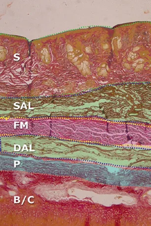 Fig 1. Nasal soft-tissue layers on histology. The skin (S, sebaceous) and the densely adherent periosteum/perichondrium (P) are hard to inject; the plane of least resistance is the fibromuscular (FM) layer sandwiched between the two areolar layers (SAL, DAL), and the vascular network sits on the surface of FM. This is why the safe deposit is deeper than FM, against B/C. · (Jones/Wu, 2019, p.148)
> Fuentes: Jones - Injectable Fillers 2019 9781119046967
Fig 1. Nasal soft-tissue layers on histology. The skin (S, sebaceous) and the densely adherent periosteum/perichondrium (P) are hard to inject; the plane of least resistance is the fibromuscular (FM) layer sandwiched between the two areolar layers (SAL, DAL), and the vascular network sits on the surface of FM. This is why the safe deposit is deeper than FM, against B/C. · (Jones/Wu, 2019, p.148)
> Fuentes: Jones - Injectable Fillers 2019 9781119046967 [C][MEDLIB].
The rule that falls out of the table: in the nose, deep is safe and intermediate is lethal. Against surgical intuition the danger is not at the floor: it is just under the skin, in layer 3 where the vessels travel. The novice who "keeps it superficial to be gentle" is injecting into the arterial plane [2][3]. The layered histology in Fig 1 documents this directly: the vascular fibromuscular plane (FM) sits above the deep safe target against bone/cartilage.
Skin by third (governs how much shows): upper third thick and mobile; middle third (mid-dorsum, over the rhinion) the thinnest, least distensible, most adherent skin, where over-correction shows first; lower third as thick as the upper with more sebaceous glands, thickest at the tip [3][6][8]. The nasal SMAS is a genuine, continuous fibromuscular sheet, not a virtual plane [5].
Nasal musculature (targets for toxin, layer 3): procerus and levator labii superioris alaeque nasi (elevators), depressor septi nasi (depressor), dilator naris anterior/posterior (dilators), transverse nasalis / compressor naris (compressor) [8]. Their layer-3 course is exactly the plane the filler injector must avoid and the toxin injector must reach.
Trampa clásica (D8.2): losing the plane. Aiming "deep" but stopping in the intermediate fibromuscular layer because the needle met soft resistance and never reached bone. If you did not feel firm bone or cartilage, you are in the arterial plane. Advance to contact or withdraw and re-enter; never deposit in the give.
Aesthetic subunits (the map for planning and for camouflage) [8][21][39]: dorsum, paired sidewalls, tip, paired soft triangles/facets, paired alae, and the columella (Burget-Menick nine-subunit scheme). Deposits and shadows are planned along subunit borders because the eye reads the highlights and valleys the cartilage casts; a straight dorsal aesthetic line reads as a better nose even when there is objectively more nose than before [21].
Skeletal thirds and the weak points [21]:
| Third | Skeleton | Injection note |
|---|---|---|
| Upper (bony pyramid) | Paired nasal bones, fused, articulating with the frontal process of the maxilla | Radix support; supraperiosteal deposits sit on bone |
| Middle (cartilaginous vault) | T-shaped upper lateral cartilages fused to the septum; keystone area (bony-cartilaginous overlap) is the structural cornerstone; the rhinion here has the thinnest skin (~0.6 mm) | The least forgiving zone: thin skin over the rhinion shows every over-elevation |
| Lower (lobule) | Paired lower lateral (alar) cartilages, medial crura, septal caudal end, anterior nasal spine; the scroll region is the upper-to-lower lateral overlap | The tip pillar builds off the spine and medial crura; the scroll unravels with age |
Skin-thickness map (governs where mistakes show): thickest at the nasofrontal groove/nasion (~1.25 mm), thinnest at the rhinion (~0.6 mm), thick again and sebaceous at the tip; thin at the columella and alar margin; at the nostril apex a soft triangle exists where lining and lobule skin meet with no subcutaneous layer, an absolute no-inject zone [6][8]. The dorsum's skin is thin and loosely adherent, so dorsal irregularity is visible; the tip's skin is thick and adherent, so it resists shape change and raises necrosis risk [3][21].
Ethnic variation (changes the goal, not the safety corridor) [3]: the Asian nose typically has a low radix, flat/under-projected dorsum, thin nostrils and a short, rounded tip, and is the classic augmentation candidate; the African nose has a flat dorsum, radix near the intercanthal line, and larger nostrils/tip with a well-developed protruding central maxilla. Augmentation goals differ by phenotype; the deep-midline safe corridor does not.
Vascular layer, restated for injection (the point of the whole block): the arterial network lies on the surface of the fibromuscular (SMAS) layer, sandwiched between skin+superficial fat above and the deep areolar fat + periosteum below [2]. Wu's classic description is five soft-tissue layers (skin, superficial fibrofatty areolar, fibromuscular, deep areolar, periosteum/perichondrium); the plane of least resistance is between the two areolar layers, which is exactly where the vessels sit, so the intended target is deeper still, against bone/cartilage [2].
Surface landmarks (the vocabulary of the injection plan) [8][21]:
| Landmark | Definition | Injection relevance |
|---|---|---|
| Radix / nasion | Root of the nose at the nasofrontal junction | P1 target; deepest point sets the nasofrontal angle |
| Rhinion | Bony-cartilaginous junction of the dorsum | Thinnest skin (~0.6 mm); over-elevation shows here first |
| Kyphion | Most prominent point of a dorsal hump | The reference for supratip camouflage (P2) |
| Supratip | Just above the tip-defining points | Break point; ⚠ excess gives a "parrot beak" |
| Tip-defining points | Paired light-reflection points of the tip | Minimal, deep, midline deposits only |
| Columella | Midline strut between the nostrils to the lip | Pillar target; columellar artery is subcutaneous here |
| Subnasale / nasal spine | Columella-lip junction / bony spine | Deep supraperiosteal base support (P4.1) |
| Alar groove / rim | Junction of ala and cheek / nostril margin | ⚠ Lateral nasal artery; no-inject zone |
| Soft triangle | Nostril apex, no subcutaneous layer | Absolute no-inject zone |
Nasal valves (do not fill an airway problem) [21][30]: the internal nasal valve (the narrowest part of the airway) is bounded by the caudal upper lateral cartilage, the septum, the head of the inferior turbinate, the pyriform aperture and the nasal floor; the external nasal valve is the alar rim/lobule region. Filler placed to "straighten" a deviation must never encroach on these; the Cottle and modified Cottle manoeuvres screen valve competence, and any true valvular/septal obstruction is a surgical referral, not an injectable target [30].
Sensory innervation (and why sudden pain is a warning) [4][8]: the dorsum and tip are supplied by the external nasal branch of the anterior ethmoidal nerve (which pierces the bony-cartilaginous junction and runs on the upper lateral cartilage), the ala by external nasal branches of the infraorbital nerve, and the base/columella by branches of the infraorbital and nasopalatine systems. Motor supply to the nasal muscles is from zygomatic/buccal branches of the facial nerve [4]. Disproportionate, sudden pain in these territories during injection is an ischaemic alarm, not a "sensitive patient"; for a formal block, the infratrochlear and external-nasal branches are targeted (Jung used an infratrochlear and internal-nasal-branch block) [17].
Keystone area and scroll region (the two structural hinges) [21]: the keystone is the overlap of the caudal nasal bones on the upper lateral cartilages, the structural cornerstone of the dorsum and the exact level of the thin-skin rhinion; the scroll is the upper-to-lower-lateral cartilage overlap that unravels with age and lets the tip drop. Both are landmarks, not injection targets: dorsal support sits deep and midline over the keystone, and tip support is built from the spine and medial crura, not from the scroll.
D8.3 · Vessels, nerves and the danger zone → J2 · Vascular Occlusion & Emergency Response
The arteries to know cold. The nose sits at the watershed between the external carotid (facial → angular, superior labial) and the internal carotid (ophthalmic → dorsal nasal, supratrochlear, supraorbital). Every one of them anastomoses across the midline and with the orbit, and that is the anatomy of blindness, mapped in Fig 2 [2][7][9].
| Artery | Origin | Course & depth | What it costs if you hit it |
|---|---|---|---|
| Dorsal nasal a. | Terminal branch of the ophthalmic a. (ICA) [4][9] | Emerges just above the medial canthal tendon, descends paramedian on the dorsum; ~1 mm calibre; supratrochlear volume ~0.085 mL to the orbit [13] | Direct retrograde route to the retina. The anatomical author of nasal-filler blindness. |
| Lateral nasal a. | Branch of the angular/facial a. | Runs just above the alar groove to supply tip and ala; superficial, bilateral [2][3] | Alar and tip necrosis. The most common nasal necrosis territory. |
| Columellar a. | Branch of the superior labial a. | Ascends the columella toward the tip, immediately subcutaneous [2][7] | Columella/tip embolism and necrosis; highest embolisation risk of the base. |
| Angular a. | Continuation of the facial a. | Ascends alongside the lateral nasal wall; anastomoses with the dorsal nasal/ophthalmic system at the medial canthus [4] | Bridge from the facial (ECA) system into the ophthalmic (ICA) system. |
| Supratrochlear / supraorbital a. | Ophthalmic a. (ICA) | Exit at the superomedial orbit; feed the radix/glabella watershed [9] | Radix/glabella injections reflux here into the ophthalmic a. |
Sensory nerves (relevant to blocks and to warning pain): external nasal branch of the anterior ethmoidal nerve (dorsum and tip) and the external nasal branches of the infraorbital nerve (ala) [4]. Sudden, disproportionate pain along these territories during injection is an ischaemic alarm, not "a sensitive patient".
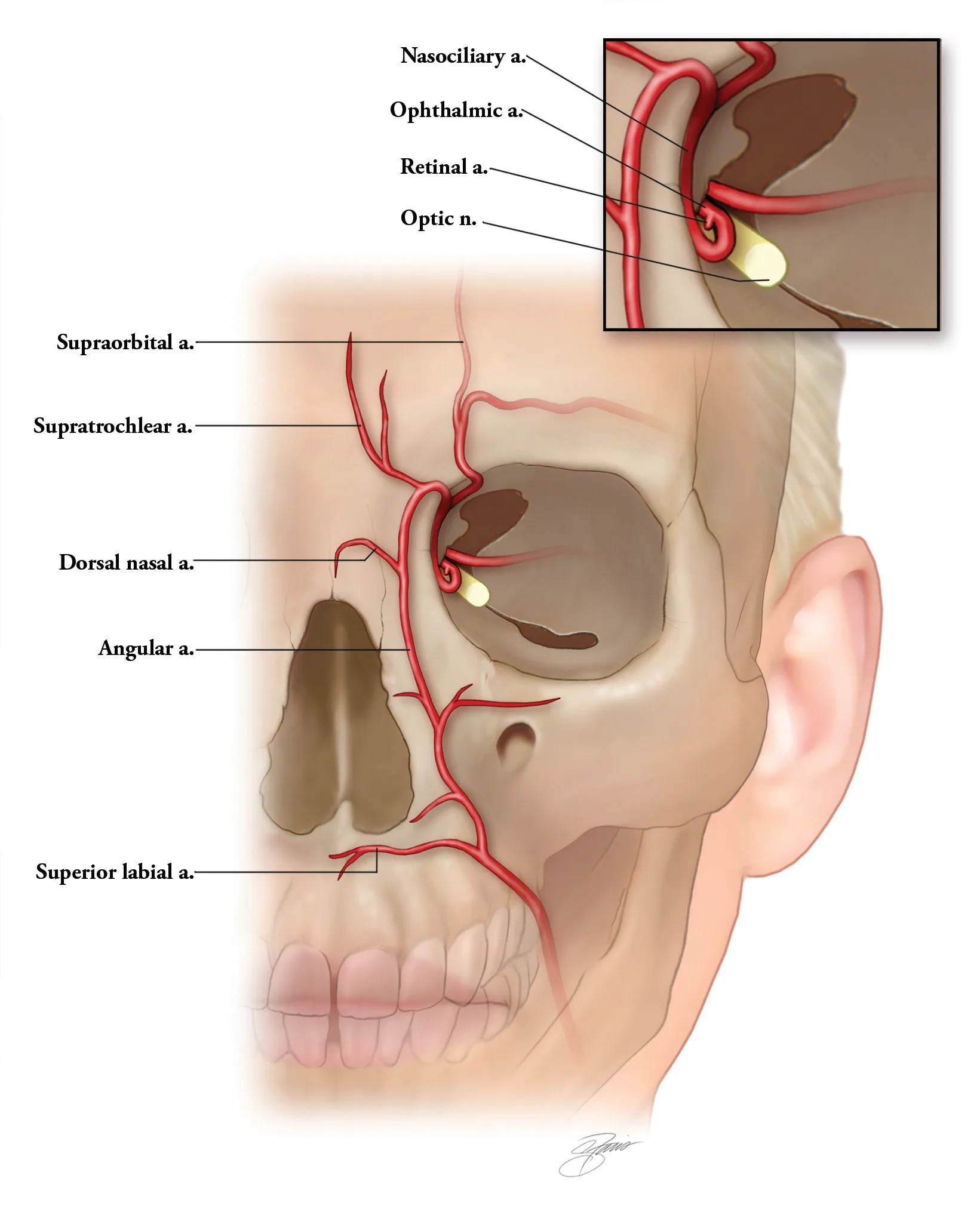 Fig 2. The dorsal nasal, angular and supratrochlear arteries connect to the ophthalmic artery; the superior labial feeds the columella. Intra-arterial injection into any terminal branch of this network can reflux retrograde into the ophthalmic and retinal arteries. · (Kontis, 2019, p.211)
> Fuentes: Kontis - Cosmetic Injection Techniques 2019 9781626234574
Fig 2. The dorsal nasal, angular and supratrochlear arteries connect to the ophthalmic artery; the superior labial feeds the columella. Intra-arterial injection into any terminal branch of this network can reflux retrograde into the ophthalmic and retinal arteries. · (Kontis, 2019, p.211)
> Fuentes: Kontis - Cosmetic Injection Techniques 2019 9781626234574 [C][MEDLIB].
Variants and calibre (why "usually" is not "always") [7][9][13]: the dorsal nasal, supratrochlear and supraorbital arteries are ~1 mm in calibre; a nasal artery can reach ~1.1 mm, which can exceed the outer diameter of a fine cannula, so "the cannula is wider than the vessel" is not guaranteed [11]. The lateral nasal artery usually runs 2-3 mm above the alar groove but its exact height varies; the columellar artery is usually a branch of the superior labial but can be duplicated or arise variably; the dorsal nasal artery may be dominant on one side. The volume needed to fill a terminal branch to the retina is small: the supratrochlear artery holds ~0.085 mL, so a sub-0.1 mL intra-arterial bolus can reach the eye [13]. The practical consequence: treat every nasal artery as present, variable and potentially larger than your instrument, and rely on plane, pressure and volume rather than on an assumed anatomy.
Zone-by-zone risk map (the mental model of the nose):
| Zone | Dominant risk | Level |
|---|---|---|
| Dorsum, strict midline, supraperiosteal/supraperichondrial | Deep midline is relatively avascular; collateral supply | Lowest of the nose |
| Radix | Proximity to dorsal nasal a. and ophthalmic anastomosis | High (blindness) |
| Lateral dorsal walls | The vessels are here; the midline is safe precisely because vessels are not medial | High |
| Alar groove / rim | Lateral nasal a., superficial | Very high (necrosis) |
| Tip | Terminal territory, tight skin, non-dissipating pressure | Very high (necrosis + embolism) |
| Columella | Columellar a., immediately subcutaneous | High |
| ⚠ Previously operated nose, any zone | Randomised, scarred vasculature; low compliance | Separate category |
Blindness mechanism (retrograde intra-arterial embolisation). When injection pressure exceeds systolic pressure at the needle tip inside an artery, product refluxes proximally along the vessel; on release it advances anterograde. From the dorsal nasal or angular artery the column reaches the ophthalmic artery and then the central retinal artery (amaurosis) and/or the posterior ciliary and cerebral arteries (infarction, stroke), the exact pathway drawn in Fig 3 [9][10][11]. The four conditions for blindness: (1) the artery injected anastomoses with the ophthalmic; (2) the tip fully perforates the lumen; (3) filler is pushed against arterial pressure; (4) the injected volume is sufficient to fill the lumen from the entry point to the central retinal artery [10]. Of 50 reported filler-blindness cases to September 2018, the nose accounted for 21 (42%) and the glabella 14 (28%) [10]; a series counting fat and filler put nasal/periforehead blindness near 100 cases, all permanent [2].
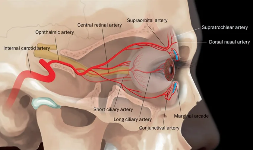 Fig 3. The internal-carotid → ophthalmic → central retinal axis. The supratrochlear, supraorbital and dorsal nasal arteries are ophthalmic branches; a retrograde column from the nasal dorsum can reach the central retinal artery and infarct the retina. · (Filler Complications, 2019, p.110)
> Fuentes: Filler Complications, Filler-Induced Hypersensitivity Reactions 2019 ISBN 978-981-13-6639-0
Fig 3. The internal-carotid → ophthalmic → central retinal axis. The supratrochlear, supraorbital and dorsal nasal arteries are ophthalmic branches; a retrograde column from the nasal dorsum can reach the central retinal artery and infarct the retina. · (Filler Complications, 2019, p.110)
> Fuentes: Filler Complications, Filler-Induced Hypersensitivity Reactions 2019 ISBN 978-981-13-6639-0 [C][MEDLIB].
Fatal case, verbatim from the literature (the reason the red lines are red): a 40-year-old received 2 mL lidocaine-containing HA to the nose via a 25G cannula by an unlicensed injector; immediate periocular pain and blurred vision, loss of consciousness within 30 minutes, coma, and death from malignant cerebral infarction with ophthalmic-artery occlusion [11]. Time windows from that literature: ~90 minutes to irreversible retinal ischaemia; ~12 hours before severe cerebral functional impairment; decompressive craniectomy only useful < 48 h [11].
Operative summary of the block: midline, deep, little, slow. Anything that departs from that (lateral, superficial, much, or fast) raises the risk, and the ala and tip raise all four at once [2]. Cross-reference the full occlusion and blindness algorithm to J2 · Vascular Occlusion & Emergency Response and hyaluronidase dosing to J3 · Hyaluronidase - Pharmacology & Clinical Protocols.
Trampa clásica (D8.3): injecting the radix as if it were a safe "high, central" point. The radix is the closest nasal point to the dorsal nasal artery and the ophthalmic anastomosis; a bolus there under pressure is the shortest path to the retina. Radix deposits stay ≤ 0.05-0.15 mL, supraperiosteal, with non-dominant-hand compression of the glabellar sides to reduce dorsal nasal flow [1].
Anastomotic anatomy and why necrosis spreads or spares (choke vs true anastomoses) [15]: adjacent angiosomes are linked by true anastomoses (normal-calibre) and choke anastomoses (reduced-calibre). After an embolus, some choke vessels dilate and compensate (limiting necrosis) while vasospasm in others acts as a barrier; this choke-vessel behaviour is the major determinant of the location and extent of necrosis [14][15]. The dorsum, with richer collateral supply, tolerates compression better than the columella and ala, which are more terminal; hence dorsal midline is the low-risk corridor and the ala/tip are the necrosis-prone poles [2].
Multi-route ECA↔ICA mapping (the argument against fixed "safe zones") [9][10]: a nasal injection can reach the ophthalmic artery by several routes: dorsal nasal, angular→dorsal nasal, supratrochlear and supraorbital, all converging on the ophthalmic. Because these routes vary between individuals and a scarred nose randomises them further, some authors argue there is no universal nasal safe zone, only a safest technique (deep midline, low pressure, small aliquots, ± ultrasound). The safe corridor reduces probability; it does not abolish the anatomy.
Depth summary (what sits where): columellar artery immediately subcutaneous in the columella; lateral nasal artery superficial just above the alar groove; dorsal nasal artery paramedian, emerging above the medial canthal tendon; the strict deep midline against bone/cartilage is the corridor with the fewest vessels [2][3][7]. A cannula does not change this map; over 75% of nasal filler-blindness cases in one large series involved a cannula, so the tool is not the safeguard, the plane and pressure are [2]. The nasal-root cannula route illustrates the trap: a long infralobular cannula that drifts into the subcutaneous layer at the radix, where the skin is not tightly bound to the SMAS, raises the embolism risk precisely where the dorsal nasal artery runs [38]. The broad facial danger-zone maps make the same point for the whole midface [40].
D8.4 · Ageing of the nose: what is lost, and in what order
Order of change (deep to superficial), the injectable logic:
| Structure | Age change in the nose | Clinical sign |
|---|---|---|
| 1 Bone (first, sets the rest) | Pyriform aperture and maxillary angle regress; pyriform, maxillary, glabellar and orbital angles decrease with age; nasal length, width and vertical dimension increase [5]. Pyriform regression more marked in Asian than Caucasian series [5]. | Loss of basal support, apparent lengthening, deepening nasolabial transition. |
| 2 Cartilage / tip support | Loss of medial-crural support and unravelling of the scroll region (upper-to-lower-lateral overlap); stretched fibrous attachments; alar cartilage weakens; tip drops and de-rotates [21]. | Tip ptosis, plunging tip on smile, increased columellar show. |
| 3 Deep fat / ligament | Scant nasal deep fat provides little volume; support is skeletal and ligamentous, so its loss reads as skeletonisation rather than deflation. | Bony dorsal step-offs become visible. |
| 4 Superficial fat / SMAS | Minimal in the nose; SMAS laxity contributes little volume but transmits mimetic pull (dynamic tip drop). | Dynamic drooping with animation. |
| 5 Skin (last) | Dorsal skin thins further, so irregularities and step-offs surface; sebaceous change at the tip; the bony dorsum does not change but its edge looks sharper as skin thins [3]. | Visible dorsal irregularity, sharper skeletal outline. |
The sequence that matters for treatment (P): in the nose the earliest and dominant age change is loss of tip support (medial crura, scroll), then skeletal/basal regression (pyriform, nasal spine), with soft-tissue atrophy a late, minor contributor because the nose is fat-poor. This inverts the mid-face logic: you rebuild support (tip pillar, nasal spine, pyriform) before you think about "volume", and toxin (depressor septi) addresses the dynamic component that filler cannot. (P) reasoning, no dose implied, no figure carried.
Why the aged nose looks longer and droopier, not deflated: unlike the cheek, the nose does not lose a fat envelope; it loses a cartilaginous strut. The tip rotates down and the dorsum skeletonises, so the corrective move is a columellar/spine pillar to re-rotate and re-project the tip, which optically shortens the nose, before any dorsal work [1][21]. General facial-ageing scaffolding (bone → ligament → deep fat → superficial fat → skin) is corpus-grounded [5][31][32][33]; its nasal application is support-first.
Young vs aged nose (what actually changes) [3][5][21]:
| Feature | Youthful | Aged | Injectable response |
|---|---|---|---|
| Tip rotation | Rotated up, projected | De-rotated, dropped | Columellar/spine pillar re-projects (filler); depressor-septi toxin for the dynamic drop |
| Nasolabial angle | 90-105° F | Narrows (tip drops) | Toxin ± spine support widens it slightly |
| Nasal length | Shorter | Apparent lengthening | Tip re-projection optically shortens |
| Dorsal line | Smooth | Skeletonised, step-offs visible | Radix/supratip camouflage, midline |
| Columellar show | Balanced | Increased | Spine/columella support |
| Skin | Thicker over dorsum | Thinner dorsally, sharper edge | Thin skin shows irregularity; deposit deep and midline |
| Pyriform/base | Full | Regressed | Deep supraperiosteal base support |
Trampa clásica (D8.4): treating the aged droopy tip by adding dorsal volume. Dorsal filler on a de-rotated tip lengthens the nose further and worsens the plunge; the correct first move is to re-project the tip from below (deep columellar pillar) and relax the depressor septi, then reassess the dorsum, which often no longer needs anything [1].
Angle changes with age (measured) [5]: the glabellar, orbital, maxillary and pyriform angles all decrease with age; the maxillary and pyriform bone and the infraorbital rim regress; nasal length, width and vertical height increase. The Lambros clockwise-rotation theory of the viscerocranium captures the net effect: the mid-face and nasal base rotate and retrude, which drops the tip and deepens the nasolabial transition [5]. Asian series show more pyriform change and less orbital/maxillary change than Caucasian series, so the ethnic starting point shifts the age trajectory [5].
What this means at the syringe (P): because the earliest structural loss is tip support and basal (pyriform/spine) regression, the youthful move is to rebuild the base and the tip pillar, not to lay volume on the dorsum. A columellar/spine pillar re-projects and re-rotates the tip, optically shortening a nose that has lengthened with age; only then is dorsal camouflage considered. Soft-tissue atrophy is a minor, late contributor in a fat-poor region, so "deflation" logic imported from the cheek misreads the aged nose. (P) reasoning, no dose implied, no figure carried.
Sequence of visible signs (early to late): dynamic tip drop on smile (depressor septi) → static tip ptosis and de-rotation (crural/scroll support loss) → increased columellar show and apparent lengthening → dorsal skeletonisation with visible step-offs → thinning dorsal skin sharpening the skeletal outline [3][21]. Each maps to a tool: toxin for the dynamic drop, a filler pillar for the static ptosis, dorsal camouflage last.
D8.5 · Assessment: measure, photograph, test dynamically, scan
What is measured (numbers to record before you touch a syringe):
| Parameter | Reference range | Why it drives the plan |
|---|---|---|
| Nasofrontal angle (radix) | ~115-135° (deeper = higher radix need) [29] | A shallow angle wants radix elevation; the commonest non-surgical request. |
| Nasofacial angle | ~30-40° | Dorsal projection relative to the facial plane. |
| Nasolabial angle | ~90-105° F, 90-95° M | Guides tip rotation and columellar/spine support; toxin can widen it ~5° (110→115°) [23]. |
| Tip projection (Goode ratio) | Goode 0.55-0.60 | Distinguishes under-projection (treatable) from over-projection (refer). |
| Dorsal aesthetic lines | Smooth, symmetric, brow-to-tip | Deviations and step-offs define camouflage points. |
| Tip recoil / support | Firm recoil | A greater than 30° divergence of medial/lateral crura reads as a bulbous tip that filler cannot narrow [30]. |
What is photographed (standardised, four views minimum): frontal, lateral (both), basal, and the top-down "bird's-eye" view that reveals dorsal deviation and that almost nobody takes [2]. Record before/after in identical lighting and head position.
What is tested dynamically: the plunging tip on smile (depressor septi overactivity, a genuine low-risk toxin indication) [22][23], nasal-valve competence (Cottle / modified Cottle manoeuvre) so an airway problem is not mistaken for an aesthetic one [30], and skin quality/thickness by palpation (thick sebaceous tip skin resists shape change and raises necrosis risk) [3].
What is scanned (ultrasound / colour Doppler), a top-yield indication in the nose:
- Pre-injection vessel mapping of the dorsal nasal and lateral nasal arteries and confirmation of the supraperiosteal/supraperichondrial plane [15][28].
- The previously operated or previously filled nose, where surface anatomy no longer predicts the vessel course: scan before adding anything [2][16].
- Post-event diagnosis: an occluded artery shows to-and-fro flow (blue-and-red in the same lumen) with a downstream anechoic HA deposit; van Loghem's "Medusa head" of dilated collaterals with the anechoic filler at its centre marks the target for US-guided hyaluronidase [16].
- Echogenicity primer: anechoic = blood/water/HA (dark), hypoechoic = muscle/fat, hyperechoic = bone/CaHA/air [15].
Consensus: ultrasound reduces anatomical uncertainty and speeds occlusion diagnosis and treatment [15][16][28]. Discrepancy: does Doppler prevent occlusion? A: pre-procedure mapping plus real-time guidance lowers events and, in the largest reported cohort, all vascular events resolved [15]. B: ⚠ a documented limitation is that in some cases the Doppler signal appeared only after the occlusion, so ultrasound is a risk-reduction and rescue tool, not a licence to inject faster, more, or more laterally [16][28]. Decide by operator skill and whether the nose is virgin or reoperated.
What the record must contain (before and after) [1][2]: the four-plus-two view photo set; the measured angles (nasofrontal, nasolabial, Goode ratio); skin thickness/quality and tip recoil; prior surgery/filler and any ultrasound map; the written plan (points, planes, product, expected result, explicit non-goals); the consent naming blindness and necrosis; and, at treatment, the product, lot, volume per point and total, and any incident. This is both a clinical and a medicolegal minimum in the one region where an event can be blinding.
Decision: when to scan before injecting. Always in a previously operated or previously filled nose; whenever the surface anatomy is ambiguous or asymmetric; when prior product is present but unknown; and at the operator's threshold for any high-risk plan. A virgin nose in expert hands with a conservative deep-midline plan can proceed on anatomic technique, but the scan is never wrong to do [15][16].
Trampa clásica (D8.5): assessing only from the lateral view because that is where you work. Widening of the dorsum shows on the frontal view and deviation shows from above; skip those two views and you finish a straighter profile with a wider, deviated dorsum you never saw [2].
Photography protocol (reproducible, so before/after are comparable): fixed focal length and distance, Frankfort horizontal, neutral expression plus a smiling frame (to capture the plunging tip), consistent background and lighting; five standard frames (frontal, right and left lateral, right and left oblique) plus basal and the top-down/bird's-eye frame that reveals dorsal deviation [2]. Repeat identically at the 2-week review and at follow-up.
Angle atlas (what "normal" is, so deviation is measurable): nasofrontal 115-135° (the radix indicator, and the commonest non-surgical request in low-bridge phenotypes) [29]; nasofacial ~30-40°; nasolabial 90-105° in women, 90-95° in men (toxin can add ~5°) [23]; Goode tip-projection ratio 0.55-0.60; a medial-to-lateral crural divergence greater than 30° predicts a bulbous tip that filler cannot narrow [30]. Record the numbers; they define both the plan and the limit of what filler can do.
Dynamic and functional tests: smile (plunging tip = depressor septi indication) [22]; Cottle and modified Cottle for internal-valve competence (do not mistake an airway complaint for an aesthetic one) [30]; skin pinch for thickness/mobility (thin dorsal skin shows everything; thick sebaceous tip skin resists and is necrosis-prone) [3]; palpation of prior surgical scars and any hard prior product [16].
Ultrasound in practice (top-yield here) [15][16][28]: a high-frequency linear probe (≥ 15 MHz) maps the dorsal nasal and lateral nasal arteries and confirms the supraperiosteal/supraperichondrial plane before injection, and diagnoses an occlusion after one (to-and-fro flow, anechoic HA deposit, dilated collaterals). Colour/power Doppler distinguishes arterial from venous flow; the "Medusa head" of dilated collaterals around an anechoic deposit marks the target for US-guided hyaluronidase, which restores flow with far less enzyme than blind flooding [16]. In the reoperated or previously filled nose, where surface anatomy no longer predicts the vessels, pre-scan is the single highest-value step [2][16].
Pre-injection ultrasound protocol (numbered, for the high-risk/reoperated nose) [15][16][28]:
- Probe and settings: high-frequency linear probe (≥ 15-18 MHz); copious gel or a standoff; low PRF/slow-flow Doppler to catch small nasal arteries; keep the probe light (pressure occludes small vessels and hides them).
- Grey-scale survey: identify skin, the fibromuscular layer, deep fat and the periosteum/perichondrium in the midline and paramedian; note skin thickness and any prior anechoic (HA) or hyperechoic (non-HA/CaHA) deposit.
- Colour/power Doppler map: trace the dorsal nasal artery paramedian at the radix, the lateral nasal artery above the alar groove, and the columellar artery in the columella; mark their depth and midline distance.
- Plane confirmation: confirm the intended supraperiosteal/supraperichondrial target is deep to the vascular (fibromuscular) plane before the first deposit.
- Reoperated/previously filled nose: map scar and any residual product; if unknown product is present, do not add until identified.
- Real-time option: for the experienced operator, inject under live visualisation, watching the needle tip and avoiding the mapped vessels; aspirate as a checkpoint, not a guarantee.
- Post-event use: if occlusion is suspected, the same probe finds the to-and-fro flow, the anechoic HA deposit and the dilated-collateral "Medusa head" that targets US-guided hyaluronidase (50-100 IU, repeated) [16].
Echogenicity crib: anechoic (black) = blood/water/HA; hypoechoic = muscle/fat; hyperechoic (bright) = bone/CaHA/air. Colour Doppler shows movement (flow or probe motion). Ultrasound reduces uncertainty and speeds rescue; it does not license higher volume, speed or a more lateral approach [15][16].
D8.6 · Goal and patient selection: who benefits, who does not, what is referred
Selection grid (decide in the first consultation):
| Presentation | Filler? | Note |
|---|---|---|
| Low radix / shallow nasofrontal angle | ✅ First-line | The highest-yield, lowest-risk indication [1][29]. |
| Small-to-moderate dorsal hump | ✅ Camouflage | Raise radix + supratip to straighten the line; the hump is masked, not removed [1][2]. |
| Droopy / under-projected tip | ✅ Support | Deep columellar/spine pillar re-projects and re-rotates; do the tip first [1]. |
| Post-traumatic saddle dorsum | ✅ | Augment the depression to restore the dorsal line [2]. |
| Flat dorsum (ethnic augmentation, Asian/African) | ✅ | Structural firm HA; frequent request in flat-bridge phenotypes [2][3]. |
| Minor post-surgical contour irregularity | ✅ Caution | Touch-up only, ⚠ scarred vasculature (see below) [9]. |
| Deviation of the appearance (not the septum) | ⚠ Selective | Asymmetric deposits can improve the look of a mild caudal deviation [17][20]; a true septal/airway deviation is surgical. |
| Wants a smaller nose | ❌ Not a candidate | Filler adds volume; a reduction goal cannot be met. Say it on day one [2][29]. |
| Large/bulbous tip, over-projected tip | ❌ Refer | Reduction and tip narrowing are surgical [30]. |
| Large hump, functional airway problem | ❌ Refer | Structural rhinoplasty [21][30]. |
| Body dysmorphic features, displaced expectation | ❌ Do not treat | The nose is the region with the most consultations of this profile; screen and refer [29]. |
Consultation red flags (stop, do not book) [2][29]:
| Red flag | Why it stops the plan |
|---|---|
| "Make my nose smaller/thinner" | Filler adds volume; the goal cannot be met |
| Multiple prior revisions or a "failed" rhinoplasty to fix | Separate risk category; expectations rarely met with filler |
| Unknown prior filler, product not documented | Assume non-HA; scan before adding anything |
| Planned surgical rhinoplasty | Residual HA distorts surgery; remove first |
| Fixation on a millimetric flaw invisible to others | BDD screen; refer, do not treat |
| Active acne, folliculitis or infection on the nose | Closed sebaceous space; defer |
| Bleeding diathesis / anticoagulation without plan | Bruising and event management complicated |
| No time or willingness for the 2-week review | The nose needs staged completion and follow-up |
| Cannot be reached for 24 h post-procedure | Time is the prognostic factor in an event |
Special situations that change the threshold, not just the plan:
- ⚠ Previously operated nose = a separate risk category. Scar randomises the vessel course, reduces envelope compliance and makes planes unpredictable; a disproportionate share of published necrosis and blindness occurs in operated noses. It is not a harder case, it is another category: higher indication threshold, ultrasound, or decline [9][11]. Compression ischaemia is more likely in fibrotic post-surgical tissue [16].
- Planning future surgical rhinoplasty: ⚠ never rinomodel a patient who intends surgery. HA is not fully resorbed; a residual fraction can persist for years and distort dissection planes and fibrosis. If HA is present, it must be removed with hyaluronidase before surgery, and the surgeon must be told what and when [1][2][17].
- Unknown prior filler: if there was previous filler and the product is unknown, assume the worst (possible non-HA) and scan before adding anything [16].
- Active acne, sebaceous inflammation, infection in the field: defer; the sebaceous, closed nasal space makes infection more frequent and worse [2][10].
- Realistic ceiling: long-term satisfaction data for NSR are high when selection is right (FACE-Q prospective cohort) [36]; a series of HA correction of surgical-rhinoplasty complications (2088 cases) documents a legitimate reconstructive niche in expert hands [37].
Who to refer, and to whom: reduction/large hump/over-projected or bulbous tip and any airway/septal problem to a rhinoplasty surgeon; suspected BDD or displaced expectation to psychology/psychiatry before any treatment; a nose with unknown or non-HA prior product, or an operated nose beyond your threshold, to an ultrasound-capable injector or complications clinic; and any acute visual symptom to ophthalmology/oculoplastics immediately [11][29][30]. Referring is a clinical decision, not a failure.
Consensus across schools: the nose is a camouflage and support procedure with limited, explicitly stated goals; ambitious volumetric remodelling produces wide dorsums and the most complications and is rejected [2][20]. An abstentionist stance (do not treat the nose with filler outside very expert hands) is defensible and coherent with the data; this atlas does not forbid, but raises the threshold explicitly [2].
Trampa clásica (D8.6): accepting the patient who says "fix my nose" without pinning down whether the wish is achievable by adding volume. Hearing "fix" and not converting it to a specific, additive, line-changing goal is how the reduction-seeker ends up dissatisfied with a technically good result [2][29].
Requirements checked aloud before injecting (if one is missing, do not inject) [2][13]:
| Requirement | Why |
|---|---|
| Hyaluronidase in the room in occlusion-protocol quantity (≥ 10 vials / > 1500 IU), not one vial | The rescue dose is not the nodule-dissolution dose [13][15] |
| Written, rehearsed occlusion protocol | Reading it during the emergency is arriving late [2] |
| Reversible product only | No therapeutic option against a non-HA product [1][20] |
| No vasoconstrictor in the anaesthetic field | ⚠ Masks the blanch sign [2] |
| Non-operated nose, or explicit acknowledgement of the higher risk if operated | Separate risk category [9][11] |
| No active inflammatory lesion or infection in the field | Closed, sebaceous space [10] |
| Consent that names blindness and necrosis in those words | A generic consent does not inform of this [2] |
| Nose not combined with another high-risk region the same day | So an event's origin is knowable [2] |
| 24-hour direct contact line explained to the patient | Time is the prognostic factor [13] |
Absolute and relative contraindications: active nasal/skin infection or acne; unrealistic or reduction-seeking goal; BDD; planned rhinoplasty with residual HA not yet removed; unknown prior filler until scanned; pregnancy/lactation (precaution); known hyaluronidase allergy limits the safety net (recombinant hyaluronidase has the lowest allergenicity but is not universally available) [16][29]. The previously operated nose is not an absolute contraindication but a threshold shift: ultrasound, expert hands, or decline [9].
Consent specifics for the nose: the standard filler consent is insufficient. Name vascular occlusion, skin necrosis, and permanent blindness explicitly; state that the nose enlarges with filler; state the effect on any future surgery (residual product, fibrosis, altered planes); and document the 24-hour contact pathway [2][29]. The nose is the one region where a generic aesthetic consent is a medicolegal and clinical failure.
D8.7 · Technique: the full grid
Consensus (all schools agree on the corridor): strict midline, deep plane (supraperiosteal on bone / supraperichondrial on cartilage), microboluses, low pressure, low flow, non-dominant-hand control, stop-and-look between deposits, small total volume, single short session, top-down sequence (radix → dorsum → tip → columella/base) [1][2][17][20]. The disagreements are instrument (needle vs cannula) and aspiration, both preserved below without averaging.
Five technique invariants that outrank every school choice. First, plane before product: the deep supraperiosteal/supraperichondrial midline is the low-risk corridor whatever the gel or the tool, and a soft product placed correctly is safer than a firm product placed in the fibromuscular plane [2][3]. Second, pressure is the embolus lever: the mechanism of intra-arterial embolisation is delivered pressure exceeding systolic at the tip, so low thumb force, an adequate-gauge needle for the gel, and a slow rate matter more than the instrument brand [10][13]. Third, volume is dose: microaliquots of ≤ 0.05 mL keep any accidental intravascular column below the sub-0.1 mL that can reach the retina, so the syringe never delivers a bolus [1][13]. Fourth, the non-dominant hand is an instrument: pinching, columellar counter-pressure and glabellar compression superficialise or occlude the arteries you are avoiding while you deposit [1]. Fifth, stop and look: reassessing between deposits from the frontal and bird's-eye views (not only the lateral) is the cheapest safety manoeuvre and the one most often skipped [2]. The grids that follow enumerate the options along each axis; these five rules decide how any option is executed.
Product grid by rheology (never averaged)
| Product | Rheology / particle | Nose use | Verdict |
|---|---|---|---|
| HA, high G′ / high cohesivity (e.g. VYC-25L / Juvederm Volux; Restylane firm family; Teosyal Ultra Deep) | High elastic modulus, high cohesivity, low hydrophilia; holds a line against the tight envelope | First-line for dorsum, radix, columella/spine pillar. VYC-25L validated in NSR (492 pts, single injector) and by global expert consensus for safe long-term use [18][19]; R4P uses high-G′ HA at the columella point [1] | ✅ Reversible, structural |
| HA, intermediate G′ | Moderate elasticity, more spread | R4P points 1-3 (radix, supratip, tip) [1]; Jung superficial-layer refinement [17] | ✅ Reversible, refinement |
| HA skin booster / low-G′ | Low elasticity, high spread | Not for structural nasal shaping (spreads, widens the dorsum) | ⚠ Wrong tool for the dorsum |
| CaHA / Radiesse (standard) | Viscosity and G′ above HA; 25-45 µm microspheres; supraperiosteal, not dermis | Historically used for dorsal augmentation, esp. Asian flat dorsum [20] | ⚠ Not reversible. No antidote for occlusion; excluded here |
| CaHA hyperdiluted | Biostimulatory dilution | Skin-quality, not nasal shaping | ⚠ Non-reversible particles; not for the nose |
| PLLA | Biostimulator, delayed collagen | Not a nasal shaping tool; nodule risk | ⚠ Non-reversible; excluded |
| PCL (polycaprolactone) | Semi-permanent microspheres | Used with HA in some NSR reports [2] | ⚠ Non-reversible; excluded here |
| PMMA / silicone / any permanent | Permanent particles 25-45 µm >> capillary 5-15 µm | Marketed for "durable" dorsum | ⚠⚠ Absolutely rejected. No antidote; late catastrophe → J8 · Biopolymer & Permanent-Filler Complications |
| Autologous fat | Graft; large particle | Dorsum/radix augmentation | ⚠ ~50% of nose/periforehead filler-blindness is fat; non-reversible; surgical domain [2] |
| Toxin | Neuromodulator | Adjunct (tip, bunny lines, flare), see D8.8 | Complementary, not a filler |
> ⚠ The nose accepts only a reversible product. This is not preference: the sole treatment for a nasal vascular occlusion is hyaluronidase, and against a non-HA product there is nothing to inject. Particle size decides occlusion severity: CaHA/PMMA/PLLA particles (25-45 µm) far exceed capillary calibre (5-15 µm) [1][20].
Instrument grid
| Instrument | Gauge / length | Where | Rationale |
|---|---|---|---|
| Sharp needle | 25-30G, short | Deep midline bolus at radix (perpendicular to bone), supratip, tip | Precise depth to bone; R4P uses needle deposits with glabellar compression [1]; Jones radix 30G perpendicular to bone [2] |
| Blunt cannula | 25G, 38-70 mm, single entry | Infratip-lobule entry, retrograde along dorsum (Jung); single-entry dorsal threading (Badia) | Theoretically parallels vessels and needs more force to penetrate one [2][17] |
| Needle + cannula combined | 25G cannula (deep) + 29-30G needle (intradermal refinement) | Badia: 25G cannula supraperiosteal/supraperichondrial + 29G needle intradermal touch-ups | Deep bulk by cannula, surface detail by fine needle [2] |
| With ultrasound guidance | Any + linear probe | Reoperated/high-risk nose, real-time vessel avoidance | Reduces uncertainty; does not license higher volume/speed [15][16] |
Plane grid
| Plane | Use in the nose | Safe? |
|---|---|---|
| Supraperiosteal (on nasal bone) | Radix, dorsum bulk, nasal spine | ✅ The safe corridor (deep, midline) |
| Supraperichondrial (on cartilage) | Mid-vault, tip pillar, columella | ✅ The safe corridor |
| Deep areolar fat | Jung deep-layer dorsal threading | ✅ Acceptable deep plane |
| SMAS / fibromuscular | none intentionally | ⚠⚠ The arterial plane; avoid |
| Superficial fat / subdermal | tip light-reflection points only, tiny | ⚠ High risk; superficial nasal deposits cause necrosis and Tyndall |
| Intradermal | fine surface irregularity, minute volume | ⚠ Blanches easily; only expert, tiny |
Movement grid
| Movement | Where it fits |
|---|---|
| Microbolus (≤ 0.05 mL) on bone/cartilage | Radix, supratip, tip, spine: the default nasal deposit [1] |
| Serial disconnected boluses | Dorsal bridge, to build a smooth line (Jones) [2] |
| Retrograde linear threading | Cannula dorsal and columellar strut (Jung, Jones) [2][17] |
| Columellar pillar (retrograde column) | Anterior nasal spine → tip, deep between medial crura [2] |
| Bolus (single large) | ⚠ Never in the nose, any point [1][2] |
| Fan / cross-hatch / tower | ⚠ Not nasal techniques; spread widens the dorsum |
Named-school grid (point-by-point, preserved, not merged)
Rino-4-Puntos (R4P) · Silikovich, Kroumpouzos 2024 [1] (retrospective, 400 patients, mean total 0.65 ± 0.17 mL, median effect 11 months, no vascular complications, 93% satisfactory, Level of Evidence IV):
| Point | Site / plane | Product | Volume | Manoeuvre |
|---|---|---|---|---|
| P1 | Radix, supraperiosteal | Intermediate G′ HA | 0.05 mL bolus (up to 0.15 mL total) | Syringe at 70° to a line tangent to the glabella; NDH compresses the glabellar sides to reduce dorsal nasal artery flow; preserve some angularity, do not over-straighten |
| P2 | Supratip (inferior border of hump), suprachondrial | Intermediate G′ HA | 0.025 mL | Pinch skin at the kyphion to superficialise the dorsal/lateral branches; skin here ~2.02 ± 0.74 mm, so a tiny bolus over-elevates; often unnecessary if P1+P3 suffice |
| P3 | Tip / interdomal, deep fat | Intermediate G′ HA | 0.05 mL bolus + 0.05 mL retrograde + 0.05 mL | Needle at 45° to the tip; NDH pushes the columella back to keep the medial crura straight |
| P4 | Columella (4.1 supraperiosteal at spine; 4.2-4.4 deep fat) | High G′ HA | up to 0.30 mL | Perpendicular to the nasal spine; the pillar that projects/rotates the tip |
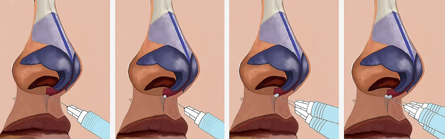 Fig 4. R4P Point 4.1: midline columella base, perpendicular to the nasal spine, 0.05 mL supraperiosteal bolus building the deep columellar/spine pillar that re-projects the tip. · (Silikovich & Kroumpouzos, 2024, p.5)
> Fuentes: Rino 4 Puntos (Silikovich & Kroumpouzos, 2024)
Fig 4. R4P Point 4.1: midline columella base, perpendicular to the nasal spine, 0.05 mL supraperiosteal bolus building the deep columellar/spine pillar that re-projects the tip. · (Silikovich & Kroumpouzos, 2024, p.5)
> Fuentes: Rino 4 Puntos (Silikovich & Kroumpouzos, 2024) [B][MEDLIB] (ref 1).
The columella-base pillar deposit (Point 4) is shown step by step in Fig 4.
Jung dual-plane · 2019 [17] (built to stop dorsal spread and the bulbous nasion): 25G, 70 mm blunt cannula, single infratip-lobule entry. (1) Highly elastic HA into the deep fatty layer, retrograde linear threading tip→radix while lifting the dorsal skin with thumb and finger; (2) moderately elastic HA into the superficial fatty layer, same threading; (3) at the tip/columella, highly elastic HA into the supracartilaginous layer to the anterior nasal spine. Total HA 0.4-1.2 mL.
Woffles Wu / Jones injection rhinoplasty [2]: seated patient; 30G needle perpendicular to bone at the midline radix, small bolus onto bone, then a series of disconnected boluses down the bridge; a columellar strut built by retrograde injection from anterior nasal spine to tip, deep between the medial crura (the columellar arteries run superficially, so stay deep); a tiny intradermal/subdermal midline tip deposit for projection; small paired radix deposits to shape the naso-orbital line. Nerve blocks 0.2 mL lidocaine 2% with adrenaline 1:200 000 for the block only (⚠ the atlas keeps the dorsal working field free of vasoconstrictor to preserve the blanch sign).
Badia [2]: blunt 25G (or wider) cannula for supraperiosteal-supraperichondrial dorsal work + sharp 29G needle for intradermal detail; constant movement, minimal volumes, tip 0.05-0.1 mL; blindness/necrosis explicitly flagged.
CaHA/Radiesse school (Bitar/Erian) [20]: 27G 0.5" needle, top-to-bottom, radix then dorsum then tip; ⚠ non-reversible, retained here only to document the historical technique, not endorsed given the no-antidote rule.
mR4P / VYC-25L nasal-grid schools: current firm-HA (VYC-25L) NSR is validated in a 492-patient series and an expert consensus [18][19]; a modified-R4P extension targets the appearance of mild-to-moderate caudal septal deviation (see D8.11 / Novedades).
Non-injectable alternatives (when the correct answer is not a filler)
| Alternative | When it is the right call |
|---|---|
| Structural rhinoplasty | Reduction of dorsum/hump/tip, airway/septal correction, over-projection, large bulbous tip, failed prior surgery [21][30] |
| Threads | Marketed for tip lift; weak evidence, not a substitute for a support pillar; not recommended over HA here |
| Skin resurfacing / EBD (laser, RF) | Rhinophyma, sebaceous tip texture, telangiectasia; a skin problem, not a shape problem |
| Observation / decline | Reduction-seeker, BDD, planned surgery, operated high-risk nose [2][29] |
Volume ladder and ceiling
| Level | Amount | Source |
|---|---|---|
| Per microbolus | ≤ 0.05 mL | [1][2] |
| Radix (P1) | 0.05-0.15 mL | [1] |
| Supratip (P2) | 0.025 mL | [1] |
| Tip (P3) | 0.05-0.15 mL | [1] |
| Columella pillar (P4) | up to 0.30 mL | [1] |
| Total per session (R4P mean) | 0.65 ± 0.17 mL | [1] |
| Dual-plane range | 0.4-1.2 mL | [17] |
| Ceiling before over-fill | The smallest total that straightens the line; if it looks wider on the frontal view, stop. Complete a deficit at the 2-week review, never the same day [1][2] |
Discrepancy · cannula vs needle (do not average): A (cannula safer): a blunt 22-27G cannula needs greater force to penetrate a vessel wall, travels parallel to the dorsal arteries and stays in a precise plane (Jones/Wu, Badia) [2]. B (cannula not risk-free): a 22G cannula has caused both necrosis and irreversible blindness, and a nasal artery (up to ~1.1 mm) can exceed a cannula's outer diameter; a deep-midline sharp-needle microbolus with aspiration is equally defensible, and in one large series over 75% of nasal filler-blindness cases involved a cannula [2][11]. Decide by: operator plane control and the specific point (radix/tip favour a controlled deep needle bolus; a dorsal threading favours a cannula). The plane, the volume and the pressure outweigh the instrument.
Discrepancy · aspiration before a nasal bolus (do not average): A (aspiration adds a layer): more reliable deep on bone/cartilage; a positive aspirate flags intravascular placement (Carruthers; Bertossi as a checkpoint) [19][27]. B (aspiration is not sufficient): false negatives with viscous filler and fine needles; alone it does not make an unsafe injection safe; rely on blunt cannula + low-pressure microaliquots + real-time ultrasound [16]. Position: aspiration is a low-sensitivity manoeuvre; if you aspirate, know what it measures; if you do not, be able to justify it from the literature, not from habit. What is indefensible is omitting it without being able to say why.
Instrument decision by point (a working compromise, not a rule) [1][2][17]:
| Point | Reasonable default | Why |
|---|---|---|
| Radix | Sharp needle, deep bolus to bone, glabellar compression | Precise depth on bone; small controlled volume [1] |
| Dorsum bridge | Cannula retrograde threading, single entry | Fewer punctures, parallels the dorsal course [17] |
| Supratip | Sharp needle microbolus | Tiny, precise, suprachondrial [1] |
| Tip | Sharp needle, deep to cartilage, minimal | Control at a terminal site [2] |
| Columella pillar | Sharp needle or cannula, deep between crura | Retrograde column to the spine [2] |
Neither instrument makes the nose safe; over 75% of nasal filler-blindness cases in one series involved a cannula [2]. Whatever the tool, the safety comes from the deep midline plane, low pressure, low volume, and stopping to look.
Trampa clásica (D8.7): "finishing it today." Seeing that a little more volume would perfect the line and adding it in the same session. The nose is the worst site for it: short session, small volume, complete at 2 weeks. Chasing perfection in one sitting is how a safe 0.5 mL becomes an over-pressurised 1.2 mL in a closed space [1][2].
Rheology reference (why firm HA and nothing soft)
| Property | What the nose needs | Reason |
|---|---|---|
| G′ (elastic modulus) | High | Must hold a projected line against the tight, recoiling envelope; a soft gel flattens and spreads [1][18] |
| Cohesivity | High | Low cohesivity fragments and spreads laterally, widening the dorsum on the frontal view [1][19] |
| Hydrophilia (water uptake) | Low | Nasal oedema is visible, persistent, and adds pressure in a closed space |
| Extrusion force / gauge match | Controlled | High-G′ gels need adequate gauge; too fine a needle raises injection pressure for the same thumb force |
| Reversibility | Mandatory | The only occlusion treatment is hyaluronidase; a non-HA gel has no antidote [1][20] |
| Particle size (if non-HA) | Excluded | CaHA/PLLA/PMMA 25-45 µm vs capillary 5-15 µm: particles that lodge and occlude [1][20] |
Named firm-HA products used for structural nasal work (private-wiki naming): VYC-25L (Juvederm Volux XC, Vycross, high cohesivity/high G′, validated for NSR in 492 patients and by expert consensus) [18][19]; Restylane firm family (Lyft/Defyne class) for dorsal support; Teosyal PureSense Ultra Deep (Jung deep layer) with PureSense Ultimate for superficial refinement [17]. Soft, low-G′ HA and skin boosters are the wrong tool for the dorsum: they spread and broaden it.
Named-product positioning for the nose (private-wiki naming, evidence separated from marketing):
| Product (brand) | Class / rheology | Nose role | Evidence |
|---|---|---|---|
| VYC-25L (Juvederm Volux XC) | Vycross HA, high cohesivity, high G′ | Structural dorsum/radix/columella pillar | 492-pt NSR series + 2025 expert consensus [18][19] |
| Restylane firm family (Lyft/Defyne class) | HA, high G′ | Dorsal support, radix | School/textbook use [2] |
| Teosyal PureSense Ultra Deep + Ultimate | HA, high + moderate G′ | Dual-plane deep + superficial dorsal refinement | Jung dual-plane [17] |
| Intermediate-G′ HA (R4P points 1-3) | HA, intermediate | Radix, supratip, tip refinement | R4P [1] |
| Radiesse (CaHA) | 25-45 µm microspheres | Historical dorsal augmentation | ⚠ Non-reversible; excluded here [20] |
| PCL / PLLA / PMMA / silicone | Semi/permanent particles | Marketed for durability | ⚠⚠ Rejected; no antidote [1][20] |
| Autologous fat | Graft | Dorsal augmentation | ⚠ ~50% of nose/periforehead blindness; surgical domain [2] |
The rule under the table: the safe product for the nose is defined by reversibility, high G′ and high cohesivity, in that priority order. Everything softer spreads and widens; everything non-HA removes the antidote.
Step-by-step protocol (single session)
- Selection consult. Confirm the goal is achievable by adding volume. If the patient wants a smaller nose, the answer is no, and the consult ends there on indication grounds [2][29].
- Screen for BDD and displaced expectation. The nose leads all regions for this profile; screen and refer as needed [29].
- Directed history: prior nasal surgery (what, when), prior filler (what), trauma, respiratory problems, recurrent skin infection, and any plan for future rhinoplasty [1][17].
- ⚠ Unknown prior filler: assume the worst until proven otherwise; ultrasound before adding anything [16].
- Examination: frontal, lateral, three-quarter, basal, and top-down views; palpate the dorsum; assess skin thickness/quality and tip recoil/support [2][30].
- Standardised photography in all views, identical lighting and position [2].
- Written plan: which point, which plane, which product, expected result, and explicitly what will not be achieved [1].
- Consent naming blindness and necrosis in those words and the effect on any future surgery. A generic consent does not inform of this [2].
- Read the requirements checklist aloud (hyaluronidase in occlusion-protocol quantity, rehearsed protocol, reversible product only, no vasoconstrictor, non-operated nose or explicit higher-risk acknowledgement, no active inflammation/infection, isolated session, 24-h contact). If one is missing, do not inject [2][13].
- Strict antisepsis (sebaceous, high bacterial load, closed space) and topical anaesthesia without vasoconstrictor [2][10].
- Inject: strict midline, deep plane to bone/cartilage contact, microboluses ≤ 0.05 mL, low pressure, slow flow, stopping and looking between deposits; non-dominant-hand glabellar compression at the radix [1][13].
- Tip/columella pillar before dorsum when both are indicated; re-project the tip, then reassess the dorsum, which often needs less than planned [1].
- Mould toward the midline with the fingers against bone, not laterally, to avoid widening the dorsum [2].
- Observe the patient in the room for several minutes before discharge; look for delayed blanching or pain [13].
- Verbal and written aftercare with a 24-h direct contact line; no pressure on the nose, no glasses resting on the dorsum for the first days, no self-massage [2].
- Review at 2 weeks. Complete any deficit then, not the same day [1].
- Record points, plane, product, lot, volume per point and total, and any incident however minor.
Session record template (fill every field) [1][2]:
| Field | Example / note |
|---|---|
| Indication | Low radix + droopy tip; camouflage of mild hump |
| Non-goals stated | No reduction, no alar narrowing, no septal correction |
| Product / lot | High-G′ HA (VYC-25L), lot number |
| Points, planes, volumes | P1 radix 0.10 mL supraperiosteal; P4 columella 0.20 mL; total ≤ 0.65 mL |
| Instrument | 27G needle radix/tip; 25G cannula dorsum |
| Anaesthesia | Topical, no vasoconstrictor |
| Ultrasound | Mapped / not indicated (virgin nose) |
| Hyaluronidase available | ≥ 10 vials / > 1500 IU confirmed in room |
| Adverse events | None / describe and time-stamp |
| Aftercare given | Verbal + written, 24-h contact |
| Review | Booked at 2 weeks |
This record is the medicolegal spine of a nasal treatment: it documents that the reversible-only, deep-midline, hyaluronidase-ready, consented process was followed.
Schools compared (position preserved, not merged)
| School | Thesis | Where it fails |
|---|---|---|
| Conservative structural ✅ | Midline, deep, minimal volume, explicit limited goals (R4P/Jung logic) | Discreet, stable results; disappoints anyone expecting a rhinoplasty [1][17] |
| Ultrasound-guided ✅ | US should be standard in the nose, above all the reoperated nose | Sound rationale; limited by availability and learning curve; ⚠ does not replace judgement [15][16] |
| Needle vs cannula | Each defends its tool as safer | Neither makes the nose safe; plane, volume and pressure outweigh the instrument [2][11] |
| Ambitious volumetric | The whole nose can be remodelled with product | ⚠ Wide dorsums, large noses, the most complications; rejected here [2] |
| Permanent-product dorsum | Longer duration, fewer touch-ups | ⚠ Rejected without nuance: no antidote for occlusion, a very difficult late picture J8 |
| Abstentionist | The nose should not be filled outside very expert hands | Defensible and coherent with the data; this atlas raises the threshold rather than forbidding [2] |
Errors and how to avoid them (technique)
| Error | Why it happens | Fix |
|---|---|---|
| Leaving the midline on the dorsum | Chasing an asymmetry | Vessels are lateral; the midline is what makes it safe [2] |
| Intermediate plane | Losing bone contact without noticing | Deep is safe, intermediate is lethal; advance to contact [3] |
| Injecting the alar rim/lobule | Patient asks to narrow the base | ⚠ Lateral nasal artery, alar necrosis; not an injection site [2] |
| A bolus anywhere in the nose | Wanting yield per puncture | Microboluses, low pressure, stop between deposits [1] |
| Vasoconstrictor anaesthesia | Habit from other regions | ⚠ Masks the blanch sign [2] |
| Soft/low-cohesivity product on the dorsum | Using whatever is open | Spreads and widens the dorsum; use high-G′ [1][19] |
| Non-reversible/permanent product | Chasing duration | ⚠ No antidote for occlusion; indefensible [1][20] |
| "Completing" volume the same day | Seeing a small deficit | Short session, small volume, complete at 2 weeks [1] |
| Excess supratip | Chasing the break point | "Parrot beak"; minimal deposit [1] |
| Combining nose with another high-risk region same day | Optimising the session | You will not know where an event came from [2] |
| Not enough hyaluronidase | "One vial just in case" | Rescue dose is not the nodule dose; ≥ 10 vials/>1500 IU [13][15] |
Risk and avoidance, point by point
| Point | Dominant risk | Avoidance |
|---|---|---|
| Radix | Dorsal nasal a. → ophthalmic (blindness) | Midline only, supraperiosteal to bone, ≤ 0.05-0.15 mL, NDH glabellar compression, low pressure [1] |
| Dorsum (bridge) | Lateral dorsal branches if off midline; widening | Strict midline serial boluses; mould toward the midline, not laterally [2] |
| Supratip | Over-elevation on thin skin ("parrot beak") | 0.025 mL only; light massage of any over-lift [1] |
| Tip | Terminal territory; necrosis and embolism | Deep to cartilage, tiny volume, low pressure; watch colour in real time [2] |
| Columella | Columellar a. (subcutaneous) | Deep between the medial crura, not on the surface; retrograde pillar to the spine [2] |
| Nasal spine / base | Support point | Supraperiosteal on the spine; a support deposit, not a superficial fill [1] |
| ⚠ Alar rim / lobule | Lateral nasal a., alar necrosis | Do not inject; alar width is not corrected with filler [2] |
| ⚠ Soft triangle | No subcutaneous layer | Absolute no-inject zone [8] |
Moulding and finishing: after each deposit, mould with the fingers against the bone/cartilage, pushing product to the midline; reassess from the frontal view for widening and from above for deviation before the next deposit; preserve a little dorsal angularity rather than an over-straight line [1][2]. Keep the field free of vasoconstrictor so a spreading blanch is visible immediately.
D8.8 · Toxin of the nose
All nasal toxin is off-label and low-volume; doses below are onabotulinumtoxinA (ona) unless a conversion is stated. Start at the low end of every range and review at 2 weeks: you can add, you cannot remove [22][24].
| Target (muscle) | Dose (ona) | Points | Plane | Antagonist / ⚠ |
|---|---|---|---|---|
| Bunny lines (transverse nasalis / compressor naris) | 1.25-5 U per side (2.5-5 U Botox/Xeomin, 7.5-15 U Dysport) [24][25] | 1-2 per side on the lateral nasal wall (± 1 dorsal point), within the "bunny-line safety zone" | Subdermal / intramuscular, needle 45-60° | Cannot be fully abolished (LLSAN fibres persist) [7]; ⚠ do not drift inferiorly onto levator labii superioris → lip ptosis |
| Drooping / plunging tip on smile (depressor septi nasi) | 2-4 U at the columella base, 1-2 mm above the labiocolumellar junction; +2-4 U mid-columella if strong [22][23] | 1 point at columella base ± mid-columella | Intramuscular, deep, single site | Only if the tip drops on smile/animation; if no dynamic drop, do not inject [22][26] |
| Nasal tip elevation (Atamoros scheme) | 6 U slight (2 U each dilator naris + 2 U depressor septi), 12 U medium, 18 U high [22][23] | dilatores naris (± depressor septi), bilateral | Intramuscular | Effect lasts 3-5 months; the alar fibres of LLSAN compensate over time [23] |
| Alar flaring (dilator naris) | 2-5 U per side [26] | 1 per side, lower nasal fibres above the lateral ala | Intramuscular | ⚠ Watch the nasal airway (do not weaken so much that the valve collapses) |
| Gummy smile component that involves the nose (LLSAN) | 1-3 U per side [23][26] | lateral to the ala, at the nasolabial origin | Very superficial | ⚠ The lowest therapeutic margin of the whole face; overdose → long, immobile, flattened upper lip for weeks; classify the smile type first (only levator-hyperactivity responds) |
Dose scheme worth memorising (tip elevation, Atamoros / Benedetto) [22][23]: slight = 6 U (2+2 dilator naris, 2 depressor septi); medium = 12 U; high = 18 U. Dayan and Kempiners: 5 U into each depressor septi + 3 U into each LLSAN widened the nasolabial angle from 110° to 115°, de-rotated less, and softened the mid-philtral crease [23]. Baran: nasal tip droop 2-6 U ona (5-10 speedywood units Dysport) at the columella base [26].
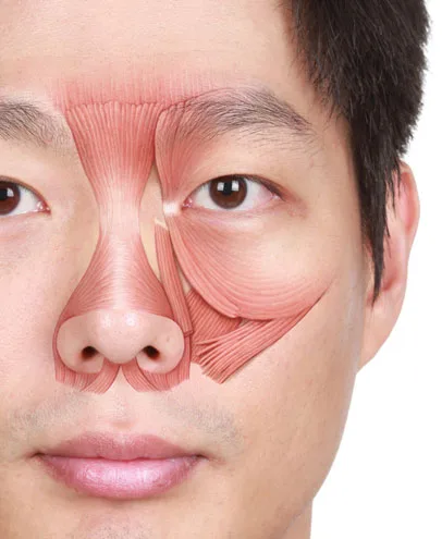 Fig 5. The muscles that a nasal toxin plan targets, all in the fibromuscular (SMAS) layer: nasalis (transverse = compressor, alar), depressor septi nasi, dilator naris, and LLSAN. Note the LLSAN runs from the medial canthus to the ala and lip, which is why a bunny-line dose that drifts down causes lip ptosis. · (Kim, 2016, p.80)
> Fuentes: Kim - Clinical Anatomy for Botulinum Toxin and Filler 2016 9789811002380
Fig 5. The muscles that a nasal toxin plan targets, all in the fibromuscular (SMAS) layer: nasalis (transverse = compressor, alar), depressor septi nasi, dilator naris, and LLSAN. Note the LLSAN runs from the medial canthus to the ala and lip, which is why a bunny-line dose that drifts down causes lip ptosis. · (Kim, 2016, p.80)
> Fuentes: Kim - Clinical Anatomy for Botulinum Toxin and Filler 2016 9789811002380 [C][MEDLIB].
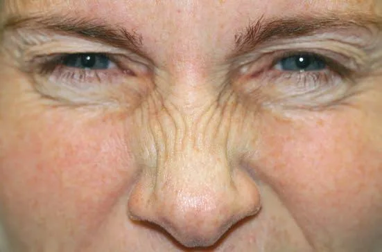 Fig 6. Bunny lines on nasalis contraction. Injection points sit on the lateral nasal wall within the safety zone (1.25-5 U/side); the dose must stay above the levator labii to avoid lip ptosis. · (Small, 2012, p.89)
> Fuentes: Small - Practical Guide to Botulinum Toxin Procedures 2012 9781609131470
Fig 6. Bunny lines on nasalis contraction. Injection points sit on the lateral nasal wall within the safety zone (1.25-5 U/side); the dose must stay above the levator labii to avoid lip ptosis. · (Small, 2012, p.89)
> Fuentes: Small - Practical Guide to Botulinum Toxin Procedures 2012 9781609131470 [C][MEDLIB].
The antagonist you must NOT touch: the levator labii superioris (superioris alaeque nasi). A bunny-line or alar dose placed too low or too heavy weakens lip elevation and produces upper-lip ptosis and an asymmetric smile, one of the most distressing toxin complications because it is visible in every social expression [22][24]. Keep bunny-line points high on the nasal wall, and confirm the depressor-septi indication (dynamic tip drop) before treating it. The target muscles and the LLSAN course that dictates this rule are shown in Fig 5.
The dynamic bunny-line creasing that defines the lateral-wall indication is shown in Fig 6.
Consensus: nasal toxin is an adjunct for dynamic problems (mimetic tip drop, bunny lines, flare); it cannot lift a statically ptotic tip, which is filler/surgical territory [22][23]. Discrepancy: none clinically decisive on nasal doses; ranges overlap and titration resolves them. Do not average across the Botox/Dysport unit systems (they are not interchangeable) [26].
Trampa clásica (D8.8): promising tip elevation from toxin when the tip does not move on smile. The depressor-septi injection only helps a dynamic drop; on a fixed, gravitationally ptotic tip it does nothing and the patient feels deceived. Test the smile first; if the tip does not plunge, the tool is filler support, not toxin [22].
Unit systems are not interchangeable (do not convert by averaging) [26]: the doses above are onabotulinumtoxinA (ona; Botox/Vistabel). Xeomin (incobotulinumtoxinA) is used at roughly the same units as ona; Dysport (abobotulinumtoxinA) runs about 2.5-3× the ona unit (bunny lines 7.5-15 U Dysport vs 2.5-5 U ona; nasal-tip droop 5-10 speedywood-equivalent Dysport vs 2-6 U ona) [24][25][26]. Choose one product's scheme and stay in it; never mix unit systems within a plan.
Bunny-line safety zone and mechanics [24]: with the nasalis contracted, place 1-2 points per side on the lateral nasal wall just medial to the nasojugal line, within the safety zone, needle 45-60° to the skin and subdermal/intramuscular; a third point over the dorsum if the transverse fibres cross the midline. Keeping the points high avoids the levator labii and the risk of lip ptosis. Note the compensation phenomenon: treating only the glabella can reveal previously masked bunny lines through compensatory nasalis recruitment, so assess the nose when planning glabellar toxin [22].
Gummy-smile classification (only the muscular type responds) [26]: classify by the area of gingival exposure before treating: anterior, posterior, mixed, asymmetric. Only the component driven by levator hyperactivity (LLSAN/levator labii) responds to toxin; exposure from vertical maxillary excess or a short clinical crown does not, and promising it is the classic indication error. If the upper lip is already long, the patient is not a candidate. Start at the lowest dose (1-3 U/side into the LLSAN, very superficial) and review at 2 weeks: overdose gives a long, immobile, flattened upper lip for weeks, the lowest-margin complication of the face [23][26].
Nasal-tip elevation mechanics (why it works and its ceiling) [22][23]: the depressor septi nasi pulls the tip down on animation; relaxing it (and, in the Atamoros scheme, the dilator naris/anterior alar fibres) lets the unopposed levator/tip-elevator vectors lift and project the tip. Effect lasts 3-5 months; over time the alar (medial) fibres of the LLSAN compensate. Dayan and Kempiners quantified the effect: 5 U into each depressor septi + 3 U into each LLSAN widened the nasolabial angle from 110° to 115° and softened the mid-philtral crease. If the tip does not move on smile, no dose will help; the tool is then filler support, not toxin.
Toxin errors and how to avoid them:
| Error | Why | Fix |
|---|---|---|
| Bunny-line dose drifts low | Chasing lateral fibres | Keep points high on the nasal wall; spare the levator labii [22] |
| Treating a static tip with depressor-septi toxin | Not testing the smile | Only a dynamic drop responds; test first [22] |
| Promising toxin for non-muscular gummy smile | Not classifying the type | Classify (anterior/posterior/mixed); only levator hyperactivity responds [26] |
| Mixing Botox and Dysport units | Habit/stock | Stay in one unit system [26] |
| Over-weakening the dilator naris | Chasing alar flare | ⚠ Watch the nasal airway/valve [26] |
D8.9 · Combination and sequence → L2 · Combination & Sequencing
Liquid rhinoplasty = HA (dorsum/radix/tip/columella) + toxin (dynamic tip and flare), inside full-face harmony. The perceived size of the nose is relative to chin and lip projection, so the nose is planned as part of a profile, not in isolation [1][29].
| Combination | Sequence & interval | Rationale |
|---|---|---|
| Toxin + filler, same visit | Toxin first, then filler; treat the depressor septi (dynamic tip drop) before building the columellar pillar | Relaxing the depressor lets the filler pillar re-project a tip that is no longer being pulled down [22][23] |
| Nose + chin / lip (profiloplasty) | Often chin/lip before or with the nose | A retruded chin and thin lips make the nose look larger; balancing them can make the nose look smaller without touching it (medical profiloplasty) [29] |
| Radix vs tip within the nose | Tip/columella pillar first, reassess dorsum after | Re-projecting the tip optically shortens the nose and often removes the need for dorsal volume [1] |
| Touch-up | At ~2 weeks, never the same day | Nose settles; complete any deficit at review to avoid same-session over-pressurisation [1][2] |
| Nose + another high-risk region same day | ⚠ Avoid | If a vascular event occurs, you must know which region it came from; isolate the nose in its own session [2] |
| After ultrasound mapping | Map first (esp. operated/filled nose), then inject | Confirms plane and vessel course before any deposit [16] |
| Filler then future surgery | ⚠ Not combinable; remove HA with hyaluronidase before surgery | Residual HA distorts dissection and fibrosis [1][17] |
Sequencing logic (P): the nose is the last thing you touch on a high-risk day and the first thing you isolate. Because it is the region where an adverse event is both most likely and most catastrophic, it earns its own session, its own consent, and its own 24-hour contact line, and it is never batched with the glabella or the tear trough on the same day. (P) reasoning, no dose implied.
Interval and longevity: R4P effect held a median of 11 months [1]; firm-HA (VYC-25L) long-term data support durability with periodic touch-ups [18][19]; toxin adjuncts last 3-5 months and are re-dosed independently of the filler [23]. Full sequencing across regions and modalities lives in L2 · Combination & Sequencing.
Trampa clásica (D8.9): injecting filler first and toxin second. If you build the columellar pillar while the depressor septi is still pulling the tip down, the result under-projects and de-rotates once you finally relax the muscle; relax the depressor first, then support [22].
Medical profiloplasty (the nose is judged relative to the profile) [29]: a retruded chin and thin lips make the same nose look larger; balancing the chin projection and lip support can make the nose read as smaller and straighter without a single nasal deposit. In a low-bridge phenotype the sequence is often chin/lip first (or together), then a conservative radix/dorsum. Treat the profile, not the isolated organ.
Interval logic (why 2 weeks, not the same day) [1][2]: nasal filler settles and any post-injection oedema resolves over ~2 weeks; completing a deficit at review avoids stacking volume and pressure in a closed space on the same day, which is precisely the setup for compression ischaemia. The nose is the region where "finish it today" is the worst instinct.
Longevity and re-dosing by modality:
| Modality | Effect duration | Re-dose logic |
|---|---|---|
| Firm HA (structural) | R4P median ~11 months [1]; VYC-25L durable with periodic touch-ups [18][19] | Reassess at 9-12 months; top up conservatively |
| Toxin (depressor septi, bunny lines, flare) | 3-5 months [23] | Independent of the filler cycle |
| Combined liquid rhinoplasty | Filler sets the shape; toxin the dynamic component | Stagger so each is judged on its own timeline |
Common combination scenarios (what to do, in order):
| Scenario | Plan and order |
|---|---|
| Low radix + droopy tip | Tip/columella pillar first (filler) + depressor-septi toxin; reassess radix, add conservatively [1][22] |
| Dorsal hump + plunging tip on smile | Depressor-septi toxin first; then radix + supratip camouflage; the straighter line masks the hump [1][22] |
| Flat dorsum + retruded chin (ethnic profiloplasty) | Chin/lip projection first (or same visit), then firm-HA dorsum; the balanced profile reads as a smaller nose [29] |
| Post-rhinoplasty minor irregularity + thin skin | Ultrasound map first; tiny deep deposits; ⚠ higher event threshold in scarred tissue [16] |
| Bunny lines + dorsal augmentation | Toxin to nasalis first (relax dynamic creasing), then dorsal filler at a later or same visit above the toxin field [24] |
| Alar flare + tip droop | Dilator-naris + depressor-septi toxin; ⚠ watch the nasal airway [26] |
What not to combine on the same day: the nose with the glabella or the tear trough (both high-risk); a large filler session with a first-ever nasal treatment; and filler with a same-day plan for surgery. Isolate risk so an event's source is knowable [2].
Cross-references: whole-face sequencing and inter-region intervals live in L2 · Combination & Sequencing; the chin/lip profiloplasty targets are in D3 · Midface & Cheeks and the lower-face chapters. Keep the nose an isolated session on any high-risk day.
D8.10 · Region-specific complications → J1 · Complications Overview · J2 · Vascular Occlusion & Emergency Response · J8 · Biopolymer & Permanent-Filler Complications
Not the generic filler complications: the ones that happen here because the nose is a terminal territory under a tight, sebaceous envelope with a direct arterial line to the eye.
Pooled epidemiology (the numbers that anchor consent) [12] (DeVictor meta, 8604 NSR patients):
| Event | Rate |
|---|---|
| Overall adverse | 2.52% |
| Bruising | 1.58% |
| Vessel occlusion | 0.35% (30 episodes) |
| Vision loss | 0.09% (8) |
| Skin necrosis | 0.08% (7) |
| Infection | 0.07% (6) |
| Haematoma | 0.13% |
Low rates, catastrophic events: nasal augmentation remains the first cause of filler-related nasal necrosis and the second cause of filler-related visual loss [2][9]. A larger 2024 NSR update reported severe vascular events (blindness/necrosis/stroke) around 0.27% with very high satisfaction, underscoring that the risk is small but never zero [18][37].
Region-specific complication grid:
| Complication | Why it is nasal | Recognition | First action |
|---|---|---|---|
| Vascular occlusion / embolism | Columella (highest embolisation risk) and dorsum (compression ischaemia, worse in fibrotic operated noses) | Immediate blanching along an arterial territory, disproportionate pain, dusky/violaceous congestion, delayed capillary refill, livedo, a transudate droplet | Stop. Full occlusion protocol → HDPH (below), J2 · Vascular Occlusion & Emergency Response |
| Blindness / stroke | Retrograde to ophthalmic → central retinal ± cerebral arteries | Sudden severe pain, blurred vision, hemianopia, decreased acuity, ptosis; may be immediate or up to ~1 h delayed | 90-minute ocular emergency: stop, ophthalmology/stroke code, retrobulbar hyaluronidase (below) [11][14] |
| Skin necrosis (tip, ala, dorsum) | Terminal territory, tight non-distensible skin, pressure that does not dissipate; more superficial + thicker/harder/tighter skin + larger volume + small-gauge = higher risk | Branching blanching → violaceous → eschar; pustules can mimic infection | Hyaluronidase flooding + supportive care [10][16] |
| Tyndall (blue-grey) | Thin nasal/dorsal skin over superficial HA | Bluish discoloration, superficial deposit | Hyaluronidase to the deposit; avoid superficial placement |
| Nodules / granuloma | Sebaceous, bacteria-rich, closed space; worse with non-HA/PMMA | Firm lump ± inflammation ± late onset | HA: hyaluronidase; non-HA → J8 · Biopolymer & Permanent-Filler Complications |
| Migration / over-correction | Spread of low-cohesivity product; widened dorsum | Broadened bridge on the frontal view | Hyaluronidase to reshape; use high-G′ next time |
| Infection / biofilm | High sebaceous bacterial load, closed nasal space | Delayed erythema, pain, fluctuance | Antibiotics; drain; consider biofilm; avoid steroid-first |
| ⚠ Post-surgical nose compression ischaemia | Fibrotic, low-compliance tissue tolerates less volume/pressure | Ischaemia at lower volumes than expected | Lower threshold to stop and treat; pre-scan [16] |
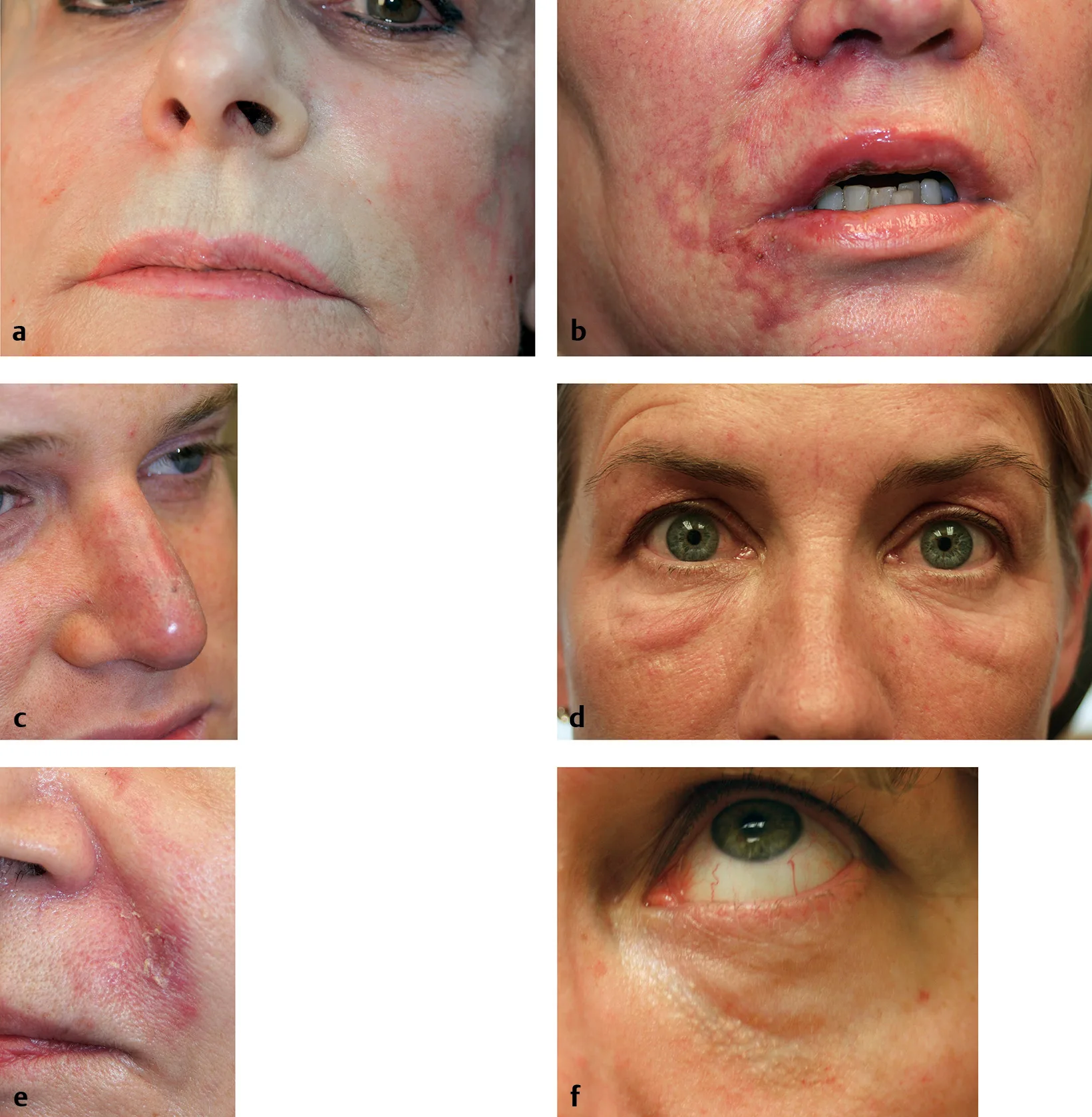 Fig 7. The signs to read in real time. Panel (a) is the acute blanch that adrenaline in the anaesthetic would mask; panel (c) is a vascular injury in a previously operated nose, the separate risk category; panel (f) is the Tyndall of superficial product. · (Kontis, 2019, p.210)
> Fuentes: Kontis - Cosmetic Injection Techniques 2019 9781626234574
Fig 7. The signs to read in real time. Panel (a) is the acute blanch that adrenaline in the anaesthetic would mask; panel (c) is a vascular injury in a previously operated nose, the separate risk category; panel (f) is the Tyndall of superficial product. · (Kontis, 2019, p.210)
> Fuentes: Kontis - Cosmetic Injection Techniques 2019 9781626234574 [C][MEDLIB].
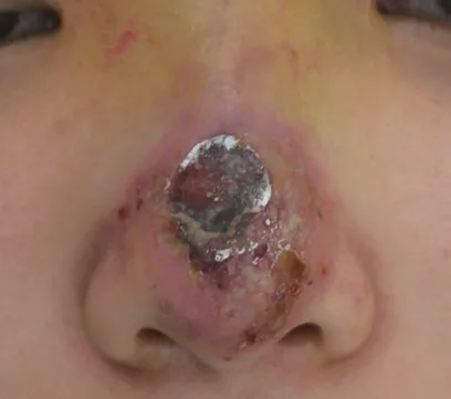 Fig 8. Nasal-tip necrosis: the terminal-territory catastrophe. Necrosis here follows pressure/embolism in the columellar and lateral-nasal territories; note the demarcated eschar. Non-HA product (as here) has no antidote, which is why the nose accepts only reversible filler. · (Filler Complications, 2019, p.85)
> Fuentes: Filler Complications, Filler-Induced Hypersensitivity Reactions 2019 ISBN 978-981-13-6639-0
Fig 8. Nasal-tip necrosis: the terminal-territory catastrophe. Necrosis here follows pressure/embolism in the columellar and lateral-nasal territories; note the demarcated eschar. Non-HA product (as here) has no antidote, which is why the nose accepts only reversible filler. · (Filler Complications, 2019, p.85)
> Fuentes: Filler Complications, Filler-Induced Hypersensitivity Reactions 2019 ISBN 978-981-13-6639-0 [C][MEDLIB].
Vascular-occlusion management (nasal specifics) → J2 · J3
Recognise early. Branching blanching, disproportionate pain, delayed capillary refill, livedo, dusky congestion, a transudate droplet at the puncture; the clinical spectrum (acute blanch, purpura, a post-surgical nasal injury, Tyndall) is documented panel by panel in Fig 7. Re-evaluate any suspicious sign at ≥ 60 minutes; do not "wait and see" [13][14][16].
High-Dose Pulsed Hyaluronidase (HDPH) · DeLorenzi protocol [13]:
| Parameter | Value |
|---|---|
| Dose | 500 IU per affected area; 1000 IU for two areas; 1500 IU for three [13] |
| Nose specifically | ~900 IU (a low-volume event of the nose, cf. ~450 IU for half a lip) [13][14] |
| Frequency | Inject every 60-90 minutes until skin colour and capillary refill normalise [13] |
| Massage | Massage to spread the enzyme and drive the embolus distally into thin-walled arterioles [13] |
| Window | Aim to complete within 72 hours of onset; soft tissue is relatively ischaemia-resistant [13] |
| Adjuncts | Warm compress, aspirin (300-650 mg) [14][15]; van Loghem: 500 IU per 5×5 cm affected skin, hourly, reconstitute in lidocaine (not epinephrine) for vasodilation [16] |
| ⚠ Nitroglycerin | Do not apply until the offending HA is dissolved (day 2-3); early dilation of adjacent pathways can propagate the embolus toward the orbit [13] |
| Ultrasound-guided | DUS-guided hyaluronidase restores flow with far less enzyme (van Loghem "Medusa head", 50-100 IU) [16] |
Ocular emergency (visual symptom = stop everything) [11][14]: ~90 minutes to irreversible retinal ischaemia. Stop, activate ophthalmology/stroke code, treat the injected field and surroundings with hyaluronidase, and consider retrobulbar hyaluronidase: Carruthers technique, 300-600 units (2-4 cm³) via a 25G needle ≥ 1 inch deep in the inferolateral orbit [15]; Fakih-Gomez cites ~800 units [14]; Pirayesh 300-1500 IU every 2-4 h, retrobulbar 8 mm below the inferior tarsal strip along the orbital floor to ~3 cm, or 1 cm above the medial canthus directed 30° medially [3]. ⚠ Retrobulbar hyaluronidase remains debated (efficacy not established); it does not replace immediate ophthalmology transfer [11][14].
Ocular-emergency step sequence (rehearse it, do not read it live) [11][14][15]:
- Stop injecting at the first visual symptom (blur, scotoma, pain, diplopia). Do not complete the deposit.
- Note the clock. ~90 minutes to irreversible retinal ischaemia.
- Flood the injected field and surroundings with hyaluronidase (it can diffuse across vessel walls) [15].
- Activate ophthalmology / oculoplastics and a stroke pathway simultaneously; arrange immediate transfer.
- Consider retrobulbar hyaluronidase if trained: 300-800 IU via a 25G needle/cannula ≥ 2.5 cm into the inferolateral orbit (or 1 cm above the medial canthus, 30° medially along the medial wall) [3][14][15]. ⚠ Do not let this delay transfer; efficacy is unproven.
- Lower intraocular pressure en route if directed: ocular massage, acetazolamide, mannitol, anterior-chamber paracentesis by the specialist [15].
- Document product, volume, site, timeline and every intervention.
Clinic emergency kit (nose-ready) [14][15]: hyaluronidase stock ≥ 10 vials / > 1500 IU, aspirin, warm compress, a written and rehearsed occlusion algorithm, ophthalmology and stroke pathways, and a 24-hour patient contact line. The rescue dose is not the nodule-dissolution dose; one vial "just in case" is not a protocol [13][15].
Consensus: flood the entire ischaemic territory with hyaluronidase, hourly, until capillary refill normalises; time is the prognostic factor [13][15][16]. Discrepancy · retrobulbar hyaluronidase for blindness: some report vision recovery, others none; the enzyme can diffuse across vessel walls but the evidence is inconsistent [11][14]. Preserve both positions; never delay ophthalmology transfer to attempt it.
Trampa clásica (D8.10): using anaesthetic with a vasoconstrictor and then waiting to see whether a pale patch recovers. Adrenaline blanches the field and hides the ischaemic sign you most need; and "waiting to see" spends the 60-90 minute window. Keep the working field free of vasoconstrictor, and treat a spreading arterial-pattern blanch as occlusion until proven otherwise [2][13].
Differential of the acute nasal event (do not confuse them)
| Sign | Occlusion / ischaemia | Benign |
|---|---|---|
| Blanching | Spreads along an arterial territory, persists | Transient pressure/volume pallor, recovers at once |
| Pain | Disproportionate, sudden | Proportionate to the puncture |
| Colour | Dusky/violaceous, livedo over hours | Normal, mild transient erythema |
| Capillary refill | Delayed in a defined area | Normal |
| Course | Worsens; eschar risk | Resolves |
| Action | Stop, occlusion protocol [13] | Observe, do not ignore |
Distinguishing the three swellings (they are treated oppositely):
| Feature | Vascular occlusion | Infection / biofilm | Delayed hypersensitivity |
|---|---|---|---|
| Onset | Immediate to ~1 h | Days (early) to delayed | Days to months |
| Pain | Severe, disproportionate | Tender, throbbing | Mild, itchy |
| Colour | Blanch → violaceous, livedo | Erythema, warmth, ± pus | Erythema, oedema |
| Pattern | Arterial territory, branching | Localised to fluctuant | Diffuse over the product |
| First move | Hyaluronidase flooding [13] | Antibiotic ± drainage; ⚠ not steroid-first [10] | Hyaluronidase ± anti-inflammatory [15] |
| ⚠ Trap | Calling it infection and giving steroid | Calling it hypersensitivity and giving steroid into pus | Calling it occlusion and missing the immune trigger |
Late and delayed complications (weeks to months): delayed-onset inflammatory nodules (immune-mediated, can appear months later, sometimes after an immunological trigger), granuloma, biofilm-related low-grade infection, migration/broadening, persistent Tyndall over thin dorsal skin, and hypertrophic scar after a necrosis episode [10][15][16]. HA nodules are managed by hyaluronidase (± anti-inflammatory or antibiotic depending on whether the picture is filler, inflammatory or infective); non-HA nodules follow the biopolymer pathway [15]. ⚠ Infection first, steroid later: injecting a steroid into an infected nodule worsens it.
Biofilm and infection (the sebaceous closed space) [10][15][35]: the nasal skin's high sebaceous bacterial load and the closed nasal space make infection more frequent and worse than expected. Suspect biofilm in a delayed, recurrent, low-grade inflammatory nodule that does not settle; treat with appropriate antibiotics (tetracyclines have anti-inflammatory value), consider drainage, and remove HA with hyaluronidase; avoid steroid-first. Strict antisepsis and avoiding injection through active acne or infection are the prevention.
Prevention bundle (the sum of the chapter's safety logic): reversible product only; strict deep midline; microboluses, low pressure, low flow; non-dominant-hand control; no vasoconstrictor (preserve the blanch sign); stop-and-look between deposits; small total volume, single session, complete at 2 weeks; ultrasound in the operated/filled nose; hyaluronidase in occlusion-protocol quantity; rehearsed protocol and 24-hour contact; consent naming blindness and necrosis; and isolate the nose on any high-risk day [1][2][13][15][16].
Trampa clásica (D8.10): calling an early ischaemic nose an infection because of pustules, and starting a steroid. Pustular, erythematous swelling can be the ischaemic picture; a steroid into an evolving occlusion loses the window. Read the pattern (arterial-territory blanching and pain point to occlusion) and treat with hyaluronidase first [13].
Necrosis staging and prognosis (nasal tip/ala) [10]
Filler necrosis in the nose usually follows pressure/compression more than a direct needle puncture (Fig 8 shows an established nasal-tip eschar); risk rises with more superficial injection, thicker/harder/tighter skin, larger volume, greater swelling and a smaller needle diameter [10]. Two patterns: extended necrosis (large plexus, wide whitish change, branching/spreading redness) versus localised necrosis (small plexus, localised whitish change, homogeneous redness) [10].
| Timeline (untreated/late) | Finding |
|---|---|
| Immediate (0-2 days) | Blanching, then branching or homogeneous redness by pattern |
| ~2-3 days | Pustules, violaceous/"red-wine" skin; infection and necrosis declare |
| ~3-7 days | Eschar at the tip/ala; demarcation |
| ~10-15 days | Yellow necrotic tissue requiring removal; debridement |
| ~3-5 weeks | Epithelialisation, reducing defect |
| ~4 months | Recovery; scar may be minimal if treated early, hypertrophic if late |
The prognosis lever is time to hyaluronidase, not the eschar. Early flooding (hours, not days) converts a would-be full-thickness loss into a mild, recoverable event; the same picture treated late leaves a hypertrophic scar [10][13]. Non-HA product (permanent filler) has no antidote, which is why the necrosis figure in this chapter is a permanent-filler case and why the nose accepts only reversible product [10].
Adjunct pharmacology in the nasal occlusion (beyond hyaluronidase)
| Agent | Role | Note |
|---|---|---|
| Aspirin | Antiplatelet, limits clot around the embolus | 300-650 mg; UFEK 300 mg then 100 mg/24 h [14][15] |
| Warm compress + massage | Vasodilation, drives enzyme distally | Continuous during treatment [13] |
| ⚠ Nitroglycerin paste | Vasodilation | Not until the HA is dissolved (day 2-3); early use can propagate the embolus toward the orbit [13] |
| Corticosteroid (IM dexamethasone / oral prednisolone) | Anti-inflammatory | Consider in evolving ischaemia; ⚠ not first-line into a suspected infection [14] |
| Antibiotic (amoxicillin-clavulanate) | Secondary infection of necrotic tissue | Start if infection supervenes [14][16] |
| LMWH / pentoxifylline / diltiazem | Rheology/vasospasm adjuncts, low evidence | Late VO adjuncts [14][16] |
| Hyperbaric oxygen | Tissue oxygenation of ischaemic skin | 2-3 atm, low evidence, adjunctive [14] |
| Collagenase ointment | Debridement of established eschar | Daily wound care in late necrosis [14] |
Aftercare and follow-up (nose-specific)
Written and verbal instructions: watch for increasing pain, colour change, blanching, blisters or any visual symptom; contact immediately at any hour (do not wait until morning); no pressure on the nose; no glasses resting on the dorsum for the first days; no self-massage; report fever or spreading redness [2].
Follow-up schedule: observe in the clinic several minutes before discharge; a 24-hour contact line stays open; formal review at 2 weeks (complete any deficit then, assess line/symmetry from the frontal and bird's-eye views); reassess durability at 9-12 months. If a vascular event occurred, follow the J2 · Vascular Occlusion & Emergency Response timeline (retreat hourly, reassess at 48 h, wound care to weeks).
Signs → action table (post-procedure):
| Sign | Interpretation | Action |
|---|---|---|
| Immediate blanching following an arterial territory | Occlusion until proven otherwise | Stop; full protocol [13] |
| Disproportionate/sudden pain | Ischaemia | Stop, evaluate |
| Any visual symptom (blur, scotoma, ocular pain, diplopia) | Ocular emergency | Protocol + urgent ophthalmology; no margin [11][14] |
| Delayed capillary refill in a defined area | Perfusion compromise | Stop, evaluate |
| Livedo reticularis over hours | Late vascular compromise | Contact and in-person review |
| Transient pressure pallor that recovers at once | Usual | Watch, do not ignore |
Coverage vs UPO
Retrieved with collection_prefixes=["Aesthetic_Medicine/UPO Sorted"]. UPO is the fastest-ageing lane; a dose resting on a single UPO slide is never_sufficient_alone and is corroborated against dated external evidence below.
| UPO teaches | State in this chapter | What the atlas adds |
|---|---|---|
| Rinomodelación: dorsum/tip filler basics (T9.4) | ✅ Full | Named-school grid (R4P, Jung, Wu, Badia) with exact points, planes and volumes [1][2][17][20] |
| Nasal danger zones (dorsal nasal artery) | ✅ Full | Depth/variant map, choke vs true anastomoses, ECA↔ICA multi-route mapping, "no safe zones" argument [9][15] |
| Acute ischaemia / vascular occlusion (Dra Tejero, T10) [34] | ✅ Full | HDPH exact dosing (500/1000/1500 IU, nose 900 IU), 72-h window, nitroglycerin caveat, US-guided flooding [13][14][16] |
| Hyaluronidase in complications | ✅ Full | Retrobulbar dosing/technique (300-800 IU), the retrobulbar controversy, emergency-kit stock (≥10 vials) [3][14][15] |
| Filler product classes | ✅ Full | Rheology grid + the reversible-only rule and the particle-size rationale for excluding CaHA/PLLA/PMMA [1][20] |
| Toxin of the mid-face/nose (bunny lines, tip) | ✅ Full | Atamoros tip-elevation scheme (6/12/18 U), depressor-septi and flare dosing, the levator-labii antagonist rule [22][23][24][26] |
| NSR-specific complication epidemiology | ➕ Added (external) | DeVictor meta 8604 pts: 2.52% overall, VO 0.35%, vision loss 0.09%, necrosis 0.08% [12] (not in UPO) |
| Current firm-HA validated for NSR (VYC-25L) | ➕ Added (external) | Jalali 492-pt series + Bertossi 2025 consensus [18][19] (post-dates most UPO material) |
| Ultrasound-guided injection and rescue | ➕ Added | Pre-injection mapping and "Medusa head" US-guided hyaluronidase [16][28] |
| Assessment angles / photography | ➕ Added | Nasofrontal/nasolabial/Goode reference ranges, five-view + basal + bird's-eye protocol [29][30] |
| Aging of the nose (order of change) | ➕ Added | Bone/pyriform regression, scroll/crural support loss, support-first logic [5][21] |
UPO topics not left uncovered: every UPO theme above is present. No UPO slide is used as the sole support for a dose; each is corroborated by a dated external source.
Self-assessment
Ten active-recall questions built only from facts already in this chapter.
- Which is the only filler class permitted in the nose, and why?
Answer
HA only, because hyaluronidase is the sole antidote to a nasal vascular occlusion; CaHA/PLLA/PCL/PMMA/silicone are non-reversible and their 25-45 µm particles far exceed capillary calibre (5-15 µm) [1][20]. - What is the safe injection plane and why is "intermediate" dangerous?
Answer
Deep, strict midline: supraperiosteal on bone, supraperichondrial on cartilage. The arterial network runs on the surface of the fibromuscular (SMAS) layer, so the intermediate plane is the arterial plane [2][3]. - State the R4P points, planes and volumes.
Answer
P1 radix 0.05-0.15 mL supraperiosteal; P2 supratip 0.025 mL suprachondrial; P3 tip 0.05-0.15 mL deep fat; P4 columella up to 0.30 mL; mean total 0.65 ± 0.17 mL [1]. - Which artery is the anatomical author of nasal-filler blindness?
Answer
The dorsal nasal artery, a terminal branch of the ophthalmic artery (internal carotid); retrograde flow reaches the central retinal artery [4][9]. - HDPH dosing and the nose-specific figure.
Answer
500 IU per area, 1000 IU for two areas, 1500 IU for three; ~900 IU for the nose; hourly (every 60-90 min) until capillary refill normalises; complete within 72 h [13][14]. - Why no vasoconstrictor in the anaesthetic field?
Answer
Adrenaline blanches the field and masks ischaemic blanching, the earliest sign of occlusion [2]. - Depressor septi nasi toxin: dose, site, and the indication test.
Answer
2-4 U at the columella base (1-2 mm above the labiocolumellar junction), only if the tip drops on smile (dynamic ptosis) [22][23]. - Which muscle must a bunny-line dose spare, and what happens if not?
Answer
The levator labii superioris (alaeque nasi); a low or heavy dose causes upper-lip ptosis and an asymmetric smile [22][24]. - The pooled NSR complication rates.
Answer
Overall 2.52%, vessel occlusion 0.35%, vision loss 0.09%, skin necrosis 0.08% across 8604 patients [12]. - Why treat the tip before the dorsum, and why never the same-day completion?
Answer
Re-projecting the tip optically shortens the nose and often removes the need for dorsal volume; same-day completion stacks volume and pressure in a closed terminal space, the setup for compression ischaemia. Complete a deficit at 2 weeks [1][2].
What's new and trends (2022-2025)
| Year | Change | Reference |
|---|---|---|
| 2024 | Rino-4-Puntos (R4P) formalised as a standardised 4-point NSR technique (400 pts): intermediate + high-G′ HA, ≤ 0.05 mL midline deep boluses, mean 0.65 mL, median 11 months, no vascular complications | [1] |
| 2024 | VYC-25L (Juvederm Volux) validated specifically for NSR in a 492-patient real-world series (single injector), consolidating firm, high-cohesivity HA as the structural nasal filler | [18] |
| 2025 | Global expert consensus on VYC-25L for safe, effective long-term structural aesthetic use, including the nose | [19] |
| 2021 | First NSR-specific complication meta-analysis (8604 pts): overall 2.52%, VO 0.35%, vision loss 0.09%, necrosis 0.08%, giving numbers for consent | [12] |
| 2023-2025 | Ultrasound moves from optional to expected in high-risk/reoperated noses: pre-injection vessel mapping and US-guided hyaluronidase ("Medusa head", 50-100 IU) reduce enzyme dose and speed reperfusion | [15][16] |
| 2024 | FACE-Q prospective data confirm durable patient-reported satisfaction and quality-of-life gains after NSR when selection is correct | [36] |
| 2024 | Large series on HA correction of surgical-rhinoplasty complications (2088 cases) defines an expert reconstructive niche | [37] |
| 2023 | Updated filler emergency kit (UFEK) codifies scenario-specific protocols (acute VO, late VO, blindness, anaphylaxis) with nose-specific hyaluronidase (900 IU) and retrobulbar dosing | [14] |
| 2023-2025 | Reversible-only doctrine hardens as case reports of non-HA and fat nasal catastrophes accumulate; permanent products increasingly treated as contraindicated in the nose | [2][10] | | 2024-2025 | Structured record and consent naming blindness/necrosis move from best practice toward standard of care in the region | [2][29] |
What did NOT change, and why the older references still stand. The core safety physiology is stable: DeLorenzi's high-dose pulsed hyaluronidase (2017) remains the backbone of occlusion management [13]; the retrograde-embolisation mechanism and the ophthalmic-artery anastomoses (described across 2016-2019 texts) are unchanged anatomy [7][9][10]; the deep-midline, low-pressure, low-volume, reversible-only doctrine predates the new products and is reinforced, not replaced, by them [2][11]. The named toxin schemes (Atamoros/Benedetto) from 2006-2018 are still the reference for nasal-tip neuromodulation [22][23]. The advances are in product (firmer, more cohesive HA), monitoring (ultrasound), and epidemiology (pooled complication rates), not in the underlying anatomy or the emergency pharmacology. UPO master material on rinomodelación is valuable but is the fastest-ageing lane; where this chapter leans on it, the claim is corroborated by a dated external source.
Maturity of each current direction (currency classification):
| Direction | Maturity |
|---|---|
| Firm high-G′ HA (VYC-25L) for structural NSR | clinically actionable now |
| Ultrasound vessel-mapping and US-guided hyaluronidase | clinically actionable now |
| High-dose pulsed hyaluronidase for occlusion | clinically actionable now |
| Real-time Doppler-guided injection as a universal standard | promising but not validated |
| Modified R4P for the appearance of caudal septal deviation | promising but not validated |
| Pressure-limited devices / anterior-ethmoidal embolic mapping | preclinical/speculative |
| "Permanent filler lasts longer and is safe in the nose" | unsupported commercial claim |
Unexplored directions (AI speculation)
> The following are AI-generated research directions, not clinical recommendations. Each is tagged [IA-ESPEC], carries no dose, product or actionable protocol, and states what would settle it. They are not [A-D] evidence.
[IA-ESPEC]Real-time Doppler-guided injection as a standard, not an adjunct. Anchor: pre-injection vessel mapping and US-guided rescue already lower enzyme dose and, in the largest reported cohort, all vascular events resolved [15][16]. Proposal: a prospective comparison of routine intra-procedural Doppler-guided nasal injection versus anatomic technique, powered for vascular events. Expected effect: fewer vascular events per 1000 procedures than anatomic technique. Confounder: operator experience and case selection differ between arms. What would settle it: an adequately powered controlled trial with a vascular-event endpoint, since events are rare and current cohorts are single-arm.[IA-ESPEC]A validated ultrasound risk score for the reoperated nose. Anchor: scarred noses randomise the vessel course and carry a disproportionate share of necrosis/blindness [9][16]. Proposal: a pre-injection Doppler score (vessel depth, midline patency, scar burden) predicting compression-ischaemia risk. Expected effect: the score stratifies compression-ischaemia risk before injection. Confounder: scar severity and residual-product presence co-vary with the score. What would settle it: a prospective cohort correlating the score with adverse events.[IA-ESPEC]Objective firmness matching between HA rheology and envelope compliance. Anchor: high-G′/high-cohesivity HA holds the dorsal line while soft gels spread [1][19]. Proposal: match measured skin-envelope compliance (elastography) to a target G′ to minimise widening and over-pressurisation. Expected effect: less frontal-view widening at equal tip projection. Confounder: injector technique and total volume vary independently of product G′. What would settle it: a trial pairing elastography-guided product selection against standard selection on frontal-view widening.[IA-ESPEC]A pressure-limited injection device for terminal territories. Anchor: the mechanism of embolism is injection pressure exceeding systolic within a lumen [10][13]. Proposal: a syringe/needle system that caps deliverable pressure below an arterial threshold. Expected effect: fewer intra-arterial embolic events. Confounder: plane and volume errors persist regardless of a pressure cap. What would settle it: bench validation of the pressure cap plus a clinical safety series.[IA-ESPEC]A modified R4P for the appearance of caudal septal deviation. Anchor: asymmetric deposits can improve the look of a mild deviation, and an R4P extension for mild-to-moderate caudal deviation has been proposed with an ~11-month effect and no reported vascular events in early reports [1][17]. Proposal: a prospective series with objective photogrammetric symmetry endpoints and safety surveillance, distinguishing cosmetic camouflage from true septal correction. Expected effect: improved photogrammetric symmetry of a mild deviation. Confounder: observer expectation and any true septal contribution confound the cosmetic change. What would settle it: a controlled series with blinded symmetry scoring and explicit exclusion of airway indications.[IA-ESPEC]Firm-HA durability head-to-head in the nose. Anchor: R4P held a median ~11 months and VYC-25L series report durable results, but no head-to-head compares firm HAs specifically in nasal tissue [1][18]. Proposal: a randomised split-cohort comparing two high-G′ HAs on the same indication for longevity and widening. Expected effect: longer median duration for the higher-G′/higher-cohesivity product. Confounder: indication mix and volume differences between the two arms. What would settle it: a randomised trial with standardised volume and blinded outcome assessment.[IA-ESPEC]Standardised anterior-ethmoidal contribution mapping. Anchor: the external nasal branch of the anterior ethmoidal nerve and vessels supply the dorsum/tip, and an anterior-ethmoidal arterial contribution to the danger map has been proposed but not confirmed [4]. Proposal: cadaveric/angiographic characterisation of any anterior-ethmoidal arterial route to the orbit. Expected effect: an additional documented ECA/ICA embolic route to the orbit. Confounder: cadaveric injection artefact and inter-individual variation. What would settle it: a dedicated anatomical study quantifying its presence and calibre.
Safety
> The nose is the highest-consequence injectable site on the face. Everything below is a red line, not a preference.
- Reversible product only. HA and nothing else in the nose. No CaHA, PLLA, PCL, PMMA, silicone or any permanent/semipermanent product. The only treatment for a nasal occlusion is hyaluronidase, and there is no antidote to a non-HA product [1][2][20].
- Deep, strict midline, low pressure, low volume, slow. Supraperiosteal/supraperichondrial to bone/cartilage contact; microboluses ≤ 0.05 mL; stop and look between deposits; never a bolus [1][13].
- No vasoconstrictor in the working field. It masks the ischaemic blanch, the earliest sign [2].
- Hyaluronidase in occlusion-protocol quantity in the room (≥ 10 vials / > 1500 IU), a written and rehearsed protocol, and a 24-hour contact line, before you inject [13][15].
- Any visual symptom is a 90-minute ocular emergency: stop, ophthalmology/stroke code, hyaluronidase to the field, consider retrobulbar hyaluronidase (300-800 IU), and do not delay transfer to attempt it [11][14][15].
- Vascular occlusion: recognise the arterial-territory blanch, disproportionate pain, delayed capillary refill and livedo; HDPH 500 IU/area (nose ~900 IU), hourly to normal refill, complete < 72 h; no nitroglycerin until the HA is dissolved [13][14].
- ⚠ Previously operated nose is a separate risk category. Higher threshold, ultrasound, or decline; compression ischaemia occurs at lower volumes in fibrotic tissue [9][11][16].
- Never rinomodel a patient planning rhinoplasty; residual HA distorts surgery and must be removed first [1][17].
- The nose enlarges with filler. A reduction-seeker is not a candidate; say it on day one [2][29].
- Isolate the nose on any high-risk day; consent must name blindness and necrosis in those words [2].
- Strict antisepsis (sebaceous, closed space); defer through active acne/infection [10].
- Not an entry procedure: anatomy, prior filler experience, a rehearsed occlusion protocol, and enough hyaluronidase, or do not perform it [owner directive].
References
Vancouver, in order of first appearance. [A-D] = source class (orthogonal to the number). DOIs/PMIDs are clickable; body carries none.
- Silikovich F, Kroumpouzos G. Nonsurgical Rhinoplasty: Results from a Retrospective Study of the Rino-4-Puntos Technique with Hyaluronic Acid. Aesthetic Plast Surg. 2024. DOI 10.1007/s00266-024-04263-z
[B] - Wu WTL. Injection Rhinoplasty. In: Jones DH, Swift A, eds. Injectable Fillers: Facial Shaping and Contouring. 2nd ed. Wiley; 2019. ISBN 9781119046967.
[C][MEDLIB] - Pirayesh A, et al. Aesthetic Facial Anatomy Essentials for Injections. 2020.
[C][MEDLIB] - Radlansky RJ. Atlas Ilustrado de Anatomia Clínica da Face. ISBN 9788578890773.
[C][MEDLIB] - Cotofana S, et al. The Anatomy of the Aging Face: A Review. Facial Plast Surg. 2016;32(3):253-260. DOI 10.1055/s-0036-1582234
[B] - Watanabe K, et al. Anatomy for Plastic Surgery of the Face, Head, and Neck. 2016. ISBN 9781626230910.
[C][MEDLIB] - Kim HJ, et al. Clinical Anatomy of the Face for Filler and Botulinum Toxin. Springer; 2016. ISBN 9789811002380.
[C][MEDLIB] - van Gijn DR, et al. Oxford Handbook of Head and Neck Anatomy. 2022. ISBN 9780198767831.
[C][MEDLIB] - Kontis TC, Lacombe VG, et al. Cosmetic Injection Techniques: A Text and Video Guide to Neurotoxins and Fillers. 2nd ed. Thieme; 2019. ISBN 9781626234574.
[C][MEDLIB] - Yi K-H, et al. Filler Complications and Filler-Induced Hypersensitivity Reactions. Springer; 2019. ISBN 978-981-13-6639-0.
[C][MEDLIB] - Yang Q, et al. Fatal Cerebral Infarction and Ophthalmic Artery Occlusion After Nasal Augmentation with Hyaluronic Acid: A Case Report and Review of Literature. Aesthetic Plast Surg. 2019. DOI 10.1007/s00266-019-01589-x
[B] - DeVictor S, Ong AA, Sherris DA. Complications Secondary to Nonsurgical Rhinoplasty: A Systematic Review and Meta-analysis. Otolaryngol Head Neck Surg. 2021;165(5):611-616. DOI 10.1177/0194599820987827 · PMID 33588622
[B] - DeLorenzi C. New High Dose Pulsed Hyaluronidase Protocol for Hyaluronic Acid Filler Vascular Adverse Events. Aesthet Surg J. 2017;37(7):814-825. DOI 10.1093/asj/sjw251
[B] - Fakih-Gomez N, Verano-Garcia A, et al. Updated Filler Emergency Kit: Next-Generation Emergency Solution. Aesthetic Plast Surg. 2023. DOI 10.1007/s00266-023-03722-3
[B] - Fabi SG, Desyatnikova S, Dayan SH. Prevention and Management of Dermal Filler Complications: A Review. Facial Plast Surg Aesthet Med. 2025. DOI 10.1089/fpsam.2024.0020
[B] - van Loghem J, et al. Soft Tissue Filler Complications. CRC Press; 2023. ISBN 9781032440460.
[C][MEDLIB] - Jung GS. Filler Rhinoplasty Based on Anatomy: The Dual Plane Technique. JPRAS Open. 2019;20:94-100. DOI 10.1016/j.jpra.2019.04.002
[B] - Jalali A. Nonsurgical rhinoplasty using the hyaluronic acid filler VYC-25L: Safety and patient satisfaction in a retrospective analysis of 492 patients. J Cosmet Dermatol. 2024;23:426-433. DOI 10.1111/jocd.15997
[B] - Bertossi D, et al. Structural Aesthetic Treatment With the Hyaluronic Acid Filler VYC-25L: Global Expert Considerations for Safe and Effective Long-Term Outcomes. J Cosmet Dermatol. 2025. DOI 10.1111/jocd.16555
[B] - Bitar GJ, Osunsade O, Devabhaktuni A. Nonsurgical Rhinoplasty with Calcium Hydroxylapatite. In: Erian A, Shiffman MA, eds. Advanced Surgical Facial Rejuvenation. Springer; 2012. ISBN 9783642178375.
[C][MEDLIB] - Cheney ML, Hadlock TA, eds. Facial Plastic and Reconstructive Surgery. CRC Press; 2015. ISBN 9781482240917.
[C][MEDLIB] - Benedetto AV, ed. Botulinum Toxins in Clinical Aesthetic Practice. 2nd ed. Informa; 2011. ISBN 9781841845098.
[C][MEDLIB] - Benedetto AV, ed. Botulinum Toxins in Clinical Aesthetic Practice. 3rd ed, Vol Two. CRC Press; 2018. ISBN 9781498716314.
[C][MEDLIB] - Small R, Hoang D. A Practical Guide to Botulinum Toxin Procedures. Wolters Kluwer; 2012. ISBN 9781609131470.
[C][MEDLIB] - Lipham WJ. Cosmetic and Clinical Applications of Botulinum Toxin and Dermal Fillers. 2015.
[C][MEDLIB] - Baran R, Maibach HI, eds. Textbook of Cosmetic Dermatology. 5th ed. CRC Press; 2017. ISBN 9781482257342.
[C][MEDLIB] - Carruthers J, Carruthers A, eds. Soft Tissue Augmentation (Aumento de tejidos blandos). 3rd ed. Elsevier; 2013.
[C][MEDLIB] - Kwon HJ, Kim BJ, Ko EJ, Choi SY. The Utility of Color Doppler Ultrasound to Explore Vascular Complications After Filler Injection. Dermatol Surg. 2017;43(12):1508-1510. DOI 10.1097/DSS.0000000000001173 · PMID 28445201
[B] - Wong V. Decision Making in Aesthetic Practice. CRC Press; 2022. ISBN 9781032046037.
[C][MEDLIB] - Farhadieh RD, Bulstrode NW, Cugno S, eds. Plastic and Reconstructive Surgery: Approaches and Techniques. Wiley; 2015. ISBN 9781118655429.
[C][MEDLIB] - Cotofana S, et al. Anatomy of the Facial Fat Compartments and their Relevance in Aesthetic Surgery. J Dtsch Dermatol Ges. 2019. DOI 10.1111/ddg.13737
[B] - Schenck TL, et al. Functional Anatomy of the Superficial Fat Compartments of the Face. Plast Reconstr Surg. 2018;141(6):1351-1359. DOI 10.1097/PRS.0000000000004364
[B] - Standring S, ed. Gray's Anatomy: The Anatomical Basis of Clinical Practice. 41st ed. Elsevier; 2016. ISBN 9780702052309.
[C][MEDLIB] - Tejero P. Complicaciones: Isquemia Aguda (Módulo II, Complicaciones de Rellenos). Máster ME, UPO Sorted.
[D][MEDLIB]never_sufficient_alone - Ko K-J, et al. Complicaciones de Rellenos Inyectables. Springer; 2019. ISBN 9789811366383.
[C][MEDLIB] - Assessing the Long-Term Impact of Non-Surgical Rhinoplasty on Patient Satisfaction and Quality of Life: A Prospective Study Using FACE-Q. Aesthetic Plast Surg. 2024. DOI 10.1007/s00266-024-04644-4
[B] - Nonsurgical Correction of Surgical Rhinoplasty Complications with Hyaluronic Acid Fillers: A Retrospective Review of 2088 Cases. J Clin Med / PubMed. 2024. PMID 39247575
[B] - Livro de Complicações de Preenchimento (Filler Complications, PT ed.).
[C][MEDLIB] - Mannan's Regional Dissection and Surface Anatomy. 14th ed. 2017. ISBN 9789386322111.
[C][MEDLIB] - Rohrich RJ, Avashia YJ, Savetsky IL. Zonas Faciais de Perigo (Facial Danger Zones). Thieme Revinter; 2020. ISBN 9786555720044.
[C][MEDLIB]
Verification: Date 2026-08-24 · plantilla REGION · region D8 (Nariz / non-surgical rhinoplasty), middle third.
- Corpus lane [MEDLIB]: full 10-subchapter corpus pass (evaluation/runs/D8.1..D8.10.jsonl, all exit 0, 0.5-0.7 MB each), plus live MCP retrieval scoped per facet (narrow: Aesthetic_Medicine for technique/product/toxin; wide: +Anatomy/+Surgery for vessels and anatomy). Grounds the layered anatomy, vessel course, R4P/Jung/Wu/Badia technique, toxin doses, hyaluronidase protocols and complication figures.
- Primary lane [B]: 15 DOI/PMID-bearing articles, each written as a DOI or PubMed URL and verified against the research-methodology refverify engine (Crossref existence + Retraction-Watch) at close: R4P [1], Cotofana [5][31], Schenck [32], Yang [11], DeVictor [12], DeLorenzi [13], Fakih-Gomez [14], Fabi [15], Jung [17], Jalali [18], Bertossi [19], Kwon [28], FACE-Q [36], 2088-case series [37].
- External lane: DeVictor meta [12], VYC-25L (Jalali [18], Bertossi [19]), FACE-Q [36] and the 2088-case HA-correction series [37] were acquired externally because the NSR-specific complication epidemiology and the current firm-HA product data are CORPUS_ACQUISITION_GAP items in the D8 scope contract (not in the own corpus); declared as added rows in Coverage vs UPO.
- Model structure and P-voice: the ageing "support-first" logic and the sequencing logic are the model's own synthesis, marked P in the body; they carry no dose, no PMID and no DOI.
- Structure declared: kept the mandated 10 subchapters D8.1-D8.10; no new subchapter was required because the region's themes (base support, ethnic augmentation, ultrasound, epidemiology) each fit an existing block; the toxin, technique and complication grids were expanded to the full exhaustiveness contract rather than split off. No content was left without a home.
- Marked ⚠: the operated nose as a separate risk category · vasoconstrictor masking the blanch · reversible-product-only · the alar/lobule no-inject zone · same-day completion · nitroglycerin timing · retrobulbar hyaluronidase controversy · aspiration limits.
- Figures: 8 nose-specific figures, each opened with Read before captioning, copied into _images/D8/ with clean names, referenced in prose (Figs 1-8); figure_pick receipt at wiki/Themes/_images/D8/figure-pick-receipt.json (606 resolved). The generic figure_pick top-15 were nose-agnostic; the embedded set was curated from the retrieval + document image links, which the brief authorises.
- Salvage: prior nose content (D4 — Non-Surgical Rhinoplasty.es, D4.1-D4.4; C3.3 nose toxin) integrated; salvage_diff --cross-lang run at close.
- Cross-refs: J1, J2, J3, J8, L2, D3, C3, A1.
- Corpus-root note: env.sh MEDLIB_CORPUS pointed at the unmounted /media/toni/PassWXD/MedlibMD; overridden to the on-disk /home/toni/Data/MedlibMD for retrieval, figure_pick and bundling (logged to AA.log).