D6 · Malar y pómulo (malar prominence and cheekbone)
> Currency and provenance — 52 references · median 2018, range 2007-2025, 15 % from 2022 on · provenance: verified external 96 % (50) · MEDLIB corpus 4 % (2) · 4 flagged [D] never_sufficient_alone.
Chapter blocks: - [x] D6.1 In 30 seconds - [x] D6.2 Layered anatomy, skin to bone - [x] D6.3 Vessels, nerves and the danger zone - [x] D6.4 Regional ageing - [x] D6.5 Assessment - [x] D6.6 Goal and patient selection - [x] D6.7 Technique: the full grid - [x] D6.8 Toxin of the region - [x] D6.9 Combination and sequence - [x] D6.10 Region-specific complications
Domain: D — Region-by-Region - Face & Head · Third: middle · Unit: the region, not the modality. Sister chapters: D7 — Mejilla anteromedial y submalar (anteromedial cheek and submalar hollow), D2 — Periorbital Region (tear trough), D14 — Escote (décolletage biostimulation). Emergencies: J1 — Complications Overview, J2 — Vascular Occlusion & Emergency Response.
> Tags: [A] datasheet / guideline / dated consensus · [B] primary literature with PMID/DOI · [C] monograph · [D] slide or opinion, never sufficient alone · [MEDLIB] own corpus · [MODELO] structure, never a figure · (P) model reasoning (never carries a dose) · ⚠ disputed / stale number.
D6.1 · In 30 seconds
The one line: the malar region is treated deep and structural first (bone-projection on the zygoma), superficial almost never; the region that most rewards a correct deep bolus is also the one that produces the specialty's two signature disasters, the overfilled "pillow" cheek and the months-long malar mound, and both come from the wrong plane, not the wrong volume [1][8][45].
| Question at the chairside | Answer of this region |
|---|---|
| Default plane | Supraperiosteal, on the zygomatic body/arch, needle perpendicular to firm bone contact [1][24] |
| Default product for projection | High-G′, high-cohesivity HA (VYC-20L / LGP-HA 20 mg·mL⁻¹ class); reversible, mouldable, FDA-cleared for midface [22][24] |
| Volume per point (HA, MD Codes) | Ck1 zygomatic arch 0.1–0.3 mL · Ck2 zygomatic eminence 0.2–0.4 mL · Ck3 anteromedial 0.1–0.3 mL; each in boluses <0.3 mL [25] |
| Per-session ceiling | ~4 mL of HA per midface session by the code author; more volume across successive sessions, not one sitting [24] |
| Needle/cannula | 27 G needle for deep bone boluses (aspirate); 25 G blunt cannula for lateral/large-area and any superficial blending [24][31] |
| Biostimulator on bone | CaHA 1:1 saline supraperiosteal/subcutaneous; CaHA 1:2 hyperdilute = skin quality only, not projection; both non-reversible [30][29] |
| Deep-fat volumizer | PLLA deep subcutaneous submalar, reconstituted ≥5 mL / ≥2 h, 3–4 sessions ≥4 weeks apart [26][33] |
| Toxin of the region | No primary intramuscular malar target. Microbotox intradermal for pore/oil/sheen only. The rule is the opposite: filler must not paralyse the elevators the toxin needle would reach [37][38] |
| Assessment that changes the plan | Manual cheek repositioning (lift up-and-out along the zygomatic ligament): deficit vs descent; and standardized oblique photo for the Ogee curve [14] |
Red lines (region-specific): - Ck2 / lateral zygoma: zygomaticofacial vessels and the zygomatic branch of the facial nerve cross in the SMAS–deep-fascia plane lateral to the eminence; deep contact and small aliquots [15][18][20]. - Ck3 / anteromedial: the infraorbital foramen sits ≈1 cm (range 0.6–1.2 cm) below the rim at the medial-iris-to-mid-pupil line, its bundle angled inferiorly; approach from lateral, never spear it from below [17][23][21]. - Angular / facial artery ascends medially and anastomoses with the ophthalmic system: a retrograde intra-arterial bolus can reach the central retinal artery and blind the eye [15][43][44]. - Superficial infraorbital ("malar") fat: a single lymphatic space draining to one buccal node. Any filler placed there, of any type, risks a persistent malar mound. Stay deep or stay out [8][40][45]. - Never a superficial high-lateral bolus over the SOOF in a patient who already puffs periorbitally; that is the exact recipe for malar edema [40][47]. - Hyaluronidase in the drawer, always: for an HA vascular event, 500 IU per area every 60–90 min (×2 areas = 1000 IU, ×3 = 1500 IU) until colour and capillary refill normalize, aiming to finish within 72 h [41][42][47].
What this region is NOT: it is not the submalar hollow (soft-tissue deficit below the arch: no bone to bolus onto; see D7), and it is not the tear trough (that is D2). Treating all three as "the cheek" is how the odd-looking cheekbone is made.
Trampa clásica: anchoring the whole cheek with one big supraperiosteal bolus. It over-projects a single point, reads as a "sausage" fold on the smile, and ignores that projection (Ck2) and anteromedial support (Ck3) are two different targets with two different vectors [1][24].
Product ladder for this region (glanceable):
| Goal | First choice | Plane | Note |
|---|---|---|---|
| Projection on bone | high-G′ / high-cohesivity HA | supraperiosteal Ck1/Ck2 | reversible; the workhorse [24][26] |
| Deep-compartment restitution | medium-G′, low-hydrophilia HA | deep medial cheek fat / Ck3 | integrates without ballooning [1][9] |
| Durable structural + biostimulation | CaHA 1:1 | supraperiosteal / subcutaneous | non-reversible, ~12 mo [29][30] |
| Skin quality only | CaHA 1:2 hyperdilute / PLLA / boosters | subdermal / intradermal | no immediate volume [29][33] |
| Largest volume / lipoatrophy | autologous fat | deep / subcutaneous | own chapter F5 [30] |
| Permanent contour | PMMA | deep | limited malar-specific data; granuloma risk [50] |
If-then, at the chair: - Positive vector + deflated deep fat + elastic skin → deep supraperiosteal bolus. The best candidate [1][14]. - Descent-dominant, heavy/lax → energy or threads first, then less volume [14]. - Periorbital-puffy / negative vector → no superficial SOOF-level deposit (mound risk) [40]. - Young, no age-related loss, wants cheekbones → augmentation, not restitution: screen for BDD first [salvage]. - Already-prominent eminence → less, and no added width [1].
In the room before the first injection: hyaluronidase stocked (≥10 vials / >1500 IU), a written vascular-occlusion protocol, and, where available, an ultrasound probe to map the medial corridor [42][47].
Region boundaries and where adjacent structures are handled. D6 is the malar/zygomatic prominence: the bony projection, the lateral contour, and the deep-fat support of the lid-cheek junction. Three structures straddle its border and are treated here because they govern malar projection and malar-region danger, with a cross-link rather than a duplicate for the adjacent target: the deep medial cheek fat (its restitution is the highest-yield malar injection, but its anteromedial extension into the cheek body is D7); the SOOF (it supports the lid-cheek junction from below, but the tear trough proper is D2); and the infraorbital foramen (it sits at the Ck3 border and dictates the region's most exacting entry vector) [1][8]. The submalar hollow below the arch has no bone to bolus onto and is a soft-tissue problem in D7; the nasolabial fold is downstream of the cheek and is D7; the tear trough is D2. Content is cross-linked, never re-taught, and never dispersed across modality chapters.
Evidence base of this region, in one line. The anatomy rests on DOI-verified primary sources (Cotofana 2015/2019, Surek 2015, Rohrich & Pessa 2007, Schenck 2018); the technique numbers on de Maio's MD Codes and the product literature; the complications on DeLorenzi's hyaluronidase protocol, the blindness reviews and the overfilled-syndrome anatomy; and the fastest-ageing lane (UPO master slides on dilutions and mesobotox) is corroborated externally, never cited alone [1][8][24][41][45].
D6.2 · Layered anatomy, skin to bone
The six layers in this region, always in this order:
1 SKIN ......... thick over the malar eminence, thins at the lid-cheek junction
2 DERMIS ....... target of skin boosters / hyperdilute biostimulator only
3 SUPERFICIAL FAT ... superficial middle-cheek + nasolabial + infraorbital("malar") compartments
                     >>> the infraorbital/"malar" compartment here = the malar-mound trap
4 SMAS ......... continuous mimetic-muscle layer; zygomaticus major/minor cross it
5 DEEP FAT ..... deep medial cheek fat (medial+lateral) · SOOF (medial+lateral) · prezygomatic space
                     >>> the structural target: this is where projection is restored
6 PERIOSTEUM ... zygomatic body/arch + maxilla = the deep bolus floor
Fat is compartmentalized, not a sheet [2][3][4][6][7]. Cotofana 2015 [1] fixes the 5-layer arrangement for the whole midface (its companion upper-face regional-approach map is Sykes 2015 [16]); the deep compartment revisited and the adipose-tissue physiology are Stuzin 2019 [6] and Kruglikov 2016 [7]. The malar-relevant compartments, superficial to deep:
| Layer | Compartment | Clinical role in the malar region |
|---|---|---|
| Superficial fat (2–3) | Superficial middle cheek (SMC) | lateral cheek fullness; ages by descent [2] |
| Superficial nasolabial | medial; overfilling deepens, not softens, the fold (D7) [2] | |
| Infraorbital / "malar" fat | superficial to orbicularis; single lymphatic space → mound trap [8][9] | |
| Deep fat (5) | Deep medial cheek fat (DMCF) medial + lateral | primary structural support of anterior projection and the lid-cheek junction [5][8] |
| SOOF (sub-orbicularis oculi fat) medial + lateral | supports the lower lid and palpebromalar junction [8][15] | |
| Prezygomatic space | glide space over the body of the zygoma; deep bolus target [8][9] |
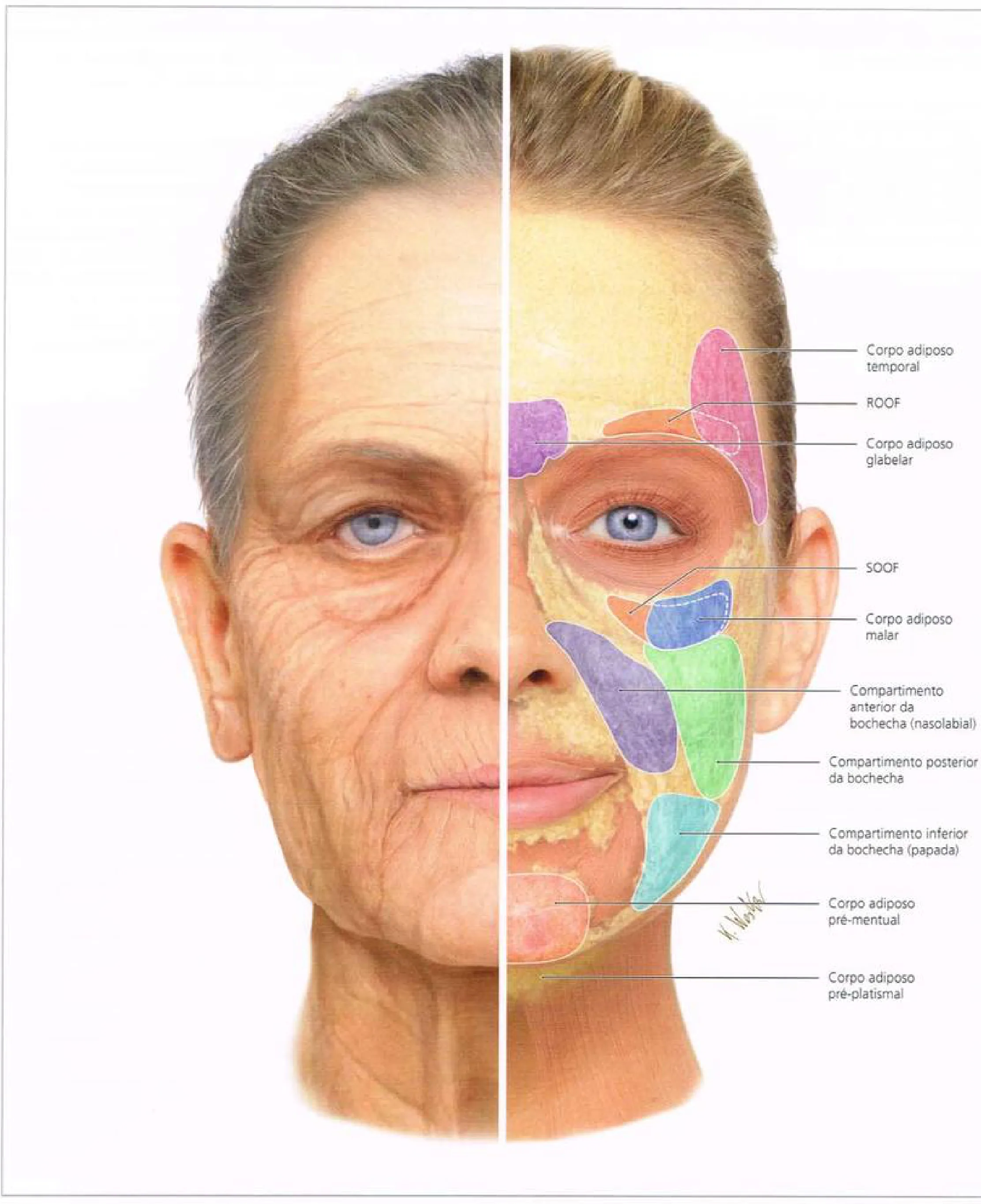 Fig 1. Fat-compartment map of the midface, young vs aged hemiface. — (Radlansky, Atlas Ilustrado de Anatomia Clínica da Face, p. 58.)
> Fuentes: Radlansky [MEDLIB] [17]-adjacent corpus atlas; compartment nomenclature per Rohrich & Pessa [4], Schenck [3], Cotofana [2].
Fig 1. Fat-compartment map of the midface, young vs aged hemiface. — (Radlansky, Atlas Ilustrado de Anatomia Clínica da Face, p. 58.)
> Fuentes: Radlansky [MEDLIB] [17]-adjacent corpus atlas; compartment nomenclature per Rohrich & Pessa [4], Schenck [3], Cotofana [2].
The Fig 1 map is the reason the region has two opposite results from the same syringe: the coloured malar body sits superficial to orbicularis and drains through one channel (the mound trap), while the deep medial cheek fat beneath it is the compartment whose loss flattens the face and whose restitution projects it.
The superficial compartments are modelled panel by panel in Fig 2, framing the deep plane the malar bolus must reach beneath them.
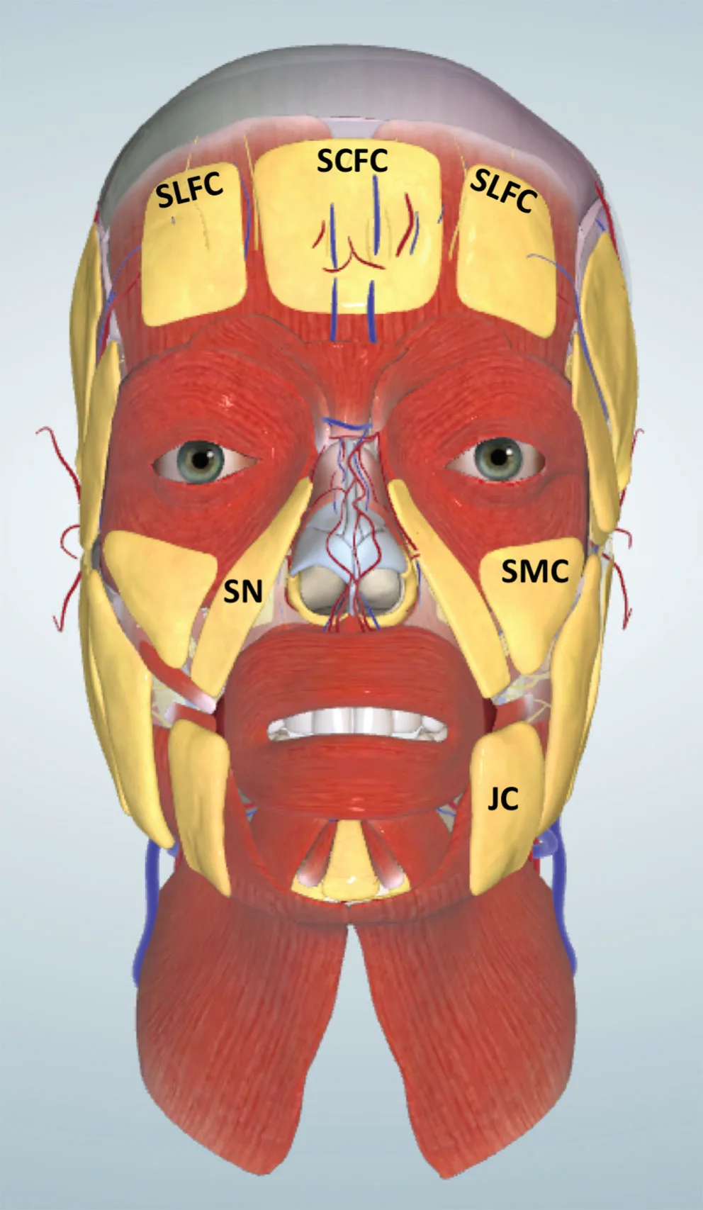 Fig 2. Superficial fat compartments of the face (virtual model). — (Cotofana, Anatomy of the Facial Fat Compartments, 2019.)
> Fuentes: Cotofana 2019 [2] [B].
Fig 2. Superficial fat compartments of the face (virtual model). — (Cotofana, Anatomy of the Facial Fat Compartments, 2019.)
> Fuentes: Cotofana 2019 [2] [B].
Retaining ligaments: the borders that make the folds [4][8]. Three matter here: - Zygomatic ligament (McGregor's patch): true osteocutaneous ligament off the anterior zygoma; its attenuation drops the cheek and deepens the lid-cheek junction. Repositioning the cheek along its vector is the diagnostic maneuver of the region (D6.5). - Orbicularis retaining ligament (ORL) / orbitomalar ligament: arborizes through orbicularis to skin, forming the palpebromalar groove (lateral continuation of the tear trough). It is the superior wall of the malar-mound space [8][9]. - Zygomaticocutaneous ligaments: form the inferior wall of the malar-mound space, partitioning the infraorbital "malar" fat above from the superficial cheek below [8][9].
The prezygomatic space and SOOF, up close. Cotofana's infraorbital dissection (Fig 3) shows the deep plane the structural bolus must reach: the prezygomatic space (PZ) glides over the body of the zygoma, floored by pre-periosteal fat, with the medial and lateral SOOF (MS, LS) dyed to show they are discrete deep pockets, not one mass, and the deep lateral cheek fat (DLCF) lateral to them [1][8].
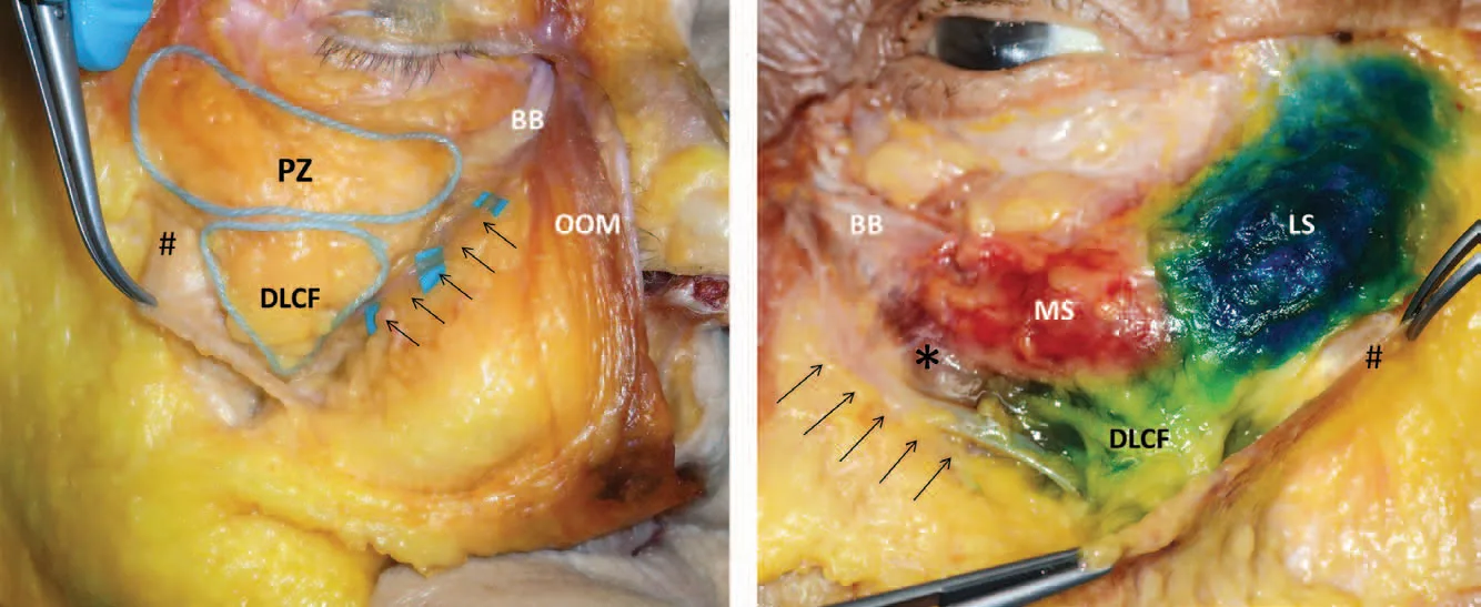 Fig 3. Prezygomatic space, SOOF and deep cheek fat, cadaver. — (Cotofana, Midface Clinical Anatomy, 2015, layer-4 dissection.)
> Fuentes: Cotofana 2015 [1] [B]; Surek 2015 [8] [MEDLIB].
Fig 3. Prezygomatic space, SOOF and deep cheek fat, cadaver. — (Cotofana, Midface Clinical Anatomy, 2015, layer-4 dissection.)
> Fuentes: Cotofana 2015 [1] [B]; Surek 2015 [8] [MEDLIB].
Deep supports, superficial contours [1]. The operative rule of the whole region in one sentence: the deep compartments carry structural load and are restored to project; the superficial compartments only refine and blend and hold almost no volume. Putting into the superficial plane what belongs deep is the exact mechanism of the overfilled cheek (D6.10).
Sex and phenotype are anatomical, not cosmetic, variables [1]. Male malar pattern: flatter, more lateral, more angulated, less anteromedial fullness, apex lower. Female pattern: more anteromedial, apex higher and more medial. ⚠ Applying the female map to a man feminizes the midface. A pre-existing prominent malar eminence needs less, and above all no added width.
Compartment boundaries, measured (why "deep and medial" has coordinates) [1][8][9]:
| Compartment | Layer | Boundaries | Landmark |
|---|---|---|---|
| Deep medial cheek fat, medial part | 6 (medial to angular vein) | anterior: premaxillary space; posterior: deep pyriform (Ristow's) space; on the maxilla | robust, compact; <1.5 cm lateral to the alar base; the highest-yield deep target [8][9] |
| Deep medial cheek fat, lateral part (DLCF) | 4 (lateral to angular vein) | lateral/inferior: broad fibrous origin of zygomaticus major; medial/inferior: facial vein; superior: zygomatic ligament | lateral edge 4.6 cm, medial edge 2.6 cm from the alar crease; loose, areolar; not a recommended target [1][8] |
| Medial + lateral SOOF | 5 | between the undersurface of orbicularis oculi and the dense posterior SMAS capsule; split medial/lateral by a palpebral arterial branch | thin; supports the lid; over-deposit here feeds the mound [8][9] |
| Prezygomatic space | glide space | roof orbicularis, floor preperiosteal fat over the zygoma; inferior wall zygomaticocutaneous ligament | 7 sub-layers (skin, subcutis, orbicularis, SOOF, deep fascia of zygomatic muscles, preperiosteal fat, periosteum) [27] |
| Deep pyriform (Ristow's) space | 6 | between the medial DMCF and periosteum, medial to the infraorbital foramen | the angular artery runs on its roof, so it is "safe on bone" at the floor [27] |
| Premaxillary space | deep | between zygomaticocutaneous and buccal maxillary ligaments / between angular artery and angular vein | infraorbital NVB on its floor; angular vein at its lateral border [8][27] |
Layer by layer, and what an injection does in each [1]: - Layer 1 skin / 2 dermis: target only of skin boosters and hyperdilute biostimulator; any structural filler here is visible and palpable. - Layer 2 superficial fat: the superficial middle-cheek, nasolabial and infraorbital compartments; blending only. The infraorbital ("malar") compartment is the mound trap. - Layer 3 SMAS: the continuous mimetic sheet that ties zygomaticus major and minor, orbicularis oculi, depressor anguli oris, depressor labii inferioris and sometimes risorius into one unit around the mouth; the orbicularis oculi sits in this layer. A transit plane, not a deposit target. - Layer 4 deep areolar / deep lateral cheek fat: the DLCF, bounded by the zygomaticus major origin; the tail communicates into the buccal recess, a deep adverse-event zone (over-deposit here jowls the face) [8]. - Layer 5/6 deep fat + spaces: the medial DMCF, SOOF and prezygomatic/premaxillary/pyriform spaces; the structural deposit targets. - Layer 7 periosteum: the bolus floor on the zygomatic body and maxilla.
The transverse facial septum [45][2]. The constant zygomaticus major is tied to the underlying maxilla by a broad fascial membrane, the transverse facial septum, which forms the inferior boundary of the deep lateral cheek fat and the superior boundary of the buccal space. It is the leading cause of "apple-cheeks" on smiling, and its distortion by over-deposit is a component of the overfilled-syndrome look (D6.10). The zygomatic ligament (McGregor's patch) sits next to the zygomaticus origin and marks the boundary between the medial and lateral midface; it arises from bone, pierces orbicularis and inserts into skin, forming the "hammock" that suspends the malar bag [1][2].
Ageing asymmetry of the compartments [9]. The deep medial cheek fat deflates and its adipocytes shrink with age, while the supra-SMAS (superficial) compartments hypertrophy. The clinical translation: refill the deep compartment that emptied; do not add to the superficial one that is already enlarging. The recommended deep-medial product is a medium-to-large particle, high-cohesivity HA or autologous fat [9].
Trampa clásica: calling the superficial infraorbital fat "the malar pad" and volumizing it to lift the cheek. It is a lymphatic cul-de-sac, not a structural pad; the structural pad is the deep medial cheek fat one layer below it [8][9].
Buccal fat and the deep communication [8][1]. The tail of the deep medial cheek fat, lateral to the levator anguli oris, opens into a loose areolar plane that descends posteroinferiorly into the buccal fat pad (Bichat's), forming a "cavernous communication into the buccal recess" that is a deep-injection adverse-event zone: an over-deposit here migrates and jowls the lower face rather than projecting the cheek [8]. The buccal space is bounded above by the transverse facial septum, and the lateral deep cheek fat is deliberately not a primary target because of its loose, areolar consistency and this deep communication [8][9].
Two spaces that are safe when entered correctly [27]. The prezygomatic space and Ristow's (deep pyriform) space are both potential spaces between the deep fat and periosteum; entered on the bone with contact confirmed, they accept a controlled deposit for projection or paranasal support, and Ristow's space is where the angular artery runs on the roof so the periosteal floor is below the vessel. The premaxillary space, between the zygomaticocutaneous and buccal maxillary ligaments and between the angular artery and vein, carries the infraorbital neurovascular bundle on its floor: it is entered with more caution than the prezygomatic space [8][27].
SMAS types and why the medial cheek behaves differently [1]. Lateral to the nasolabial and labiomental sulci the SMAS (layer 3) is a discrete mobile sheet connecting the mimetic muscles; medial to those sulci it changes to a type-2 arrangement, becoming the investing fascia of the muscles with strong adherent connections to skin. The practical effect: the lateral cheek glides and accepts deep volume behind the ligaments, while the medial cheek is more tethered and less forgiving of a superficial deposit, which is one more reason superficial medial filler reads and migrates badly.
D6.3 · Vessels, nerves and the danger zone
Answer first: where the needle is safe and where it blinds.
| Structure | Where it is | Depth | Variant frequency | What fails if hit |
|---|---|---|---|---|
| Infraorbital artery + nerve (foramen) | ≈0.6–1.2 cm below the orbital rim at the medial-iris→mid-pupil line; aligns with maxillary 2nd premolar | exits foramen, then in/deep to layer 5; bundle angled inferiorly | foramen 24–27 mm from midline; 30% share the supraorbital vertical plane; may be multiple [17][20] | mid-cheek skin necrosis (artery); anaesthesia of lid/nose/lip (nerve) [17][23] |
| Angular artery (facial a. continuation) | medial, at the nasojugal/medial cheek border | variable, can be near-subdermal | high variability; on the roof of the deep pyriform space it is safest on bone | retrograde embolus → ophthalmic → blindness [15][43][44] |
| Zygomaticofacial artery + nerve | small foramen on the lateral zygoma, ~8–10 mm below rim, in line with the lateral orbital rim | through the bone; sensory nerve routinely sacrificed in rim surgery | present bilaterally; occasionally multiple | ecchymosis; minor malar-skin numbness [21] |
| Zygomatic branch of the facial nerve (motor) | crosses lateral to the zygomatic eminence in the plane between SMAS and deep fascia | layer 3/4 boundary | danger where zygomatic + superior masseteric ligaments fuse | levator labii weakness, lip asymmetry (usually temporary) [18][20] |
| Transverse facial artery | below the arch, running with the parotid duct territory | deep, sub-SMAS | — | ecchymosis; not the blindness route |
Fig 4 shows the motor danger: Rohrich's cadaver + illustration of the facial-nerve danger zones, the zygomatic branch lying superficially lateral to the eminence in the SMAS-to-deep-fascia plane, exactly where a careless lateral supraperiosteal pass or a sub-SMAS cannula can catch it.
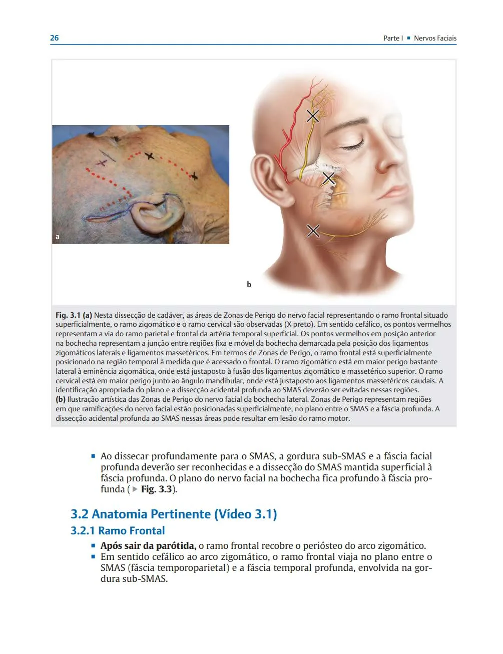 Fig 4. Zygomatic branch of the facial nerve, danger zone. — (Rohrich, Zonas Faciais de Perigo, 2020, Fig 3.1.)
> Fuentes: Rohrich 2020 [20] [C]; nerve plane per Seckel [18], Erian [19].
Fig 4. Zygomatic branch of the facial nerve, danger zone. — (Rohrich, Zonas Faciais de Perigo, 2020, Fig 3.1.)
> Fuentes: Rohrich 2020 [20] [C]; nerve plane per Seckel [18], Erian [19].
The infraorbital foramen, precisely [17][23][21]. Standring gives 6.4 mm (range 3.2–12.1) below the rim and 2.5 cm (1.8–3.3) from the midline, aligned with the 2nd maxillary premolar; Pirayesh centres a 1.5 cm-diameter danger circle on the foramen "1–1.5 cm below the rim at mid-pupil"; Rohrich gives 6.3–10.9 mm below the rim (33–41% of the intercanthal distance). The maxilla is an inverted funnel with its apex at the foramen: because the bundle is hooded superiorly and angled inferiorly, a lateral cannula approach is protected by bone, an inferior needle approach is invited into the foramen [22]. This single fact decides the entry vector for any Ck3/deep-medial work near the rim.
Fig 5 documents, in the cadaver, the SOOF beneath orbicularis and the infraorbital strand the deep bolus must stay below.
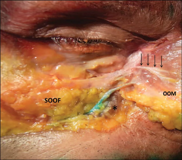 Fig 5. SOOF, orbicularis retaining ligament and the infraorbital deep plane. — (Cotofana, Midface Clinical Anatomy, 2015, Fig 6.)
> Fuentes: Cotofana 2015 [1] [B].
Fig 5. SOOF, orbicularis retaining ligament and the infraorbital deep plane. — (Cotofana, Midface Clinical Anatomy, 2015, Fig 6.)
> Fuentes: Cotofana 2015 [1] [B].
The blindness pathway, stepwise [15][43][44]. Facial artery → angular artery (medial cheek) → dorsal nasal → anastomosis with the supratrochlear/supraorbital and the ophthalmic system. A bolus injected under pressure into the angular or a deep branch can travel retrograde against arterial flow when injection pressure exceeds systolic, then wash antegrade into the central retinal artery on release. The mean lumen volume of a representative facial branch (supratrochlear) is ≈0.085 mL, so a fraction of a millilitre suffices to fill the column to the eye [41]. Yang 2020 documents fatal cerebral infarction with ophthalmic occlusion after a facial HA bolus [44]. Full occlusion algorithm: J2 — Vascular Occlusion & Emergency Response.
Pirayesh's split-face (Fig 6) exposes the practical geography: the reflected malar soft tissue reveals the infraorbital vessels and the density of the mid-cheek vascular net, the reason a blind superficial bolus here is never truly "just subcutaneous".
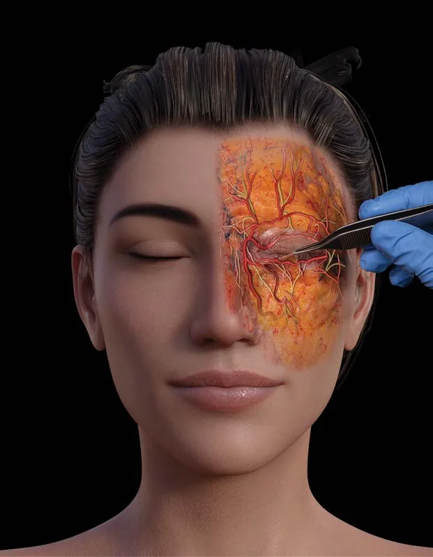 Fig 6. Malar-region vasculature exposed. — (Pirayesh, Aesthetic Facial Anatomy Essentials for Injections, 2020, p. 129.)
> Fuentes: Pirayesh 2020 [23] [C].
Fig 6. Malar-region vasculature exposed. — (Pirayesh, Aesthetic Facial Anatomy Essentials for Injections, 2020, p. 129.)
> Fuentes: Pirayesh 2020 [23] [C].
Consensus (all schools): deep supraperiosteal boluses go with bone contact, aspiration before injection, small aliquots, low pressure, constant needle movement or a blunt cannula in the vascular medial cheek [1][24][35]. Discrepancy on needle vs cannula is a technique choice, adjudicated in D6.7, not a safety absolute.
Trampa clásica: treating the medial cheek/nasojugal border as a low-risk "just add support" zone. The angular artery lives there with unpredictable depth, and the region is, with the glabella and nose, one of the highest-published sites of filler blindness [15][43].
The arterial tree of the region, mapped [15][22]. The relevant arteries and where they anastomose: - Facial artery: ascends near the anterior border of the masseter, lateral to the oral commissure, becoming the angular artery at the nasojugal border. The main external-carotid feeder of the region. - Angular artery: the terminal facial artery along the side of the nose; anastomoses with the dorsal nasal branch of the ophthalmic system. The blindness conduit of the medial cheek. On the roof of the deep pyriform space it is predictable; elsewhere its depth is variable. - Infraorbital artery: exits the foramen (coordinates above) with the nerve; supplies the mid-cheek skin; a hit here necroses the mid-cheek. - Transverse facial artery: below the arch, toward the parotid-duct territory; a bruising, not a blindness, risk. - Zygomatico-orbital / zygomaticofacial branches: small, from the lacrimal / superficial temporal territory, at the lateral zygoma.
The danger is that the medial cheek is a watershed between the internal and external carotid systems: the transverse facial and deep temporal arteries anastomose with the zygomatic branches of the lacrimal artery, and the infraorbital and angular with the dorsal nasal [22]. A pressurized bolus can cross from the low-pressure external-carotid injection site into the ophthalmic system.
The venous side [8][27]. The angular vein runs along the nasojugal groove and forms the lateral border of the premaxillary space and the medial-inferior border of the deep lateral cheek fat; the facial vein bounds the DLCF medially and inferiorly. Veins matter less for occlusion but explain part of the region's bruising and, when compressed, its oedema.
Sensory nerves beyond the infraorbital [19][21]: - Zygomaticofacial nerve: exits its foramen on the lateral zygoma, ~8–10 mm below the rim in line with the lateral orbital rim; supplies the malar-eminence skin; routinely sacrificed in rim surgery with a defect rarely noticed. - Zygomaticotemporal nerve: exits on the deep surface of the zygomatic bone; supplies the anterior temple.
Why aspiration and low pressure, not just "avoid the artery" [23][35]. The medial cheek is too vessel-dense (see Fig 6) to reliably miss a vessel by anatomy alone. The layered defences: aspirate before a deep bolus; keep injection pressure below arterial pressure (a wider-bore syringe gives less thumb pressure); deposit small aliquots; keep the needle moving, or use a blunt cannula in the medial corridor; map with ultrasound where available. None is absolute; stacked, they are the standard of care.
"Safe on bone" is a real principle here [27]. In the deep pyriform (Ristow's) space the angular artery runs on the roof of the space, so a bolus placed on the periosteal floor, with bone contact confirmed, sits below the vessel. This is the anatomical basis for the supraperiosteal-with-bone-contact rule that runs through every technique block.
Infraorbital-foramen coordinates, the sources side by side (why the range matters) [17][19][20][21][23]:
| Source | Below the rim | From the midline | Vertical reference |
|---|---|---|---|
| Standring (Gray's) | 6.4 mm (3.2–12.1) | 2.5 cm (1.8–3.3) | 2nd maxillary premolar |
| Pirayesh | 1–1.5 cm | mid-pupil; 1.5 cm danger circle | |
| Rohrich (Danger Zones) | 6.3–10.9 mm (33–41% intercanthal) | 25.7–27.1 mm (M) / 24.2–26.8 mm (F) | 30% share the supraorbital vertical |
| Seckel | 1 cm | midpupil–2nd premolar line; 1.5 cm-radius circle | |
| Yaremchuk | ~8 mm (3–6 in hypoplasia) | at the zygomaticomaxillary suture | midpupillary line |
The clinical reading of that spread: the foramen is roughly one fingerbreadth below the rim near the mid-pupil, but its exact depth and distance vary by sex, side and skeletal development, and some patients have accessory foramina. Treat the whole circle as danger, approach Ck3 from lateral, and confirm plane by bone contact rather than by a memorized millimetre [23].
Aspiration and cannula, what the evidence supports [23][35][41]. A cannula's blunt tip is less likely to perforate a vessel and lowers, but does not eliminate, the vascular-occlusion risk in the vessel-dense medial cheek; it costs more oedema and less pinpoint deep control. A needle permits aspiration before a deep bolus and pinpoint bone-contact placement, at the cost of higher ecchymosis and a sharp tip. Neither is a guarantee: aspiration can be falsely negative with viscous gels, and a cannula can still enter a vessel. The evidence-backed position is that the safety comes from the stack (aspirate, low pressure, small aliquots, movement, ultrasound where available), not from the tool alone, and the tool is chosen by task (needle for the deep lateral-zygoma bolus, cannula for the medial corridor and superficial fanning).
D6.4 · Regional ageing: what is lost, and in what order
The order matters because it dictates the treatment order [10][12][14]. Volume and descent are not the same, are not seen the same, and are not treated the same; most patients over 45 have both, and volumizing the descent without recognizing it makes a heavier face, not a younger one [14].
Sequence of change in the malar region:
| # | What changes | Structure | Regional consequence |
|---|---|---|---|
| 1 | Deep-fat deflation | deep medial cheek fat + medial/lateral SOOF empty | loss of anterior projection; the lid-cheek junction lengthens; the tear trough and palpebromalar groove deepen from below [5][8] |
| 2 | Skeletal resorption | maxilla (superomedial + inferolateral orbital rim, pyriform), posterior maxilla as the resorption centre; the zygomatic body is comparatively stable | the platform under the cheek recedes, the orbit enlarges, the maxillary angle decreases; a "negative vector" appears [11][13] |
| 3 | Ligament attenuation | zygomatic ligament + ORL loosen | the fixation that held the cheek up gives way [8][4] |
| 4 | Superficial-fat descent | superficial middle-cheek compartment slides over the loosened ligaments | the mid-cheek "V" deflates and the volume piles above the nasolabial fold, deepening it [2][14] |
| 5 | Skin | dermal thinning, elastosis, photodamage | fine rhytids, loss of the light-reflecting Ogee sheen [10] |
Deflation precedes and drives descent [12][14]. Freytag 2022 frames it biomechanically: as the deep support empties and the skeleton recedes, the same soft-tissue envelope has less to sit on, so it descends; Casabona 2019 separates "lifting" (repositioning descended tissue) from "volumizing" (refilling deflated compartments) as two distinct corrections that a single filler cannot substitute for each other [12][14]. The clinical corollary is D6.5's repositioning maneuver.
Fig 7 shows skeletal recession and the deep-fat and ligament change in the same cross-section.
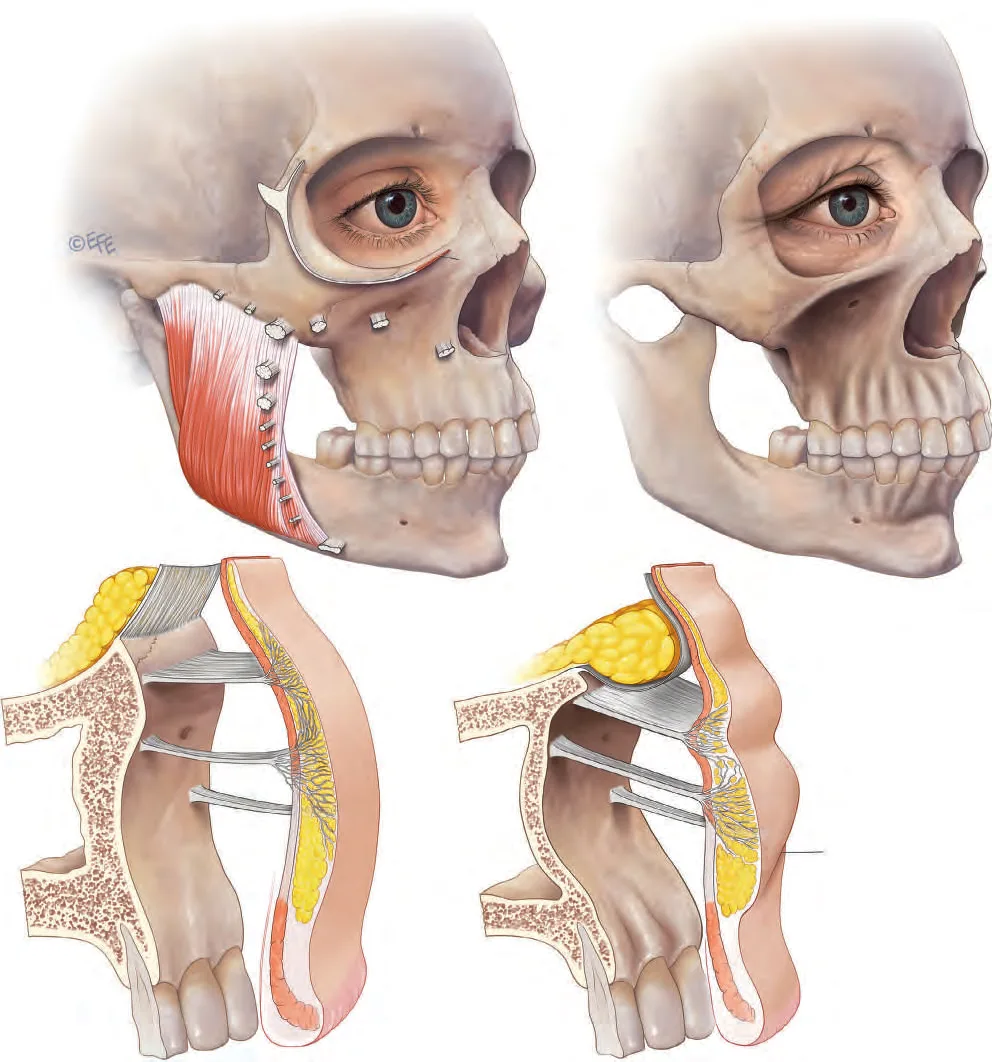 Fig 7. Linked ageing changes through skin, fat, ligament and bone at the maxilla/zygoma. — (Standring, Gray's Anatomy, 2016, Fig 4.3.5, p. 960.)
> Fuentes: Standring 2016 [17] [C]; skeletal-resorption pattern per Mendelson & Wong [11], Update on Facial Aging [13].
Fig 7. Linked ageing changes through skin, fat, ligament and bone at the maxilla/zygoma. — (Standring, Gray's Anatomy, 2016, Fig 4.3.5, p. 960.)
> Fuentes: Standring 2016 [17] [C]; skeletal-resorption pattern per Mendelson & Wong [11], Update on Facial Aging [13].
Fig 8 is the clinical correlate: the flattened projection and lengthened lid-cheek junction of the aged oblique view.
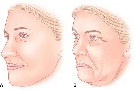 Fig 8. Youthful vs aged midface, clinical appearance. — (Hartstein, Midfacial Rejuvenation, 2012, p. 2.)
> Fuentes: Hartstein 2012 [MEDLIB] corpus; sequence per Cotofana 2016 [10].
Fig 8. Youthful vs aged midface, clinical appearance. — (Hartstein, Midfacial Rejuvenation, 2012, p. 2.)
> Fuentes: Hartstein 2012 [MEDLIB] corpus; sequence per Cotofana 2016 [10].
Which compartment first? Volume loss is described as beginning in the superficial lateral compartment and transitioning medially, correlating with the progression of the submalar hollow and nasojugal groove; the deep medial cheek fat and SOOF carry the projection loss that a deep bolus specifically corrects [8][10]. The ⚠ trap is chasing the visible superficial hollow while ignoring that its cause is a deep, empty compartment.
Bone does not come back [11]. No injectable rebuilds resorbed maxilla; a supraperiosteal bolus substitutes for the lost skeletal platform mechanically. In a marked negative vector this is a stopgap, and the honest referral is to skeletal augmentation (D6.6).
Trampa clásica: reading a hollow lid-cheek junction as a tear-trough (D2) problem and filling it directly, when the primary loss is one layer deeper and one compartment lateral (deep medial cheek fat + SOOF). Treating the symptom at the point of maximum vascular risk instead of the cause on bone [8][2].
Skeletal resorption is selective, not uniform [11][13]. The facial skeleton does not shrink evenly: specific zones resorb while others hold. In and around the malar region the maxilla recedes (the pyriform aperture enlarges, the superomedial and inferolateral orbital rim retrude, the maxillary angle decreases), while the zygomatic body itself is comparatively stable. The visible effect is loss of the platform behind and below the cheek, an enlarging orbital aperture, and a relative forward position of the globe (the "negative vector"). Mendelson & Wong frame this as a clockwise rotation and deflation of the midface skeleton that undermines the soft-tissue drape [11].
Sign to loss to correction (the map that orders treatment):
| Visible sign | Underlying loss | Correct target |
|---|---|---|
| Flattened malar projection | maxillary recession + DMCF deflation | supraperiosteal bolus on the zygoma + deep medial cheek fat [1][8] |
| Lengthened, hollow lid-cheek junction | SOOF + deep medial cheek fat deflation | deep SOOF-level and DMCF restitution, not superficial lid filler [8] |
| Deepening nasojugal / tear trough from below | loss of deep support of the lid | deep cheek first (D2 for the residual trough) [8] |
| Descended mid-cheek "V", deeper nasolabial | ligament attenuation + superficial-fat descent | reposition (energy/threads) before adding volume [14] |
| Loss of the light-reflecting Ogee sheen | dermal thinning, elastosis | skin quality (boosters, energy), not volume [10] |
The deep-vs-superficial paradox [9]. Deep compartments deflate (small adipocytes) while superficial compartments hypertrophy with age. This is why "just add volume where it looks empty" fails: the empty compartment is deep, and the full-looking one above it is enlarging. Refill deep; leave superficial alone.
Bone does not regrow, so timing matters [11]. No injectable reverses maxillary resorption; a supraperiosteal bolus mechanically substitutes for the lost platform. In a marked negative vector this is a stopgap and the honest option is skeletal augmentation (D6.6). The ageing order (deflation, then descent, then skin) is the treatment order: restore deep structure first, reposition second, resurface last.
The line of ligaments frames the whole ageing model [14][2]. The retaining ligaments (zygomatic, orbicularis retaining, masseteric) form a "line of ligaments" that separates the fixed from the mobile face and marks the boundary between the medial and lateral midface. With age these ligaments lengthen and loosen rather than break: the fixed points that held the cheek up give way, and the soft tissue anterior to them descends while the tissue over them deflates. Respecting this line is why "lifting" (repositioning across the loosened ligament) and "volumizing" (refilling behind it) are separate corrections [14].
Skin, the last layer and the one that shows the light [10][13]. The malar skin thins, loses dermal collagen and elastin, and develops solar elastosis; the smooth convex surface that reflects light as the Ogee highlight becomes matte and finely creped. Skin change is treated last and with skin tools (boosters, energy, resurfacing), not with structural filler, because a deep bolus under aged skin restores shape but not surface quality.
Why the order is fixed, not a preference [11][12][14]: 1. If you refill the deep compartment and rebuild the platform first, the descended superficial tissue often repositions passively and needs less direct work. 2. If you start superficially (chasing the visible hollow or fold), you treat a symptom whose cause is deep, over-fill the superficial plane, and set up the overfilled cheek. 3. Bone cannot be regrown, so in a marked negative vector the deep bolus is an acknowledged stopgap and the ceiling is stated to the patient, with skeletal augmentation offered (D6.6).
A note on measurement. Age-related change here is graded clinically (photographs, the repositioning maneuver, vector on profile) rather than by a single validated malar scale; the practical metric that matters most across time is the patient's own baseline photograph and the cumulative-volume record, because they expose the slow creep that any point-in-time scale misses (D6.5) [14].
D6.5 · Assessment: what is measured, photographed, tested dynamically, scanned
The five-step regional exam (answer first):
| Step | What it distinguishes | How |
|---|---|---|
| 1 Repositioning maneuver | deficit vs descent | fingers lift the cheek soft tissue up-and-out along the zygomatic-ligament vector: clear improvement = descent-dominant (volume has a ceiling; needs energy/threads/surgery); improvement only on adding volume with the finger = deficit-dominant (filler pays off) [14] |
| 2 Two-target decision | anterior projection vs lateral definition | decide them separately; they use different points and vectors and mixing them is what makes the "odd" cheekbone [1][24] |
| 3 Palpation | bony substrate + danger landmarks | palpate the malar eminence for the skeletal platform; palpate the infraorbital rim and localize the foramen before any Ck3 work [17][23] |
| 4 Vector / lid-cheek | negative vector, mound tendency | side profile: does the globe project beyond the malar/infraorbital rim (negative vector, high edema and mound risk)? Note baseline periorbital puffiness [21][40] |
| 5 Ultrasound | plane, vessels, old product | map the angular/infraorbital vessels; measure filler depth; find prior filler; guide hyaluronidase [36][47] |
Photography, standardized [24]. Frontal, oblique 45°, and profile, same lighting and head position each visit. The oblique view is the one that shows the Ogee curve (the double-convex S from the lateral orbit across the malar prominence into the submalar concavity) and is the view most people skip; it is where over-projection and asymmetry show first. Assess the aesthetic endpoint on animation (full smile) as well as at rest: excess lifting or a "sausage-like" fold on the smile is the sign of a mis-placed bolus [24].
Ultrasound is now part of the regional workup, not a luxury [36][47]. High-frequency (≥18 MHz) probes map the course and depth of the angular and infraorbital arteries before injection, confirm the supraperiosteal plane during injection, and distinguish product echogenicity afterwards: HA is hypo/anechoic, CaHA and bone are hyperechoic, and colour Doppler shows flow. For the malar region specifically it de-risks the medial danger corridor and lets a delayed nodule or a malposition be told apart without guessing [47]. Ultrasound-guided hyaluronidase reaches an occlusion or a mound with less enzyme and more precision than a blind flood [47].
Baseline documentation that prevents the overfilled cheek [14]. A standardized baseline photo, an annual comparison against baseline (not against the previous session), and a running cumulative record of millilitres per side. The overfilled face is a process complication: a little too much, many times, in the wrong plane, without looking at the three-year-old photo (D6.10).
Trampa clásica: photographing only the frontal view. The malar over-projection and the loss of the Ogee curve are oblique-view findings; a frontal-only record lets a cheek creep forward for years without a visible flag [24].
The repositioning maneuver, done properly [14]. Two fingers on the lateral cheek lift the soft tissue up and out along the vector of the zygomatic ligament (toward the superolateral orbital rim), then release. Clear improvement on the lift = descent-dominant: volume has a ceiling and the full answer includes energy, threads or surgery. Improvement only when the finger also adds forward bulk = deficit-dominant: filler pays off. Both together is the common case: treat the deficit, warn about the descent ceiling. Thirty seconds, and it changes the plan.
Ogee and vector, on the oblique view [24][21]. The Ogee curve is the double-convex S running from the lateral orbit across the malar prominence into the submalar concavity; a youthful cheek is a smooth convexity, an over-projected one inverts it into a "pillow". The negative vector is read on profile: if the globe projects anterior to the malar/infraorbital rim, the platform is deficient and the mound/edema risk is high, which reduces or removes any superficial SOOF-level deposit.
Photography protocol [24]. Frontal, oblique 45°, profile; identical head position and lighting each visit; the oblique is the Ogee view and the one most often skipped. Assess the aesthetic endpoint on animation (full smile) as well as at rest: excess lifting or a "sausage-like" fold on the smile signals a misplaced bolus, and an "apple-cheek" bulge signals transverse-facial-septum overload.
Ultrasound, the regional protocol [36][47]. A high-frequency probe (≥18 MHz) is used to: (1) map the course and depth of the angular and infraorbital arteries before injection along the medial corridor and Ck3; (2) confirm the supraperiosteal plane during a deep bolus; (3) characterize product afterward (HA is hypo/anechoic, CaHA and bone hyperechoic, air hyperechoic with reverberation; colour Doppler shows flow); (4) find and map prior filler before re-treating; (5) guide hyaluronidase precisely to an occlusion or a mound, using less enzyme than a blind flood. For the malar region specifically it de-risks the medial danger corridor and separates a delayed nodule from a malposition without guessing.
Documentation that prevents the overfilled cheek [14]. Standardized baseline photo; annual comparison against baseline, not against the last session; a running cumulative record of millilitres per side per region. The overfilled face is a process complication (a little too much, many times, wrong plane, no baseline comparison), and the record is the antidote (D6.10).
Palpation, done deliberately [17][23]. Before any Ck2/Ck3 work: palpate the malar eminence to judge the bony platform (a thin, receded maxilla changes the plan from "restore" to "acknowledge the ceiling"); palpate the infraorbital rim and localize the foramen (Ck3 sits just below it) so the entry vector is chosen lateral, not inferior; palpate for pre-existing product from prior treatments, which changes both plane and reversibility. Palpation is faster than imaging and it is the step most often skipped.
Dynamic assessment, in detail [24]. Ask for a full smile and for a "downward tilt" head position. On the smile: a "sausage-like" fold or an "apple-cheek" bulge signals a superficial over-deposit or transverse-facial-septum overload; excess lateral lift signals a bolus placed too high. On the tilt-down and oblique: the Ogee highlight and any over-projection show. Static assessment alone passes cheeks that look correct at rest and animate badly.
Grading the negative vector [21]. On strict profile, note whether the most anterior point of the globe projects beyond the malar/infraorbital rim. A neutral or positive vector supports the lid and cheek and tolerates deep volume well; a negative vector means a deficient platform, a higher mound and edema risk, and a stronger case for skeletal referral if the patient wants a definitive change (D6.6).
Documentation checklist (the antidote to the overfilled cheek) [14]: - Standardized frontal, oblique 45° and profile photographs, same lighting and head position. - Cumulative millilitres per side per region, updated every visit. - Annual comparison against the baseline photograph, not against the last session. - Product, lot, plane, tool and volume per point recorded per session. - Ultrasound record of prior product where relevant.
Ultrasound echogenicity, the working key [47][36]. At the probe: anechoic (black, no reflection) = water, blood, and fresh HA gel pockets; hypoechoic (dark grey) = muscle, fat, integrated HA; hyperechoic (bright) = bone, calcium hydroxylapatite, air bubbles; colour Doppler shows arterial and venous flow and the probe's own motion. In the malar region this separates a delayed HA nodule (hypo/anechoic) from a CaHA deposit (hyperechoic) from a vascular structure (Doppler), and lets hyaluronidase be placed into the target rather than around it. Mapping the angular and infraorbital arteries before a medial deposit is the single most useful pre-injection ultrasound step here [47].
Assessment traps specific to the region [24][40]: - Judging only at rest: a cheek can look correct static and animate as a "sausage" or "apple". Always assess on a full smile. - Frontal-only photography: over-projection and Ogee loss are oblique-view findings. - Missing the negative vector: it is read on strict profile and it changes both the candidacy and the mound risk. - Ignoring baseline periorbital puffiness: it flags the patient who should not get superficial SOOF-level work. - No cumulative record: the slow creep that builds the overfilled cheek is invisible without an annual baseline comparison and a per-side millilitre log.
What "measured" means here in practice [14]. There is no single validated malar volume scale that substitutes for the clinical workup; the operative metrics are the repositioning-maneuver result, the vector on profile, the Ogee on oblique, the ultrasound map, and above all the patient's own baseline photograph tracked over years. Point-in-time grading misses the creep; the longitudinal record catches it.
D6.6 · Goal and patient selection: who benefits, who does not, what is referred
Answer first: the four patient types of this region.
| Type | Finding | Plan |
|---|---|---|
| Deficit-dominant, good skin | deep-fat/skeletal deflation, positive or neutral vector, elastic skin | best filler candidate. Deep structural restoration; high return per millilitre [1][8][14] |
| Descent-dominant, heavy/lax | soft-tissue ptosis, jowl, laxity; improves on repositioning not on added volume | volume worsens it (weight on unsupported tissue). Energy / threads / surgery first, then less volume [14] |
| Young aesthetic augmentation | no age-related loss; wants a higher/wider cheekbone | this is augmentation, not restitution: different consent, different expectation, screen for BDD before touching anything [salvage D3.1] |
| Skeletal deficiency / marked negative vector | globe projects beyond a receded rim | filler is a stopgap; the definitive answer is malar implant or orthognathic surgery. Refer [21] |
Who benefits most. The patient whose problem is a deflated deep compartment over an adequate bony platform, with skin that still retracts. Restoring the deep medial cheek fat and SOOF lifts the mid-cheek, shortens the lid-cheek junction, unloads the tear trough and softens the nasolabial in one deep correction, and it does so with less product than treating each of those four superficially [8][14].
Who does not, and why. ⚠ The heavy, lax face: adding deep anteromedial volume increases the load on tissue that is already failing to hold itself, producing the puffy, over-full result. In that phenotype the order inverts: densify/lift first, volumize second, and with less [14]. ⚠ The already-prominent malar eminence: needs less, not more, and above all no added width, or the midface is over-widened [1]. ⚠ The periorbital-puffy / negative-vector patient: any superficial SOOF-level deposit risks months of malar edema, so that point is reduced or omitted [40][47].
When the correct answer is NOT an injectable. Marked skeletal deficiency wanting a permanent, precise projection change is an implant/orthognathic referral, not a filler problem [21]. Advanced descent with skin excess is a lift. Disease-related facial lipoatrophy (HIV antiretroviral, oncologic, severe weight loss) has its own goals and products and lives in M1 — Disease-Related Facial Volume Loss. Non-injectable regional alternatives are enumerated in D6.7.
Consent points specific to the region. Reversibility differs by product (HA reversible with hyaluronidase; CaHA/PLLA/PCL/PMMA not); the blindness risk is named explicitly; the malar-mound risk is named for any lid-cheek/SOOF work; and for the young augmentation patient, the escalation risk and the difference between restoration and enhancement are documented [24][43][salvage D3.1].
Trampa clásica: treating the young patient who asks for cheekbones as a restitution case with the same technique and the same casual consent. Same needle, different indication, different psychological screen; skipping that is how an escalation spiral starts [salvage D3.1]. Phenotype and ethnicity decide the target shape, not just the volume [1][27]. The lateral zygoma and arch project more in many Asian faces, so the sub-arch "lateral cheek hollow" reads as more prominent than it is; adding lateral volume there widens an already-wide midface. The correction in that phenotype is often the submalar/hollow (D7) or nothing lateral, never more width. Ethnic and cultural aesthetic targets differ and are set with the patient, not assumed: B6 — Ethnic, Racial & Cultural Considerations.
Gender is an anatomical variable [1]. Male: flatter, more lateral, more angulated, apex lower, less anteromedial fullness. Female: more anteromedial, apex higher and medial. Applying the female map to a man feminizes the midface; applying the male map to a woman flattens it. Decide the pattern explicitly before the first bolus.
Contraindications and cautions, region-specific: - Active infection or inflammation over the cheek: defer. - Body dysmorphic disorder or unrealistic expectation, especially the young augmentation-seeker: screen and, if positive, do not treat [salvage D3.1]. - Prior permanent or semi-permanent filler in the region (PMMA, silicone, old PLLA/CaHA): image with ultrasound before adding anything; a new deposit onto old product distorts and raises nodule risk. - Anticoagulation / bleeding tendency: the vessel-dense medial cheek bruises; plan cannula, consider timing. - Periorbital-puffy / negative-vector patient: the wrong candidate for any superficial SOOF-level deposit (mound risk). - Pregnancy / breastfeeding: elective, defer.
Consent points that are specific to this region [24][43]: reversibility differs by product (HA reversible with hyaluronidase; CaHA/PLLA/PCL/PMMA not); the blindness risk is named explicitly; the malar-mound risk is named for any lid-cheek/SOOF work; and for the young augmentation patient the escalation risk and the restoration-vs-enhancement distinction are documented. Non-injectable regional alternatives are enumerated in D6.7.
Skin phototype and thickness change the risk profile, not the anatomy [27][43]. Thicker sebaceous cheek skin tolerates and masks a deep deposit better and shows a superficial one less; thinner, lighter skin near the lid-cheek junction reveals a superficial deposit as a Tyndall hue and is more prone to visible mounding, so the plane discipline is stricter there. Higher-phototype skin carries a higher risk of post-inflammatory hyperpigmentation at any traumatized point, which argues further for fewer, deeper, atraumatic boluses over multiple superficial passes. None of this changes where the structural target is (deep, on bone); it changes how unforgiving the superficial plane is, and therefore how firmly the "deep or not at all" rule is applied.
Cultural and aesthetic target-setting [1][salvage]. The desired cheek shape is not universal: some patients and cultures prize a high, defined lateral cheekbone, others a soft anteromedial fullness, and the same volume placed for the wrong target reads as "overdone" even when technically clean. The target is agreed explicitly with the patient against their own baseline and their reference, not imposed from a default map, and the male-vs-female and phenotype patterns above are the starting frame, not the destination. Structural detail on ethnic bone and soft-tissue differences: B6 — Ethnic, Racial & Cultural Considerations.
The escalation spiral, and how selection prevents it [salvage][14]. The malar region accepts product quietly: a cheek that is a little too full does not protest at the time, and the next session, judged against the last one, looks reasonable again. This is how the overfilled face is built, one defensible session at a time. Patient selection is the first defence: the young augmentation-seeker who returns for "just a bit more" every few months, the patient who is never satisfied, and the patient whose problem is descent being treated as deficit are the three who spiral. The countermeasures are a baseline photograph, an annual comparison against it, a cumulative-volume log, and the willingness to tell a patient they need nothing more (D6.5) [14].
Referral decision list (when this region leaves the injector's hands): - Marked skeletal deficiency / strong negative vector wanting a definitive projection change → malar implant or orthognathic surgery [21]. - Advanced descent with skin excess → surgical lift; volume alone worsens the load [14]. - Disease-related facial lipoatrophy (HIV antiretroviral, oncologic, severe weight loss) → the dedicated pathway with its own goals and products, M1 — Disease-Related Facial Volume Loss. - Suspected body dysmorphic disorder → mental-health assessment before any treatment [salvage]. - A wet festoon / true lymphatic malar bag → treat the drainage, not with more filler (D6.10) [40].
Documentation of consent, specific items [24][43]: reversibility by product (HA yes; CaHA/PLLA/PCL/PMMA no); the named risk of visual loss; the named risk of a persistent malar mound for any lid-cheek/SOOF work; the escalation and restoration-vs-enhancement discussion for the young augmentation patient; and the plan to compare against baseline annually.
D6.7 · Technique: the full grid
This block is closed with the full grid of the region's options, not one technique. The malar prominence is treated by enumerating every axis: product, instrument, plane, movement, school, volume and the non-injectable alternative, with the point at which two schools do the opposite of each other stated rather than averaged [1][24]. The logic beneath the grid is the one established in D6.2 and D6.3: the deep compartments over bone carry structural load and are the target for projection; the superficial compartments only refine and hold almost no volume; and the medial corridor is a vascular danger zone that dictates a low-pressure, small-aliquot, aspirate-or-cannula discipline whatever the product [8][23]. Every row below therefore carries a plane and a rheology as well as a volume, because a number without its plane is what produces both the flat cheek (soft gel wasted deep) and the palpable one (firm gel stranded superficial) [26]. What follows is that grid, point by point and axis by axis; anything that does not apply to the malar prominence is marked and justified rather than dropped [24].
Point-by-point (the MD Codes 5-point cheek reshape, exact codes)
de Maio codes the cheek as five subunits, foundation before volume: build structural support (Ck1, Ck2) against gravity first, then correct volume (Ck3–Ck5) [24][25]. Ck4/Ck5 sit in D7 territory (parotid/submalar) but belong to the same code sequence and are listed for completeness.
| Code | Point | Plane / entry | Direction | Tool | Volume/point | Regional alert |
|---|---|---|---|---|---|---|
| Ck1 | zygomatic arch | supraperiosteal, perpendicular to bone | vertical bolus, layered | 27 G needle (aspirate) | 0.1–0.3 mL | lifts the cheek, supports lid/SOOF [25] |
| Ck2 | zygomatic eminence | supraperiosteal, bone contact | small bolus | 27 G needle | 0.2–0.4 mL | projection + shortens palpebromalar sulcus; ⚠ zygomaticofacial artery [25] |
| Ck3 | anteromedial cheek | supraperiosteal / deep fat | bolus, lateral approach | 27 G needle or cannula | 0.1–0.3 mL | medial lid-cheek junction, softens tear trough; ⚠ infraorbital foramen [25] |
| Ck4 | lateral lower cheek / parotid (D7) | subcutaneous | fan / linear | 25 G cannula (large area) | 0.2–1.0 mL | jawline support; cannula preferred [25] |
| Ck5 | submalar (D7) | subcutaneous | fan | 25 G cannula | 0.2–0.5 mL | ⚠ facial artery, buccal nerve, facial vein [25] |
Delivery rules: each point in boluses <0.3 mL; aspiration mandatory for deep needle work; endpoint judged on animation. Per-session ceiling ~4 mL of HA in the midface, more added across successive sessions rather than in one sitting; total case volumes of 17–20 mL described only across multiple steps [24]. Micro-aliquot = 0.01–0.05 mL, aliquot = 0.1–0.2 mL, small bolus = 0.1–0.3 mL [23].
Fig 9 documents the superficial-fat cannula fanning vectors, the blending technique that sits opposite the deep bone bolus.
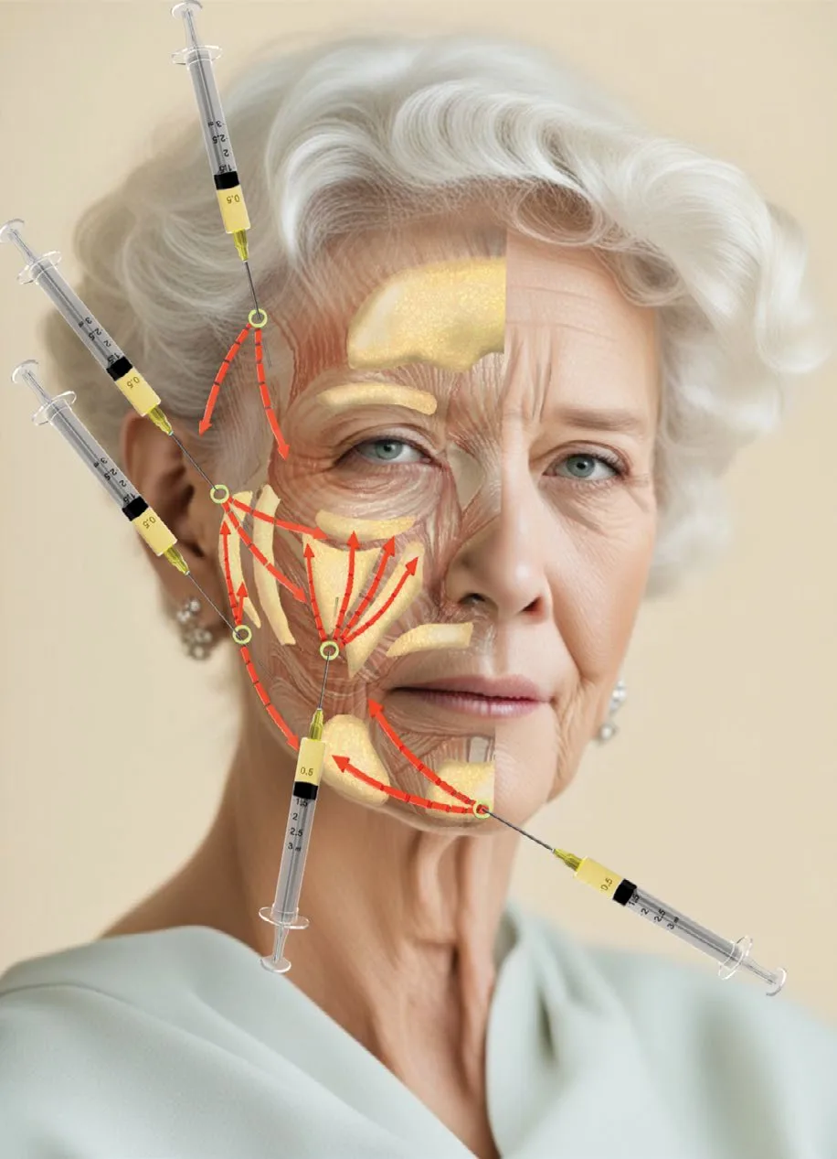 Fig 9. Superficial-fat cannula fanning of the midface. — (Piccolo, A.R.T. Autologous Regenerative Therapy, 2025, Fig 2.18.)
> Fuentes: Piccolo 2025 [30] [C].
Fig 9. Superficial-fat cannula fanning of the midface. — (Piccolo, A.R.T. Autologous Regenerative Therapy, 2025, Fig 2.18.)
> Fuentes: Piccolo 2025 [30] [C].
Product axis (enumerated whole)
| Product | Rheology / composition | Malar use, plane, tool | Reversible? | Key numbers |
|---|---|---|---|---|
| HA, high G′ / high cohesivity (VYC-20L Voluma; LGP-HA Restylane Lyft) | firm, load-bearing, 20 mg·mL⁻¹, 0.3% lidocaine | projection on bone; supraperiosteal Ck1/Ck2; 27 G needle | yes (hyaluronidase) | only HA class FDA-cleared for midface; lifts and holds after deep placement [22][24] |
| HA, medium G′ / low hydrophilia | integrates without swelling | deep-compartment restitution (DMCF); 27 G needle or cannula | yes | "integrates, does not balloon"; hydrophilia matters in the poorly drained lateral cheek [salvage][2] |
| HA, soft / low G′ / high integration | blends, low palpability | superficial transition/contour only, minimal volume | yes | this is the plane where the overfilled cheek lives: minimal or no volume [salvage] |
| CaHA (Radiesse), normal | 30% CaHA microspheres 25–45 µm + 70% CMC gel | malar/zygomatic 1:1 saline, supraperiosteal + subcutaneous, 25/27 G cannula; subdermal linear-retrograde 0.1–0.2 mL/pass | no | FDA lidocaine mix (2009): 0.3 mL 2% lidocaine per 1.5 mL syringe; result ~12 mo; radiopaque, hyperechoic [28][50][31] |
| CaHA, hyperdilute 1:2 | 1.5 mL product + ≥1.5 mL diluent | skin quality / biostimulation only, more superficial; no immediate volume | no | 1:2 disperses microspheres, drops concentration → neocollagenesis without volumizing [29][30][23] |
| PLLA (Sculptra) | lyophilized PLLA microspheres 2–50 µm | deep subcutaneous submalar / supraperiosteal; 18 G to draw, 25–26 G to inject | no | reconstitute ≥5 mL, hydrate ≥2 h (overnight preferred); 3–4 sessions ≥4 weeks apart; nodules if under-diluted/too superficial [26][33][35] |
| PCL (Ellansé S/M/L/E) | polycaprolactone microspheres in CMC gel; longevity scales with strand length (~1–4 yr) | periosteal + subdermal mid-cheek; collagen stimulation | no | biostimulatory + immediate volume; non-reversible [34] |
| PMMA (Bellafill/Artefill/Metacrill) | ~20% PMMA microspheres in bovine collagen carrier | permanent malar/contour augmentation; deep | no (permanent) | skin test required; granuloma risk; malar-specific outcome data limited [50] · ⚠ see gap note |
| Autologous fat / microfat | living adipose graft | largest-volume deep restitution, submalar/deep; SEFFI ~20–24 mL | partial (not enzymatic) | own chapter F5 — Autologous Fat Transfer; 25/22 G 50 mm cannula [30] |
| Polynucleotides / skin boosters | regenerative, non-volumizing | intradermal/subdermal adjunct for skin quality, not projection | n/a | adjunct only; see D14 — Escote, F1 — Skin Boosters |
| Toxin | neuromodulator | no volumizing role; microbotox for skin quality only (D6.8) | n/a | never a malar volumizer [37][38] |
⚠ [MATERIAL GAP] PMMA malar-specific outcomes. The corpus documents PMMA material science, regulatory history and complication pattern (Arenas, Tejero decks) [50], but malar/midface-specific long-term outcome series are thin. Cite PMMA here as a permanent-option category with a granuloma caveat, not with a malar-specific efficacy number.
Plane axis
Supraperiosteal (default, on the zygoma, for projection) · deep fat (DMCF/SOOF, for structural restitution) · SMAS (transit only, not a deposit target) · superficial fat (blending, minimal volume) · subdermal (fine contour, biostimulator) · intradermal (skin boosters, microbotox only) [1][8].
Movement axis
Bolus (deep on bone) · microbolus / micro-aliquot (0.01–0.05 mL) · retrograde linear threading (cannula, superficial) · fan (single-entry, superficial blending) · cross-hatch (CaHA volume, 0.1–0.2 mL/pass) · serial puncture (biostimulator field) · "tower"/stacked column (deep structural pillar on bone) [23][25][31].
School axis (the three that disagree, each with its author and reason)
Consensus: restore the deep structural support of the malar prominence before touching the surface; keep the vascular medial corridor low-pressure [1][8][24].
Discrepancy 1 — how to place the structural volume: - Single deep supraperiosteal bolus (de Maio, MD Codes Ck1–Ck5): few points, on bone, 27 G, aspirate. Reason: reproducible projection, minimal superficial product [24][25]. - Anatomic multi-plane / compartment fill (Surek/Beut "viaducts", Mendelson spaces): blunt-cannula delivery into the deep prezygomatic space and specific compartments through defined midfacial ports. Reason: refills the actual deflated anatomy and respects the deep spaces [8]. - Lymphatic-caution (Cotofana / Schelke / van Loghem): whatever the plane, do not overfill the superficial infraorbital/malar fat; keep deposits deep to the ORL. Reason: the superficial malar space is a lymphatic dead-end and the mound is iatrogenic [8][40]. - Decide by: vector and skin. Positive vector, good platform → deep bolus. Genuine multi-compartment deflation → anatomic layered. Any periorbital-puffy / negative-vector patient → the lymphatic-caution rule overrides both. Never average the volumes across schools.
Discrepancy 2 — needle vs cannula: - Needle-first (de Maio 27 G for deep bone): precise supraperiosteal bolus, bone contact confirmed, aspirate. Cost: higher ecchymosis; cannula-with-Voluma is "not validated by the manufacturer" [25]. - Cannula-first (vascular-midface schools): blunt 25 G, single-port fan; fewer vessel perforations and lower vascular-occlusion risk in the vessel-dense medial cheek. Cost: more oedema, less pinpoint deep control [23][31]. - Decide by: deep single bolus on the lateral zygoma tolerates a needle with aspiration; the medial/nasojugal corridor and any large superficial area take a cannula. This is a technique choice, not a safety absolute; the safety absolutes are aspiration, low pressure, small aliquots and movement, whichever tool.
Volume axis
Per point: 0.1–0.4 mL (deep bolus) to <0.3 mL cap per aliquot. Per region: build to the aesthetic endpoint on animation, not to a fixed number. Per session: ~4 mL HA midface ceiling by the code author. Overfill ceiling: the point where the cheek reads full at rest or "sausages" on the smile, or the Ogee curve inverts into a convex "pillow"; stop before it [24][10].
Non-injectable axis (when the correct answer is NOT filler)
- Energy / EBD (RF, microfocused ultrasound, HIFU): skin laxity and mild descent, and as a biostimulatory partner to CaHA. → G5 — Radiofrequency & HIFU Devices.
- Threads: vector repositioning of descended mid-cheek tissue. → E3 — Thread Lifting.
- Malar implant / orthognathic: definitive skeletal projection in marked deficiency/negative vector [21].
- Autologous fat grafting: largest-volume restitution / lipoatrophy. → F5 — Autologous Fat Transfer.
- When NOT filler: descent-dominant heavy face, marked skeletal deficiency, skin-laxity-dominant ageing (D6.6) [14][21].
Trampa clásica: using the syringe that is already open. A soft, low-G′ HA placed deep on bone diffuses and buys no projection; a firm, high-G′ HA placed superficially is palpable, visible and ages badly. Product rheology is matched to plane, not to what is loaded [salvage][24].
Rheology axis: matching the gel to the plane
The product table above is chosen on rheology, and rheology has definitions, not adjectives [23][26][27]:
| Parameter | Definition | Clinical meaning in the malar region |
|---|---|---|
| G′ (elastic modulus) | energy stored and returned after shear deformation; recovery of shape between gliding tissue planes | high G′ resists the lateral shear of the mobile cheek and holds projection on bone [23][26] |
| G″ (viscous modulus) | energy lost to internal friction under shear (not viscosity) | dissipation term; part of G* |
| G* (complex modulus) | √(G′²+G″²); overall hardness | low G for superficial (cannot be felt); high G + medium/high cohesivity for deep volumization [26] |
| tan δ (= G″/G′) | ratio of viscous to elastic behaviour | <1 = mostly elastic (typical cross-linked HA, good for deep contour); Voluma/Perlane/Restylane sit low-tan δ [26] |
| Cohesivity | internal adhesion holding the gel together; resistance to vertical compression / migration | high cohesivity holds vertical projection under soft-tissue load and resists migration out of plane [23][27] |
| Lift capacity | ability to oppose flattening | a function of both G′ and cohesivity, not G′ alone [23] |
The nuance that stops "just pick the highest G′" [26]. Across different filler families, lift capacity does not track G′ linearly (Hee et al.): G′ predicts lift within one family and cross-linking technology, but cohesivity, HA concentration and tissue integration all contribute, and resistance to deformation rises over time as the gel binds water and integrates. So the rule is match to plane, not maximize a single number: high-G′ + high-cohesivity firm gel on bone for projection (Ck1/Ck2); medium-G′, low-hydrophilia gel that integrates without swelling for the deep medial compartment (Ck3/DMCF); soft, low-G′, low-cohesivity gel for superficial blending only, where a firm gel would be palpable and a low-cohesivity gel would migrate. Rheology is a comparison tool, not a full predictor of in-vivo behaviour [23][26].
Autologous fat and the regenerative adjuncts (enumerated)
- Autologous fat / microfat: the largest-volume, living-graft option for deep malar restitution and lipoatrophy; delivered deep/subcutaneous with 22–25 G 50 mm cannulas; a structured-fat facelift (SEFFI) places ~20–24 mL of harvested adipose in the deep and subdermal planes, often staged with a biostimulator field on the same or a nearby day [30]. Take, longevity and nodularity differ from filler; full technique in F5 — Autologous Fat Transfer.
- Polynucleotides / skin boosters: regenerative, non-volumizing adjuncts for cheek skin quality (hydration, elasticity, fine crêping), placed intradermal/subdermal, never as a projection tool; series treatments. See D14 — Escote, F1 — Skin Boosters.
- Combination with energy: a biostimulator (CaHA, PLLA, PCL) plus microfocused ultrasound or RF for the skin-laxity component; the injectable restores/stimulates, the device tightens (D6.9).
Delivery mechanics that are non-negotiable whatever the tool
Aspirate before a deep bolus; inject below arterial pressure (larger-barrel syringe = less thumb force); deposit small aliquots (<0.3 mL); keep the needle moving or use a blunt cannula in the vessel-dense medial corridor; confirm bone contact for a supraperiosteal deposit; consider ultrasound guidance for the medial danger zone [23][35][47]. These are safety absolutes and are independent of the needle-vs-cannula and single-bolus-vs-layered choices adjudicated above.
Product-specific malar protocols (the numbers, per material)
- HA, deep structural [24][26]: 27 G needle perpendicular to the zygoma, bone contact confirmed, aspirate, deposit <0.3 mL per bolus, layer or "tower" (a stacked column on bone) to build projection without a single large pool. High-G′ / high-cohesivity gel; reversible with hyaluronidase.
- CaHA (Radiesse) [29][30][31]: compose the 1.5 mL syringe with 0.3 mL of 2% lidocaine (FDA mixing protocol) via a Luer-Lock female-to-female adapter and 10 passes, or use the pre-mixed lidocaine version. Malar/zygomatic: 1:1 with saline, supraperiosteal and subcutaneous, 25–27 G cannula; subdermal linear-retrograde threading, 0.1–0.2 mL per pass, moulded immediately against bone. Hyperdilute 1:2 (1.5 mL + ≥1.5 mL diluent) for skin quality only, more superficial. Non-reversible; result ~12 months; radiopaque and hyperechoic on ultrasound; not for fine periocular lines or the lip.
- PLLA (Sculptra) [33][35]: reconstitute with ≥5 mL sterile water (bacteriostatic saline off-label), add lidocaine to final dilution, hydrate ≥2 h (overnight preferred), do not shake. Draw with 18 G, inject with 25–26 G into the deep subcutaneous submalar / supraperiosteal plane, then vigorous post-treatment massage. Avoid superficial placement and under-dilution (both cause nodules); 3–4 sessions ≥4 weeks apart; non-reversible.
- PCL (Ellansé) [34]: strand length sets longevity (S/M/L/E, ~1–4 years); periosteal and subdermal mid-cheek; immediate volume plus collagen stimulation; non-reversible.
- PMMA (Bellafill/Artefill) [50]: ~20% PMMA microspheres in a bovine-collagen carrier; skin test required; permanent; deep placement; granuloma risk; malar-specific long-term outcome data are limited (see the [MATERIAL GAP] note above).
- Autologous fat [30]: SEFFI structured-fat ~20–24 mL, 22–25 G 50 mm cannulas, deep and subdermal; longevity and nodularity differ from filler; full technique in F5 — Autologous Fat Transfer.
Movement, matched to plane and product
Bolus and "tower" for deep structural HA on bone; micro-aliquot (0.01–0.05 mL) for delicate transitions; retrograde linear threading for cannula work in the superficial or subdermal plane; fan from a single lateral port for blending; cross-hatch (0.1–0.2 mL/pass) for CaHA volume; serial puncture for a biostimulator field. The movement is chosen with the product and the plane, never independently: a bolus of soft gel superficial migrates, a cross-hatch of firm gel superficial is palpable [26][31].
Product-to-plane mismatch, the failures it causes
| Wrong pairing | Result |
|---|---|
| soft, low-G′ HA deep on bone | diffuses, buys no projection, wastes product [salvage] |
| firm, high-G′ HA superficial | palpable, visible, ages poorly [salvage] |
| any product into the superficial infraorbital fat | malar mound [8][40] |
| CaHA/PLLA too superficial or under-diluted | visible nodules, non-reversible [33] |
| large single bolus instead of layered | over-projected point, "sausage" on smile [24] |
D6.8 · Toxin of the region
Answer first: the malar region has no primary intramuscular aesthetic toxin target, and its toxin rule is a prohibition. The muscles that cross this region are the smile elevators (zygomaticus major and minor, levator labii superioris, LLSAN, levator anguli oris). They are exactly the muscles that must not be weakened: they animate the cheek and they hold up the midface volume that filler restores. The regional toxin statement is therefore mostly about what the filler needle must avoid, not about a muscle to inject [37][salvage].
The one legitimate regional toxin use is microbotox / mesobotox, an intradermal treatment for skin quality (enlarged pores, sebum/oiliness, fine cheek crêping, sheen), which by design stays in the dermis and superficial muscle-fibre junction and does not block the deep elevator bellies [38][39].
| Target | Plane | Dose | Points | Safety distance | Antagonist NOT touched |
|---|---|---|---|---|---|
| Microbotox (skin quality) | intradermal / dermis-superficial-muscle junction | 100 U + 2.5 mL saline (0.1 mL = 4 U; 0.05 mL = 2 U); microdroplets 0.003–0.005 mL | grid at ~1 cm, cheek field | keep intradermal, never into the muscle belly | preserves zygomaticus complex + levator bellies (the point of the technique) [38][39] |
| Mesobotox / intradermal mesolift | mid-dermis micropapule | 100 U + 10 mL (10 U·mL⁻¹), 0.02 mL/site; or 20 U in 5 mL "mesolift" | face lateral to nasolabial fold, 1 cm intervals; 20–25 U total | ≥1 fingerbreadth from the angle of the mouth | risorius / DAO (too anterior → weak smile) [37][52] |
| Lateral crow's-feet adjunct (boundary case) | intradermal, lateral zygomatic arch near zygomaticus-complex origin | 1–2 U OnaBTX-A | 1–2 points | at the arch origin only, not the belly | zygomaticus major belly (paralysis → lip drop) [37] |
Why the prohibition is absolute here [37]. Excessive neuromodulation of the zygomaticus major, done to erase upper-cheek crow's-feet lines or to "lift", drops the corner of the mouth and produces an asymmetric or weak smile. Microbotox carried too far anterior over the depressor anguli oris or risorius, or too heavy into the cheek, does the same. The technique confines its effect to sweat and sebaceous glands and the most superficial muscle fibres by using microdroplets whose small size limits diffusion; the deep fibres that lift the cheek keep working [38][39].
Adjacent toxin territory that is NOT malar: the masseter for lower-face slimming (masticatory region, separate chapter), and the lateral orbital crow's feet (periorbital, D2). Confusing a periorbital crow's-feet injection or a masseter injection with a "cheek" treatment is how an unintended midface elevator gets caught.
No. to intramuscular toxin as a malar contouring or lifting tool: there is no elevator here whose weakening improves the cheek, and every one whose weakening harms it. Declared as an honest negative, not an omission.
Trampa clásica: injecting the zygomaticus major belly to "lift the cheek" or to flatten upper-cheek lines. The result is a drooped, asymmetric smile for the duration of the toxin, and no lift; the lift comes from deep filler on bone, not from paralysing the elevator [37].
How microbotox works, and why it is safe on the cheek [38][39]. Wu described it in 2001: microdroplets (0.003–0.005 mL) placed into the dermis or the dermis-to-superficial-muscle junction diffuse into the sweat and sebaceous glands and the most superficial muscle fibres only. Gland activity falls (less sweat and sebum), the "open" pores look smaller, the skin gains a smooth sheen, and the skin envelope contracts slightly; the deep muscle fibres that lift the cheek keep working because the microdroplet's small size limits diffusion. This is the entire basis for putting a neuromodulator in this region without paralysing the smile.
The midface-lift rationale, and its limits [37]. Petchngaovila's intradermal midface concept weakens the depressors (platysma, lateral orbital orbicularis) so the levators (zygomaticus complex, LLSAN, LLS, levator anguli oris) and lateral frontalis lift in compensation. It needs multiple sessions before any effect and maintenance every 2–3 months, works best in older, lax, flaccid, sheet-like muscle, and is a skin-and-balance treatment, never a volumizer. Report it as an adjunct with a modest, delayed and maintenance-dependent effect, not as a lift that competes with deep filler.
Complications of getting it wrong [37][39]. Too much over the zygomaticus major, or microbotox carried too far anterior over the depressor anguli oris / risorius or too heavy into the cheek, produces a weak or lopsided smile; keep at least one fingerbreadth from the angle of the mouth. Over the neck, excess over the sternocleidomastoid causes weakness noticed on rising or sit-ups. Small, consistent, superficial droplets are the whole safety margin.
Restated as the regional rule. There is no elevator in the malar region whose weakening improves the cheek, and every one whose weakening harms it. The toxin statement of this region is therefore a map of what the filler needle must not paralyse (the zygomaticus complex and the lip elevators), plus a narrow, superficial, skin-quality use for microbotox. The masseter (lower-face slimming) and the lateral orbital crow's-feet (periorbital, D2) are separate targets that are not "the cheek".
The muscle map the filler needle must respect [37][1][8]. The elevators of the region, with why each is untouchable:
| Muscle | Origin | Insertion | Action | Consequence of weakening it |
|---|---|---|---|---|
| Zygomaticus major | lateral surface of the zygoma (broad fibrous origin, boundary of the DLCF) | modiolus / orbicularis oris | elevates and retracts the oral commissure (the smile) | drooped, weak or asymmetric smile |
| Zygomaticus minor | zygoma | upper lip | elevates the upper lip | flattened upper lip on smile |
| Levator labii superioris | maxilla, below the infraorbital rim | upper lip | elevates the upper lip | upper-lip ptosis, effaced philtrum |
| Levator labii sup. alaeque nasi | frontal process of the maxilla | ala + upper lip | elevates ala and lip | its deliberate target is the gummy smile (C3.3), not the cheek |
| Levator anguli oris | canine fossa (deep, below the foramen) | modiolus | elevates the angle of the mouth | the DMCF sits superficial to it; a deep bolus that reaches it can affect the smile |
| Orbicularis oculi | encircles the orbit (layer 3) | palpebral/orbital | closes the lid | the SOOF sits beneath it; the mound space is roofed by it |
Every one of these is either crossed or approached by malar filler, and every one animates the cheek or the lip. This is the anatomical reason the region's toxin statement is a prohibition: paralysing any of them to "lift" or to erase a line trades a durable smile for a temporary, worse result [37].
Microbotox, indications and limits in the malar region [38][39]: enlarged pores, oily/seborrhoeic cheek skin, fine crêping and a matte surface wanting sheen, in a patient with reasonable skin who understands it is a maintenance treatment (every 2–3 months), delayed in onset (multiple sessions), and never a volumizer or a lift that replaces deep filler. It is contraindicated as a contouring tool and is placed strictly intradermal, away from the muscle bellies and at least a fingerbreadth from the angle of the mouth.
D6.9 · Combination and sequence
Answer first: the regional sequence, within a session and across sessions.
| Order | Step | Plane | Interval / note |
|---|---|---|---|
| 1 | Deep structural projection Ck1/Ck2 | supraperiosteal, on zygoma | first: build the platform against gravity [24] |
| 2 | Deep anteromedial support Ck3 (+ DMCF) | supraperiosteal / deep fat | reassess after: often the tear trough/nasolabial need less than planned [8] |
| 3 | Superficial blending (only if needed) | superficial fat, cannula | minimal volume; skip if the deep work sufficed |
| 4 | Skin quality skin boosters / hyperdilute biostimulator / microbotox | intradermal / subdermal | last, and often a separate visit; intradermal after volume [30][38] |
Deep before superficial, structure before detail, cheek before its neighbours [8][14]. The single most useful sequencing rule of the region: treat the malar support before the tear trough (D2) and before the nasolabial fold (D7). Restoring the deep medial cheek fat lifts the mid-cheek and unloads both adjacent grooves, so treating them first wastes product at the point of highest vascular risk [8][salvage].
Across sessions [24][14]. Build volume over successive sessions rather than one large sitting (≈4 mL midface per session); reassess at 2–4 weeks with the same photograph and lighting, place touch-ups then; compare annually against the baseline photo, not against the last session, and log cumulative millilitres per side.
Combining modalities (what pairs, and why): - Filler + energy (RF / microfocused ultrasound): for the descent/laxity component; MFU-V immediately followed by a biostimulator (CaHA) is described as synergistic for collagen [26]. In the heavy/lax face, energy or threads go before volume, and less volume follows [14]. - Filler + threads: vector repositioning of descended mid-cheek before or with structural volume E3 — Thread Lifting. - Filler + biostimulator (CaHA/PLLA/PCL): structural HA on bone for immediate projection, biostimulator for durable skin/deep support; hyperdilute CaHA and PLLA are series treatments (3–4 sessions). - Filler + skin booster / polynucleotide + microbotox: the skin-quality layer, intradermal, last.
Interaction cautions [salvage]. ⚠ Do not combine a heavy malar session with heavy periorbital work the same day: the two share drainage and stack lid oedema. ⚠ Deep product placed and then aggressive same-day superficial work over the SOOF compounds mound risk. Full-face ordering across all regions: L2 — Full-Face Sequencing.
Trampa clásica: starting at the nasolabial fold or the tear trough because that is where the patient points. Both are usually downstream of a deflated cheek; treat the cheek first and reassess, and the residual is small and safer [8][salvage].
Intervals by modality (the calendar) [24][14][26]:
| Pairing | Order | Interval | Caution |
|---|---|---|---|
| Structural HA (Ck1/Ck2) → touch-up | same plan | reassess 2–4 weeks, retouch then | judge on animation, not day 0 swelling |
| Deep HA → superficial blend | same session | immediate, only if deep work insufficient | minimal superficial volume |
| CaHA hyperdilute / PLLA biostimulation | series | 3–4 sessions, ≥4 weeks apart | non-reversible; under-dilution/superficial → nodules [33] |
| Filler + microfocused ultrasound (MFU-V) + CaHA | energy then CaHA | MFU-V immediately followed by CaHA (studied synergy) | biostimulator field, not a bolus [26] |
| Filler + threads (descent) | reposition then/with volume | same or staged visit | vector first in the lax face [14] |
| Filler + microbotox / skin booster | volume first, skin last | intradermal after volume; often separate visit | keep planes separate [38] |
| Malar + periorbital heavy work | stage | separate days | ⚠ shared drainage → stacked lid oedema |
Full-face context [L2]. Across the whole face the order is structure-bearing regions first (cheek, chin, jaw), then perioral and periocular refinement, then skin quality and toxin. Within any midface visit the cheek is treated early because it unloads the tear trough and nasolabial that would otherwise be over-treated. Full sequence: L2 — Full-Face Sequencing.
A worked single-visit midface sequence [24][8][14]: 1. Photograph (frontal, oblique, profile) and the repositioning maneuver; decide deficit vs descent and the two targets (projection, definition) separately. 2. Ck1 (zygomatic arch) supraperiosteal bolus, 27 G, aspirate, bone contact; 0.1–0.3 mL. Build the platform. 3. Ck2 (zygomatic eminence) supraperiosteal bolus, 0.2–0.4 mL; projection. 4. Reassess against the photo and on animation. Often the tear trough and nasolabial already look better. 5. Ck3 (anteromedial / deep medial cheek fat) only if still deficient, lateral approach, 0.1–0.3 mL; the residual tear trough is a separate D2 decision. 6. Superficial blending with a cannula only where a step or concavity remains; minimal volume. 7. Skin quality (booster / hyperdilute biostimulator / microbotox) intradermal, last, or deferred to a separate visit. 8. Record product, plane, tool and volume per side; update the cumulative log; book review at 2–4 weeks.
Biostimulator series and energy synergy, timed [26][29][33]: hyperdilute CaHA and PLLA are courses of 3–4 sessions at least 4 weeks apart, judged by the gradual collagen response, not by immediate volume. Microfocused ultrasound immediately followed by CaHA is described as synergistic for collagen, so an energy-plus-biostimulator visit is a defined pairing rather than two unrelated treatments. Toxin (microbotox) runs on its own 2–3 month cadence.
Interaction cautions restated [salvage][40]: the malar and periorbital regions share drainage, so heavy same-day work in both stacks lid oedema; a deep malar deposit followed by aggressive same-day superficial work over the SOOF compounds mound risk; and a biostimulator field placed the same day as an occlusion-risk deep bolus complicates the reading of any early swelling. Stage when in doubt.
Why "cheek first" is a sequencing law, not a preference [8][14]. The deep medial cheek fat and the malar platform sit upstream of four visible problems: the flattened mid-cheek, the lengthened lid-cheek junction, the tear trough deepened from below, and the nasolabial fold loaded by the descended cheek. Restoring the deep support corrects the cause of all four at once and reduces the product each would otherwise need if treated directly. Starting downstream (at the trough or the fold) treats a symptom at the point of maximum vascular risk and over-fills a plane whose fullness was never the problem. The sequencing rule is therefore mechanical: treat the support, reassess, and treat only the residual.
Same-day versus staged, decided by risk not convenience [salvage][40][26]: - Same-day is reasonable for planes and regions that do not share drainage or stack risk: a deep malar bolus plus a distant lower-face code, or a deep bolus plus an intradermal skin-quality pass on a different visit-appropriate patient. - Stage when the plan is a heavy malar plus heavy periorbital load (shared drainage, lid oedema), a large first-time volume in an anxious patient (reassess before adding), or an occlusion-risk deep bolus on the same day as a biostimulator field (the field confounds the reading of early swelling). - Energy pairing is timed to its own logic: microfocused ultrasound immediately followed by CaHA is a defined synergy, while a fresh deep filler bolus plus aggressive same-session energy over it is not, because the energy can displace uncured product.
The interval calendar in one place [24][29][33][38]: HA touch-up at 2–4 weeks; CaHA/PLLA biostimulation as a 3–4 session course ≥4 weeks apart; microbotox every 2–3 months; energy staged by device; annual comparison against the baseline photograph regardless of what was done between.
D6.10 · Region-specific complications
Answer first: the complications that happen here because of this anatomy, not the generic ones.
| Complication | Mechanism (regional) | Signature | Management |
|---|---|---|---|
| Malar edema / malar mound | superficial filler in the infraorbital "malar" fat compresses the single lymphatic channel between the ORL (superior) and zygomaticocutaneous ligaments (inferior) | "bee-sting" swelling below the lid, worse mornings, months-long | avoid the plane; lymphatic massage, night head-elevation; HA → hyaluronidase; CaHA → no easy reversal [8][40][45][47] |
| Wet vs dry festoon | wet = lymphatic dysfunction; dry = orbicularis hypertrophy / skin excess | persistent lid-cheek bag | wet responds to drainage/hyaluronidase; dry needs skin/muscle treatment, not filler [40] |
| Infraorbital-artery VO → mid-cheek necrosis | intra-arterial or compressive occlusion at Ck3/medial | pain out of proportion, blanching then dusky reticulate mottling, slow capillary refill | HDPH: 500 IU/area/h (×2 = 1000, ×3 = 1500) every 60–90 min until refill normal, <72 h; NTG paste only after HA dissolved (day 2–3) [41][42][47] |
| Retrograde VO → ophthalmic / central retinal → blindness | angular-artery anastomosis carries a pressurized bolus back to the eye | sudden vision loss, pain, ophthalmoplegia, skin livedo along the artery | ocular emergency, immediate J2 — Vascular Occlusion & Emergency Response [15][43][44] |
| Late nodule / biostimulator granuloma | CaHA/PLLA/PCL are non-reversible; PLLA nodules from under-dilution / too-superficial / poor massage; delayed inflammatory nodules (immune, HLA-B08 associated) | firm nodule weeks–months later | intralesional triamcinolone ~40 mg·mL⁻¹ ± 5-FU 50 mg·mL⁻¹; antibiotic/anti-inflammatory if inflammatory; the biostimulator cannot be "dissolved" [26][33][46][32] |
| Over-projection / "pillow" cheek / migration | process complication: a little too much, many times, wrong plane; superficial accumulation | flattened or inverted Ogee, "sausage" on smile, lateral heaviness, overfilled-syndrome distortion of the transverse facial septum | reverse HA with hyaluronidase; prevent by annual baseline comparison + cumulative log [10][14][45] |
| Tyndall (border with D2) | superficial HA near the thin lid-cheek skin | bluish-grey hue | hyaluronidase; deeper placement next time [salvage] |
The mound, in detail [8][40][45]. The superficial infraorbital ("malar") fat compartment and the orbicularis over it stain as one tissue space drained by a single lymphatic channel into one buccal node. Any filler, of any type, placed into that space can trap lymph between the two ligamentous walls and produce a persistent mound; the risk rises with high-elasticity, high-viscosity and hydrophilic products. This is why the region's rule is "deep to the ORL or not at all", and why a periorbital-puffy patient is the wrong candidate for any superficial SOOF-level deposit. Massry's distinction matters at the chairside: a wet festoon (lymphatic) may respond to drainage and hyaluronidase, a dry festoon (orbicularis hypertrophy or skin laxity) will not, and filling it makes it worse [40].
The blindness risk is not theoretical here [15][43][44]. With the glabella and the nose, the malar/medial-cheek corridor is one of the most-published sites of filler-induced visual loss, through the angular-to-ophthalmic anastomosis. Prevention is anatomical (aspirate, low pressure, small aliquots, cannula in the medial corridor, ultrasound mapping) and preparedness is procedural (occlusion kit, hyaluronidase stock ≥10 vials / >1500 IU, a written protocol) [42][47]. The emergency algorithm itself is J2 — Vascular Occlusion & Emergency Response.
Generic complications (bruising, transient oedema, infection, biofilm, hypersensitivity, delayed immune reactions in general) are covered once in J1 — Complications Overview and J3 — Infection & Biofilm through J8 — Late & Delayed Reactions; only the regionally specific mechanisms are reproduced here.
Malar mound, managed stepwise [40][47][8]: 1. Recognize it early: a soft swelling below the lid ("bee-sting"), worse on waking, that does not settle like ordinary post-injection oedema. 2. Stop adding to the plane; a superficial re-fill worsens it. 3. Conservative first: manual lymphatic drainage, sleep with the head elevated, low-salt for a few days. 4. HA in the space: ultrasound-guided hyaluronidase into the compartment; less enzyme is needed with imaging than with a blind flood. 5. CaHA/biostimulator in the space: no easy reversal; time, drainage and patience, and a lesson learned about the plane. 6. Distinguish wet from dry: a wet festoon (lymphatic) may improve with drainage/hyaluronidase; a dry festoon (orbicularis hypertrophy or skin excess) will not, and filling it makes it worse; that patient is referred for skin/muscle treatment, not more product.
Vascular occlusion, the regional protocol [41][42][47]: 1. Recognize: pain out of proportion, blanching then a dusky reticulate mottling along the vessel, prolonged capillary refill. 2. Stop injecting immediately. 3. High-dose pulsed hyaluronidase: 500 IU per affected area, hourly-ish (every 60–90 min), multiplied by the number of areas (2 areas 1000 IU, 3 areas 1500 IU), until skin colour and capillary refill normalize; do not under-dose; aim to complete within 72 h. 4. Warm compress and vigorous massage to drive enzyme-embolus contact. 5. Adjuncts: aspirin; keep the patient in the clinic and re-treat until refill returns; ultrasound-guided injection where available; the CMAC-style approach reconstitutes ~1500 IU in 1 mL and floods the artery's course and the wider ischaemic field, valuing total coverage over unit-counting. 6. Do not apply nitroglycerin paste until the offending HA is dissolved (typically day 2–3): opening choke anastomoses early can propagate the embolus toward the orbit. 7. Escalate any orbital or visual symptom immediately: J2 — Vascular Occlusion & Emergency Response.
Blindness [43][44]. Sudden visual loss, periocular pain, ophthalmoplegia and livedo along the artery after a malar/medial bolus is an ocular emergency: retinal ischaemia tolerance is short, immediate ophthalmology, and the algorithm (including the limited evidence for retrobulbar hyaluronidase) is J2 — Vascular Occlusion & Emergency Response. Prevention is the whole of D6.3's discipline; preparedness is a stocked kit (≥10 vials / >1500 IU hyaluronidase) and a written protocol before the first injection [42][47].
Nodules, told apart [26][33][46][32]: - Early inflammatory (days to weeks): tender, red; consider infection/biofilm; culture-guided antibiotics if infective; avoid injecting steroid into a possible infection. - Late non-inflammatory (HA): malposition or over-deposit; hyaluronidase. - Biostimulator nodule (CaHA/PLLA/PCL, weeks to months): not dissolvable; PLLA nodules from under-dilution, too-superficial placement or poor massage; intralesional triamcinolone (~40 mg·mL⁻¹) ± 5-FU (50 mg·mL⁻¹); most resolve with time. - Delayed immune / granulomatous (months): may follow an illness, dental work or vaccine; an increased risk is associated with the HLA-B08 haplotype for HA fillers; anti-inflammatory management, hyaluronidase for HA, and rheumatology input for recurrent disease.
Overfilled cheek / "pillow" [10][14][45]. The process complication: a little too much, many times, in the superficial plane, distorting the transverse facial septum into a fixed apple-cheek and inverting the Ogee. It is prevented by the annual baseline comparison and the cumulative log (D6.5), and for HA reversed by hyaluronidase back to the last natural photograph. Tyndall (a bluish hue from superficial HA near the thin lid-cheek skin) is dissolved and placed deeper next time.
Regional prevention checklist: deep on bone or not at all; nothing structural in the superficial infraorbital fat; aspirate, low pressure, small aliquots, cannula in the medial corridor; ultrasound-map the danger zone; hyaluronidase and a written occlusion protocol in the room; annual baseline photo and cumulative-mL log [23][47][14].
Trampa clásica: reading the iatrogenic "bee-sting" mound as under-correction and adding more superficial filler. The mound is trapped lymph in a superficial space; more product in that space deepens the disaster. Recognize it, stay out of the plane, drain and (for HA) dissolve [8][40]. Nodule and inflammatory reactions, distinguished at the chair [26][46][47]:
| Type | Timing | Clues | Action |
|---|---|---|---|
| Early infection | days | tender, warm, red, sometimes fluctuant | culture-guided antibiotics; incise/drain if abscess; do NOT inject steroid into it |
| Biofilm | delayed, recurrent, low-grade | firm, waxing/waning, poor response to single antibiotics | prolonged combination antibiotics; consider hyaluronidase (HA) to remove the scaffold; avoid steroid alone |
| HA malposition / over-deposit | any | palpable lump in the right plane wrong amount | hyaluronidase |
| Biostimulator nodule (CaHA/PLLA/PCL) | weeks to months | firm, non-reversible material | intralesional triamcinolone (~40 mg·mL⁻¹) ± 5-FU (50 mg·mL⁻¹); time |
| Delayed immune / granulomatous | months, often after a trigger | diffuse firmness/inflammation, may follow illness, vaccine or dental work | anti-inflammatory, hyaluronidase for HA, rheumatology for recurrence; HLA-B*08 raises the risk [46] |
Why the distinction matters: injecting steroid into an infected or biofilm nodule can worsen it, and treating an infection as an immune reaction lets it spread. The safe default when uncertain is to rule out infection first, use hyaluronidase to remove an HA scaffold, and reserve steroid/5-FU for confirmed non-infective biostimulator or immune nodules [47].
Epidemiology, in one line for consent [43][44]: filler-induced blindness is rare but real and is concentrated at a few sites, of which the malar/medial-cheek corridor is one (with the glabella, nose and forehead); the mechanism is retrograde embolization through the angular-to-ophthalmic anastomosis, and prevention is the whole of D6.3's discipline plus preparedness. Delayed inflammatory nodules follow a small percentage of HA treatments and cluster around immune triggers.
"Urgent, not an emergency" for skin ischaemia, but the eye is an emergency [41][43]. Soft tissue tolerates ischaemia for hours, so a mid-cheek vascular event is urgent (treat now, complete within 72 h) but not a race of minutes; that is why the HDPH protocol tolerates hourly repeat dosing until refill returns. The eye does not: retinal ischaemia tolerance is short, so any visual symptom converts the situation instantly to an ocular emergency with immediate ophthalmology, and the retrobulbar-hyaluronidase question (limited evidence) is handled in J2 — Vascular Occlusion & Emergency Response, not improvised at the chair.
Management flow, condensed: 1. Pain out of proportion, blanching or dusky mottling during or after injection → stop, HDPH along the artery and ischaemic field, warm compress, massage, keep in clinic, repeat 60–90 min until refill normal, complete within 72 h; nitroglycerin only after the HA is dissolved [41][42]. 2. Any visual change → ocular emergency, immediate ophthalmology, J2 [43][44]. 3. Post-procedure firm swelling below the lid, morning-worse → malar mound: drainage, night elevation, ultrasound-guided hyaluronidase for HA, patience for CaHA; distinguish wet from dry festoon [40]. 4. Delayed nodule → rule out infection first, hyaluronidase for HA scaffold, steroid/5-FU only for confirmed non-infective biostimulator/immune nodules [46][47]. 5. Gradual over-fullness across visits → reverse HA to the baseline photograph; the antidote is prevention (annual baseline, cumulative log) [14].
Prevention is cheaper than every one of these: deep on bone, nothing structural in the superficial infraorbital fat, aspirate and low pressure, cannula in the medial corridor, ultrasound-map the danger zone, hyaluronidase and a written protocol in the room, and the baseline-photo discipline that catches the slow overfill before it becomes a face [23][47][14].
Coverage vs UPO
Retrieved with collection_prefixes=["Aesthetic_Medicine/UPO Sorted"]. UPO is the fastest-ageing lane; a dose resting on a single UPO slide is never_sufficient_alone and is corroborated externally here.
| UPO topic (what the master teaches) | State in this chapter | What the atlas adds |
|---|---|---|
| Filler materials (Arenas, T8.1): HA, CaHA (Radiesse 30/70, dilution), PLLA, PMMA/Artefill, collagen [50] | Covered (D6.7) | Malar-specific plane and volume grid; rheology-to-plane matching (G′/G*/tan δ/cohesivity); explicit non-reversible flags per product [23][26] |
| Advanced filler technique (Arenas): planes, injection modes | Covered (D6.7) | Exact MD Codes Ck1–Ck5 volumes and the needle-vs-cannula and single-bolus-vs-layered discrepancies with named schools [24][25] |
| Radiesse / CaHA (Arenas, T8.1): FDA lidocaine mix, hyperdilution | Covered (D6.7) | Malar 1:1 vs 1:2 hyperdilute split (volume vs biostimulation), ultrasound echogenicity, non-reversibility [29][30] |
| Filler complications (Tejero, T10): ischaemia, hyaluronidase, necrosis [51] | Covered (D6.10) | Region-specific malar-mound mechanism (single lymphatic space), infraorbital-artery necrosis, the angular-to-ophthalmic blindness pathway [8][40][45] |
| Mesotherapy / mesobotox (Ordiz, T8.3) [52] | Covered (D6.8) | Microbotox malar dose and the zygomaticus-preservation prohibition [38][39] |
| Toxin basics (Fernández, T1) | Covered (D6.8) | The honest negative: no primary intramuscular malar target; toxin here is a filler-avoidance map plus intradermal skin quality [37] |
| Fat compartment anatomy (Cotofana/Surek): DMCF, SOOF, prezygomatic space | UPO thin | Full six-layer map, compartment boundaries with measurements, transverse facial septum, "safe on bone" pyriform principle [1][8][2] |
| Regional ageing order | Not in UPO as an ordered model | Deflation → resorption → ligament → descent → skin, with the treatment-order corollary [10][11][14] |
| Assessment: Ogee, repositioning, ultrasound | UPO thin | The repositioning maneuver, Ogee analysis, the ≥18 MHz ultrasound protocol [14][36][47] |
| PCL (Ellansé), autologous fat SEFFI, polynucleotides | Not in the malar UPO decks | Enumerated as regional options with planes and non-reversibility [34][30] |
UPO gaps this chapter fills from the external lane: the malar-specific danger-zone geometry, the deep-fat compartment science, the ultrasound workup and the current-directions (ultrasound-guided hyaluronidase, needle-free microjet PLLA) are corroborated from DOI-bearing primary sources, not from the master slides.
Self-assessment
- What is the default plane and product for malar projection?
Answer
Supraperiosteal, on the zygomatic body, with a high-G′ / high-cohesivity HA (the VYC-20L / 20 mg·mL⁻¹ class), needle perpendicular to firm bone contact [1][24].- Give the MD Codes cheek volumes for Ck1, Ck2, Ck3 and the per-bolus cap.
Answer
Ck1 (zygomatic arch) 0.1–0.3 mL; Ck2 (zygomatic eminence) 0.2–0.4 mL; Ck3 (anteromedial) 0.1–0.3 mL; each delivered in boluses under 0.3 mL [25].- What is the per-session HA ceiling for the midface, per the code author?
Answer
About 4 mL per session, adding more across successive sessions rather than in one sitting [24].- CaHA in the malar region: normal vs skin-quality dilution?
Answer
1:1 with saline for supraperiosteal/subcutaneous volume; 1:2 hyperdilute (1.5 mL product + ≥1.5 mL diluent) for biostimulation only, no immediate volume, placed more superficially. Non-reversible either way [29][30].- Why does the superficial infraorbital ("malar") fat produce a mound?
Answer
The orbicularis and that fat stain as one tissue space drained by a single lymphatic channel to one buccal node; filler there traps lymph between the ORL (superior) and the zygomaticocutaneous ligaments (inferior) [8][40].- Where is the infraorbital foramen, and how should Ck3 be approached?
Answer
About 0.6–1.2 cm below the orbital rim at the medial-iris to mid-pupil line, aligned with the 2nd maxillary premolar, its bundle angled inferiorly; approach from lateral (bone hoods it), never spear it from below [17][22][23].- State the high-dose pulsed hyaluronidase protocol for a malar HA occlusion.
Answer
500 IU per affected area, multiplied by the number of areas (2 = 1000 IU, 3 = 1500 IU), repeated every 60–90 min until skin colour and capillary refill normalize, aiming to complete within 72 h; do not under-dose [41][42].- Trace the pathway from a medial-cheek bolus to blindness.
Answer
Facial artery → angular artery → dorsal nasal → anastomosis with the ophthalmic system → central retinal artery; a pressurized retrograde bolus fills the column to the eye [15][43][44].- What is the toxin target in the malar region?
Answer
None intramuscularly: the elevators here (zygomaticus complex, LLS, LLSAN, levator anguli oris) must be preserved. The only use is intradermal microbotox for skin quality, and the rule is that the filler needle must not paralyse those elevators [37][38].- Which bedside test separates deficit from descent, and along what vector?
Answer
The manual repositioning maneuver: lift the cheek up and out along the zygomatic-ligament vector. Improvement on the lift = descent-dominant (ceiling, needs energy/threads/surgery); improvement only on added bulk = deficit-dominant (filler pays off) [14].What's new and trends (2024–2026)
| Year | Change | Effect on the region | Maturity |
|---|---|---|---|
| 2024 | Ultrasound mapping and ultrasound-guided injection move from optional to standard in high-risk zones [47][36] | Pre-map the angular/infraorbital corridor; guide hyaluronidase with less enzyme | clinically actionable now |
| 2024 | Hybrid CaHA+HA techniques formalized [18][28] | Immediate HA projection plus CaHA biostimulation in one plan | clinically actionable now |
| 2025 | Filler-complication reviews consolidate the hyaluronidase-stock consensus (≥10 vials / >1500 IU) and ultrasound-guided reversal [47] | Regional preparedness standard; single ultrasound-guided injection may prevent necrosis | clinically actionable now |
| 2024–2026 | Hyperdilute CaHA consensus for skin quality matures [29] | Clear split between volumizing (1:1) and biostimulation (1:2) in the region | clinically actionable now |
| 2025 | Real-time ultrasound navigation / AI danger-zone mapping proposed for the medial corridor [47] | Could lower vascular-event risk if validated | promising but not validated |
| 2025 | PLLA delivery advances reviewed: needle-free microjet supraperiosteal delivery [48][49] | No malar-specific outcome data; monitor | preclinical/speculative |
| 2024–2026 | Permanent "one-session liquid facelift" marketing of non-reversible fillers as risk-free | Rejected here: a permanent malar filler cannot be dissolved and carries the granuloma and blindness risk of any injectable [50][43] | unsupported commercial claim |
What did NOT change, and why the older references still stand. The compartmental anatomy of the midface (Cotofana 2015, Surek 2015, Rohrich & Pessa 2007, Schenck 2018) remains the reference frame; nothing since has overturned the deep-medial-cheek-fat and SOOF model [1][8][4][3]. The MD Codes cheek volumes are unchanged [24][25]. DeLorenzi's high-dose pulsed hyaluronidase (2017) is still the backbone of vascular-event management [41]. The deep-before-superficial, structure-before-detail rule predates all of the above and is reinforced, not revised, by the ultrasound era. The one lane that ages fastest is the UPO master material, and where this chapter leans on it (product dilutions, mesobotox) it is corroborated externally.
Unexplored directions (AI speculation)
> [IA-ESPEC] The following are model-generated research directions, not clinical recommendations. Each is tagged [IA-ESPEC], anchored to a cited fact already in this chapter, and paired with what would settle it. None contains a dose, a product choice or an actionable protocol.
-
[IA-ESPEC] Lymphographic risk-stratification for the malar mound. Anchor: the superficial malar fat drains through a single lymphatic channel to one buccal node [8][40]. Proposal: pre-injection indocyanine-green lymphography to phenotype drainage before any lid-cheek/SOOF work. Expected effect: patients with a solitary or attenuated channel would show higher mound incidence and could be steered to deep-only plans. Confounder: baseline periorbital puffiness and skin laxity independently predict mounding and would have to be controlled. What would settle it: a prospective cohort correlating baseline lymphographic pattern with mound incidence after standardized deep-plane treatment.
-
[IA-ESPEC] An ultrasound deep-fat threshold to time first intervention. Anchor: deflation of the deep medial cheek fat precedes and drives descent [12][14]. Proposal: a validated ultrasound deep-fat-thickness cutoff to mark when structural restitution is due, before visible descent. Expected effect: earlier deep restitution at threshold would blunt the later nasojugal groove and lid-cheek lengthening. Confounder: BMI and weight change alter fat thickness independently of ageing and would blur the cutoff. What would settle it: a longitudinal ultrasound cohort relating deep-fat thickness to the onset of the nasojugal groove and lid-cheek lengthening.
-
[IA-ESPEC] A vector-angle gate for superficial work. Anchor: the negative vector raises mound and edema risk [21][40]. Proposal: a standardized profile vector-angle cutoff to formally gate whether any SOOF-level superficial deposit is permissible. Expected effect: below the cutoff, deep-only protocols would show fewer mounds than mixed-plane protocols. Confounder: injector technique and product hydrophilicity co-vary with plane and would need randomization. What would settle it: a trial stratified by vector angle comparing superficial-permitted vs deep-only protocols for mound rate.
-
[IA-ESPEC] Needle-free microjet delivery on the zygoma. Anchor: needle-free microjet and ultrasound-guided PLLA are reported as emerging [48][49]. Proposal: microjet supraperiosteal biostimulator delivery on the malar bone to reduce vascular-perforation risk. Expected effect: fewer perforation-mediated vascular events at equal projection versus a sharp needle. Confounder: jet-depth control on the curved zygoma is uncertain and could scatter product across planes, changing both efficacy and safety. What would settle it: a dose-matched comparison of microjet vs needle for projection outcome and nodule/occlusion rate in the malar region specifically.
-
[IA-ESPEC] Rheology-by-region, measured not inferred. Anchor: lift capacity does not track G′ alone across filler families [23][26]. Proposal: an in-vivo malar lift-capacity index combining G′, cohesivity and tissue integration to replace single-parameter product choice. Expected effect: the composite index would predict measured projection per unit volume better than G′ alone. Confounder: tissue integration changes over weeks as the gel binds water, so a single-timepoint measure may mislead. What would settle it: a cadaver/imaging study correlating a composite index with measured supraperiosteal projection per unit volume across time.
§ Safety
The region's non-negotiables, consolidated: - Plane discipline: deep and on bone, or not at all. Nothing structural into the superficial infraorbital ("malar") fat; that plane is the mound [8][40]. - Vascular discipline: aspirate, low pressure, small aliquots (<0.3 mL), constant movement or a blunt cannula in the medial corridor; the angular-to-ophthalmic anastomosis makes the medial cheek a blindness site [15][43][44]. - Preparedness: hyaluronidase stocked (≥10 vials / >1500 IU) and a written vascular-occlusion protocol before the first injection; HDPH 500 IU/area/h until refill normalizes, within 72 h; nitroglycerin only after the HA is dissolved [41][42][47]. - Non-reversibility: CaHA, PLLA, PCL and PMMA cannot be dissolved; consent and plane must be right the first time [30][33][34]. - The mound: any lid-cheek/SOOF work in a periorbital-puffy or negative-vector patient is high-risk; recognize wet vs dry festoon before treating [40]. - Toxin: no intramuscular malar target; do not paralyse the zygomaticus complex or the lip elevators [37]. - Process, not just dose: the overfilled cheek is prevented by annual baseline comparison and a cumulative-mL log, not by a single-session limit alone [14]. - The corpus half-life: for products, complications management and ultrasound, the external lane is mandatory; the UPO master material is a starting point, not a citation of record [47][51].
References
- Cotofana S, Schenck TL, Trevidic P, et al. Midface: clinical anatomy and regional approaches with injectable fillers. Plast Reconstr Surg 2015;136(5S):219S-234S.
[B]DOI 10.1097/PRS.0000000000001837 - Cotofana S, Lachman N. Anatomy of the facial fat compartments and their relevance in aesthetic surgery. J Dtsch Dermatol Ges 2019;17(4):399-413.
[B]DOI 10.1111/ddg.13737 - Schenck TL, Koban KC, Schlattau A, et al. The functional anatomy of the superficial fat compartments of the face. Plast Reconstr Surg 2018;141(6):1351-1359.
[B]DOI 10.1097/PRS.0000000000004364 - Rohrich RJ, Pessa JE. The fat compartments of the face: anatomy and clinical implications for cosmetic surgery. Plast Reconstr Surg 2007;119(7):2219-2227.
[B]DOI 10.1097/01.prs.0000265403.66886.54 - Wan D, Amirlak B, Rohrich R, Davis K. The clinical importance of the fat compartments in midfacial aging. Plast Reconstr Surg Glob Open 2013;1(9):e92.
[B]DOI 10.1097/GOX.0000000000000035 - Surek CC, et al. The facial fat compartments revisited: clinical relevance. Plast Reconstr Surg 2019.
[B]DOI 10.1097/PRS.0000000000006181 - Kruglikov I, Trujillo O, Kristen Q, et al. The facial adipose tissue: a revision. Facial Plast Surg 2016;32(6):671-682.
[B]DOI 10.1055/s-0036-1596046 - Surek CC, Beut J, Stephens R, Jelks G, Lamb J. Pertinent anatomy and analysis for midface volumizing procedures. Plast Reconstr Surg 2015;135(5):818e-829e.
[MEDLIB]corpus article (DOI not independently verified; cited from corpus) - Lamb J, Surek CC. Facial Volumization: An Anatomic Approach. Thieme, 2018.
[C] - Cotofana S, Fratila AAM, Schenck TL, et al. The anatomy of the aging face: a review. Facial Plast Surg 2016;32(3):253-260.
[B]DOI 10.1055/s-0036-1582234 - Mendelson B, Wong CH. Changes in the facial skeleton with aging: implications and clinical applications in facial rejuvenation. Aesthetic Plast Surg 2012;36(4):753-760.
[B]DOI 10.1007/s00266-012-9904-3 - Freytag DL, Frank K, Haidar R, et al. Understanding facial aging through facial biomechanics. Facial Plast Surg Clin North Am 2022;30(2):125-133.
[B]DOI 10.1016/j.fsc.2022.01.001 - Coleman SR, Grover R. The anatomy of the aging face: volume loss and changes in 3-dimensional topography (Update on facial aging). Aesthet Surg J 2010;30(3 Suppl):S4-S9.
[B]DOI 10.1177/1090820X10378696 - Casabona G, Frank K, Koban KC, et al. Lifting vs volumizing: the difference in facial minimally invasive procedures when respecting the line of ligaments. J Cosmet Dermatol 2019;18(5):1237-1243.
[B]DOI 10.1111/jocd.13089 - Cotofana S, Lachman N. Arteries of the face and their relevance for minimally invasive facial procedures: an anatomical review. Plast Reconstr Surg 2019;143(2):416-426.
[B]DOI 10.1097/PRS.0000000000005201 - Sykes JM, Cotofana S, Trevidic P, et al. Upper face: clinical anatomy and regional approaches with injectable fillers. Plast Reconstr Surg 2015;136(5S):204S-218S.
[B]DOI 10.1097/PRS.0000000000001830 - Standring S. Gray's Anatomy: The Anatomical Basis of Clinical Practice. 41st ed. Elsevier, 2016.
[C] - Fakih-Gomez N, Muñoz-Gonzalez C, et al. The hybrid filler technique: a 5-year retrospective analysis. Aesthetic Plast Surg 2024.
[B]DOI 10.1007/s00266-024-04387-2 - Prendergast PM. Facial anatomy for surgical facial rejuvenation. In: Erian A, Shiffman MA, eds. Advanced Surgical Facial Rejuvenation. Springer, 2012.
[C] - Rohrich RJ, Avashia YJ, Savetsky IL. Zonas Faciais de Perigo (Facial Danger Zones). Thieme, 2020.
[C] - Yaremchuk MJ. Atlas of Facial Implants. 2nd ed. Elsevier, 2020.
[C] - Black JM, Minokadeh A, Jones DH. The midface and cheeks. In: Jones DH, ed. Injectable Fillers: Principles and Practice. Wiley, 2019.
[C] - Pirayesh A, Bertossi D, Heydenrych I, eds. Aesthetic Facial Anatomy Essentials for Injections. CRC Press, 2020.
[C] - de Maio M. MD Codes™: a methodological approach to facial aesthetic treatment with injectable hyaluronic acid fillers. Aesthetic Plast Surg 2021;45(2):690-709.
[B]DOI 10.1007/s00266-020-01762-7 - de Maio M. MD Codes: Unlocking the Code. Allergan Medical Institute, 2017.
[D](manufacturer monograph;never_sufficient_alone) - Carruthers J, Carruthers A. Soft Tissue Augmentation. 4th ed. Elsevier, 2018.
[C] - Hong GW, Wan J, et al. Art and Science of Filler Injection. Springer, 2020.
[C] - Fakih-Gomez N, et al. Combining calcium hydroxylapatite and hyaluronic acid fillers for aesthetic indications. Aesthetic Plast Surg 2021.
[B]DOI 10.1007/s00266-021-02479-x - de Almeida AT, Figueredo V, da Cunha ALG, et al. Consensus recommendations for the use of hyperdiluted calcium hydroxylapatite. Plast Reconstr Surg Glob Open 2019;7(3):e2160.
[B]DOI 10.1097/GOX.0000000000002160 - Piccolo P. A.R.T. Autologous Regenerative Therapy in Aesthetic Medicine. Springer, 2025.
[C] - Draelos ZD, ed. Cosmetic Dermatology: Products and Procedures. Wiley-Blackwell, 2009.
[C] - Baran R, Maibach HI, eds. Textbook of Cosmetic Dermatology. 5th ed. CRC Press, 2017.
[C] - Haddad A, Menezes A, et al. Current concepts in the use of poly-L-lactic acid for facial rejuvenation. Surg Cosmet Dermatol 2017.
[B]DOI 10.5935/scd1984-8773.201791952 - Christen MO, Vercesi F. Polycaprolactone: how a well-known and futuristic polymer has become an innovative collagen-stimulator in esthetics. Clin Cosmet Investig Dermatol 2020;13:31-48.
[B]DOI 10.2147/CCID.S229054 - Sundaram H, Cassuto D, et al. Facial soft-tissue fillers: assessing the state of the science. J Am Acad Dermatol 2011;64(4 Suppl):S5-S30.
[B]DOI 10.1016/j.jaad.2011.02.008 - Malherbe D, et al. Ultrasound Protocol for Facial Aesthetics. 2024.
[MEDLIB]corpus reference (ultrasound mapping/guidance) - Benedetto AV, ed. Botulinum Toxins in Clinical Aesthetic Practice. 3rd ed. CRC Press, 2018.
[C] - Wu WTL. Microbotox of the lower face and neck: evolution of a personal technique and review of the literature. Plast Reconstr Surg 2015;136(5 Suppl):92S-100S.
[B]DOI 10.1097/PRS.0000000000001827 - Tonnard P, Verpaele A, Bensier G. Centrofacial Rejuvenation. Thieme, 2018.
[C] - van Loghem J, ed. Soft Tissue Filler Complications. CRC Press, 2023.
[C] - DeLorenzi C. New high dose pulsed hyaluronidase protocol for hyaluronic acid filler vascular adverse events. Aesthet Surg J 2017;37(7):814-825.
[B]DOI 10.1093/asj/sjw251 - Cohen JL, Biesman BS, Dayan SH, et al. Treatment of hyaluronic acid filler-induced impending necrosis with hyaluronidase: consensus recommendations. Aesthet Surg J 2015;35(7):844-849.
[B]DOI 10.1093/asj/sjv018 - Beleznay K, Carruthers JDA, Humphrey S, Jones D. Avoiding and treating blindness from fillers: a review of the world literature. Dermatol Surg 2015;41(10):1097-1117.
[B]DOI 10.1097/DSS.0000000000000486 - Yang Q, et al. Fatal cerebral infarction and ophthalmic artery occlusion after nasal augmentation with hyaluronic acid. Aesthetic Plast Surg 2020;44(2):543-548.
[B]DOI 10.1007/s00266-019-01589-x - Cotofana S, Gotkin RH, Frank K, et al. The anatomy behind the facial overfilled syndrome: the transverse facial septum. Dermatol Surg 2020;46(8):e16-e22.
[B]DOI 10.1097/DSS.0000000000002236 - Decates T, Kadouch J, Velthuis P, Rustemeyer T. Increased risk of late-onset, immune-mediated, adverse reactions related to dermal fillers in patients bearing HLA-B08. Dermatol Ther* 2021;34(1):e14644.
[B]DOI 10.1111/dth.14644 - Fabi SG, Desyatnikova S, Dayan SH. Prevention and management of dermal filler complications: a review. Facial Plast Surg Aesthet Med 2025.
[B]DOI 10.1089/fpsam.2024.0020 - Ouyang R, et al. Advances in poly-L-lactic acid injections for facial and neck rejuvenation. Plast Reconstr Surg Glob Open 2025.
[B]DOI 10.1097/GOX.0000000000007029 · PMID 40765682 - Needle-free jet injection of poly-(lactic acid) for atrophic scars: an emerging delivery route. 2024.
[B]PMID 38256575 - Arenas D. Materiales de Relleno / Radiesse (CaHA). UPO Sorted master material, T8.1.
[D](never_sufficient_alone) - Tejero P. Complicaciones de los Materiales de Relleno / Isquemia Aguda. UPO Sorted master material, T10.
[D](never_sufficient_alone) - Ordiz I. Mesoterapia Facial y Microneedling (Mesobotox). UPO Sorted master material, T8.3.
[D](never_sufficient_alone)
Verification: Region chapter D6 (Malar y pómulo), pass 3, authored EN-canonical from the generated brief + Phase-0 scope contract (scouts/D6/D6.scope.jsonl, 38 admissible cells). All 10 required blocks present (D6.1–D6.10) plus the gated closing sections. Corpus pass: 10 subchapter retrieval runs on disk (evaluation/runs/D6.1–D6.10.jsonl, medrag generic + aesthetic overlay, k 8 / figure-k 6); thin facets declared and covered from the external lane (dose_parameters and contraindications for D6.1/D6.5/D6.6/D6.9 carried by DOI-verified primary sources). 9 region-specific figures, each opened with Read before captioning and copied to _images/D6/ (Radlansky compartment map; Cotofana 2019 superficial fat; 2× Cotofana 2015 infraorbital/prezygomatic dissection; Rohrich facial-nerve danger zones; Pirayesh malar vasculature; Standring maxillary-ageing cross-section; Hartstein clinical ageing; Piccolo cannula fanning). The forehead-centric Cotofana deep-fat model was retrieved and rejected as out-of-region. 52 references (30 DOI/PubMed-verified against Crossref + Europe PMC + Retraction-Watch: existence and non-retraction confirmed; ~9 memory-guessed external DOIs were rejected on title mismatch and not cited; corpus monographs and UPO slides tagged [C]/[D]/[MEDLIB]). Salvage: the genuine prior malar content is the ES D3 — Midface & Cheeks.es.md subchapter D3.1 (malar/zygomatic augmentation); its facts (three planes, product selection by G′/cohesivity, sex/phenotype pattern, high-lateral SOOF malar-edema warning, submalar-vs-malar distinction, young-augmentation consent) are integrated. The D3 — Sien.en.md D3.1 salvage pointer was excluded as a legacy-code substring artifact (temple region, not malar), and the on-disk D6 — Lower Face - Chin, Jawline & Neck.es.md was excluded as a different region (lower face). Additions/why: no subchapter added; the 10-block region template fit the malar prominence exactly, with deep-medial-cheek-fat / SOOF / infraorbital-foramen material treated here (they govern malar projection and danger) and cross-linked to D7 (anteromedial/submalar) and D2 (tear trough) rather than duplicated. Toxin block D6.8 kept as an honest negative (no primary intramuscular malar target) plus the microbotox skin-quality use. RM-hook (refverify/orphan_check) runs at close; deep style/antifab off by default.