D0 · Hub de regiones: rejillas comparadas — the table nobody makes
Domain: D — Region-by-Region - Face & Head · Unit: cross-region hub · Rows: the 13 D-regions (D1–D13) · Job: put the seven decisions side by side so the discrepancy between two regions is visible in one glance.
Subchapters
- [x] D0.1 — Which plane per region (one row per region; each cell carries the source its region cites)
- [x] D0.2 — Which product / rheology per region
- [x] D0.3 — Which instrument per region: needle or cannula, gauge and length
- [x] D0.4 — Ceiling volume per region: per point, per session, before overfill
- [x] D0.5 — Dangerous vessel per region: which one occludes and at what depth
- [x] D0.6 — Toxin per region: target muscle and units per point
- [x] D0.7 — Classic trap per region and its clinical signature
> Tags: [A] label/guideline/consensus with year · [B] primary literature with identifiers · [C] monograph/narrative review · [D] slide or opinion, never sufficient alone · [MEDLIB] own corpus · [MODELO] structure only, never a number · ⚠ disputed or stale figure. (P) marks model reasoning and never carries a number. Attribution in the body is a token (Author year, bracketed number); identifiers live only in References.
> Currency and provenance — 44 references · median 2020, range 2007-2026, 36 % from 2022 on · provenance: verified external 100 % (44) · MEDLIB corpus 0 % (0) · 1 flagged [D] never_sufficient_alone.
What this chapter is. Not a content chapter: the table nobody makes. Seven grids, one question each, thirteen rows each — which plane · which product · which instrument · ceiling volume · the vessel that kills · the toxin dose · the classic trap. It carries zero new claims: every cell is a value already published in its own region chapter (D1–D13), reprinted so it can be compared. When two regions disagree on a number, the grid makes the disagreement visible — and the rule is to go and check the region, not to average the two here. This is the layer that turns thirteen deep chapters into one point-of-care lookup.
How to read a cell. Each cell states the value and the bracketed reference [n] that supports it. Where the same fact rests on the same source across regions, the number repeats. Where a value is ⚠, it is disputed or fast-ageing and the region chapter holds the discussion. [MATERIAL GAP] marks a value the corpus does not standardise (temple and submalar per-point maxima are the recurring examples) — the region declares the gap, and so does this table, rather than inventing a number to fill the column.
The one rule that orders the whole hub. Deep first, and the deep plane is usually the safe plane — the facial vasculature runs mostly in the superficial and subcutaneous planes, so a supraperiosteal deposit on bone is, region by region with named exceptions, both the structural foundation and the low-risk corridor [4][2]. The exceptions are the whole reason the table is worth building: the nose (deep midline is the only corridor, but the territory is terminal), the nasolabial fold (no reliably safe plane at all), the temple (a lethal intermediate plane between two safe ones), the lip (safety is superficial and paramedian, not deep), and the neck (the killer is not a vessel but the depth of the platysma over the swallowing muscles). Read the master box, then the seven grids underneath it.
In 30 seconds
The seven decisions, one line each. Doses in onabotulinumtoxinA (ona) units unless a brand is named; brand units are not interconvertible. Filler volumes are per side unless stated. Every number is reprinted from its region chapter — verify against the region before copying.
| Decision (block) | The cross-region default | The named exceptions that break it |
|---|---|---|
| Plane (D0.1) | supraperiosteal / deep-fat first, on bone, medial to the line of ligaments [4][5][9] | nose = deep midline only [11]; NLF = no safe plane [26]; temple = two safe planes, lethal middle [6]; lip = superficial paramedian [24]; tear trough = on rim, 3–4 mm below edge [5] |
| Product (D0.2) | high-G′ / high-cohesivity HA deep for lift; soft low-G′ HA superficial for fill [9][12][13] | nose = HA only, reversible, no permanent [11]; tear trough = low-hydrophilicity or firm NASHA (Eyelight) [16]; chin/angle = "bone-mimic" high-G′ (Volux) [17][18] |
| Instrument (D0.3) | 25–27G needle to bone for deep bolus; 25G blunt cannula for layering/large area [9][33] | avoid cannulas < 25G (majority of severe occlusions) [32]; nose = rigid 25–30G needle or single-entry cannula [11]; DCA neck = needle on a 1-cm grid [—] |
| Ceiling volume (D0.4) | small staged deposits; the overfilled face is reached by a little too much, many times [9] | midface ~4 mL/session [9]; jawline VYC-25L ~3–4 mL total [18]; chin 1.3–1.5 mL severe; temple/submalar per-point maxima [MATERIAL GAP] |
| Killer vessel (D0.5) | a facial-artery tributary anastomosing to the ophthalmic system → retrograde blindness [26][27] | temple adds a venous route (middle temporal vein, valveless → lung) [6]; chin/jaw = no blindness route, the risk is the mental/marginal-mandibular nerve [8]; neck = not a vessel, dysphagia [—] |
| Toxin (D0.6) | glabella 20 U in 5 points; distances, not doses, keep it safe [38][9] | midface/tear-trough/temple = no primary toxin target; masseter 20–30 U ×3; platysma bands ≤30–40 U/session, never below the thyroid cartilage [44] |
| Classic trap (D0.7) | treating the wrong compartment / the wrong phenotype for the region [3] | frontalis crutch (D1); pigment-not-hollow (D5); submalar-not-cheek (D7); nose-like-cheek (D8); start-at-the-fold (D9); one-protocol-for-every-neck (D13) |
The three questions to answer before any needle, in every region: (1) is the deficit in the layer I am about to enter? — the plane follows the layer that is missing, not the syringe you picked up; (2) what vessel lives in that plane here, and where does it drain? — if the answer is "the ophthalmic system," the deposit is small, slow, retrograde and reversible; (3) is this even an injectable problem? — dermatochalasis, true ptosis, skeletal Class II, festoons, gland ptosis and skin excess are surgical, and the commonest region-specific trap is treating them with a syringe [3][8].
Hyaluronidase is in the room for every HA injection in every region on this page. It is the single cross-region safety constant, and the one product rule that survives every regional exception: reversibility is why HA is the defensible default almost everywhere, and the loss of it is the argument against a permanent product in any of these thirteen regions [29][31].
D0.1 · Which plane per region
The grid. One row per region. The "default plane" is where the structural deposit goes; the "secondary plane" is the superficial or lifting option; the "forbidden plane" is the one that either shows the product or finds the killer vessel. Each cell reprints the plane its region chapter defends.
| # | Region | Default (structural) plane | Secondary / superficial plane | Plane NOT to use | Src |
|---|---|---|---|---|---|
| D1 | Forehead & glabella | forehead deep supraperiosteal, ≥2 cm above the orbital rim [2][5] | none routine; glabella, if ever, intradermal just below the rhytide, microdroplets | glabella bolus / deep / midline (max-blindness zone); forehead mid-plane where SO/STr change depth 15–20 mm above the rim [26][39] | [2][5][26] |
| D2 | Brow & tail | temple support supraperiosteal on bone or interfascial (US) [6][14] | lateral orbicularis subdermal for the chemical brow lift | direct brow bolus (adds weight to an unanchored structure, drops the tail) | [6][14] |
| D3 | Temple | ① deep supraperiosteal on bone, cranial to the middle temporal vein; ② interfascial (US only) [12][40] | subdermal / superficial temporal fat, cannula | free-hand mid-depth loose-areolar / intermediate plane (sentinel + middle temporal vein) [12] | [12][6][40] |
| D4 | Upper periorbital & eyelid | deep sulcus supraperiosteal / deep pre-septal under orbicularis [5][9]; periorbital MD Codes algorithm [10] | ROOF deep on periosteum; crow's-feet toxin superficial subcutaneous | superficial in lid skin (thinnest of the body → Tyndall) [3] | [5][9][10][3] |
| D5 | Tear trough | supraperiosteal on the infraorbital rim, 3–4 mm below the palpable edge, no medial to the inner limbus [16] | subdermal microsheeting over orbicularis to mask a shadow | behind the orbital septum (recurrent periorbital swelling); superficial hydrophilic gel (malar edema) [16] | [16][5] |
| D6 | Malar / zygoma | supraperiosteal on the zygomatic body/arch, needle perpendicular to firm bone [1][9] | deep medial cheek fat; 25G cannula lateral/superficial blending | superficial over the SOOF / malar septum (persistent malar mound) [1] | [1][9] |
| D7 | Anteromedial cheek & submalar | deep supraperiosteal on the anterior maxilla, medial to the line of ligaments (projection) [6][7] | submalar = subcutaneous lamina (no bone floor) | mid-depth bolus; a firm structural gel in the submalar subcutaneous plane [6] | [6][7][9] |
| D8 | Nose | deep, strict midline: supraperiosteal on bone, supraperichondrial on cartilage [11][19] | none — the deep midline is the only corridor | any lateral / superficial plane (fibromuscular vessels); the previously operated nose [11][28] | [11][19][28] |
| D9 | Nasolabial fold | pyriform / deep-cheek support supraperiosteal on bone (treat the cause) [7][9] | soft residual subdermal pass in the fold, low volume | no reliably safe plane in the sulcus — the facial artery depth is variable and unpredictable [25][26] | [7][25][26] |
| D10 | Lip & perioral | subcutaneous, paramedian (between skin and orbicularis oris); < 3 mm from skin at the vermilion body [24] | white roll / border superficial retrograde thread | midline / submucosal bolus (artery submucosal in 78.1 %); a deep or commissure bolus [24] | [24] |
| D11 | Chin | deep supraperiosteal to bone, midline, needle vertical, aspirate [9][17] | optional superficial fat above mentalis for surface | superficial product in the mentalis belly (inserts into dermis → visible on movement) [8] | [9][17][8] |
| D12 | Jawline & angle | angle/prejowl supraperiosteal bolus on bone; body subdermal / supra-SMAS [9][18] | body retrograde subdermal cannula | the intermediate deep-fat plane (facial artery + marginal mandibular nerve both live there) [25] | [9][18][25] |
| D13 | Neck & cervicomandibular | there is no volumising plane — the frame (chin, mandibular border) is rebuilt, the neck is not filled [1][43] | platysma bands superficial subdermal (pinch); DCA mid-subcutaneous | deep or high platysma below the thyroid cartilage (dysphagia); DCA between border and submental crease [44] | [1][43][44] |
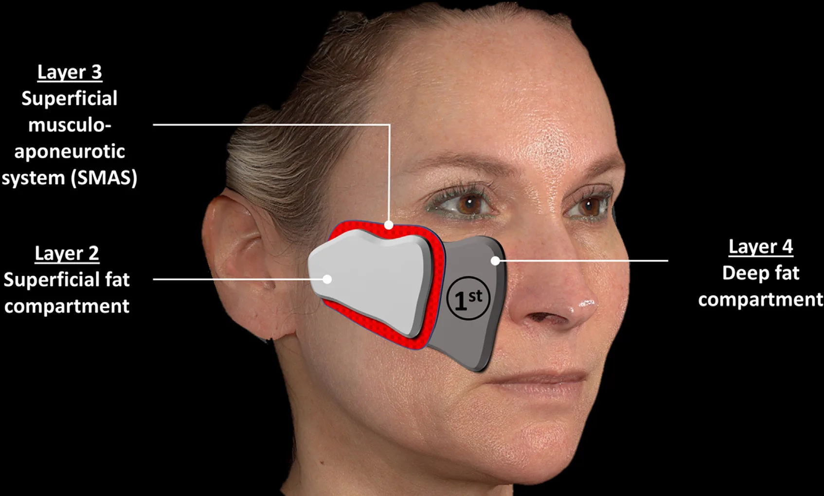 Fig 1. Deep-first as the cross-region default. Layer 4 (deep fat) is the structural fundament built before the mobile superficial soft tissue; the facial vasculature runs mainly in the superficial planes, so the supraperiosteal/deep-fat plane is generally both the support layer and the low-risk corridor — the rule the plane grid obeys except in the nose, NLF, temple, lip and neck. — (Freytag/Cotofana, 2022, p.6).
> Fuentes: [4].
Fig 1. Deep-first as the cross-region default. Layer 4 (deep fat) is the structural fundament built before the mobile superficial soft tissue; the facial vasculature runs mainly in the superficial planes, so the supraperiosteal/deep-fat plane is generally both the support layer and the low-risk corridor — the rule the plane grid obeys except in the nose, NLF, temple, lip and neck. — (Freytag/Cotofana, 2022, p.6).
> Fuentes: [4].
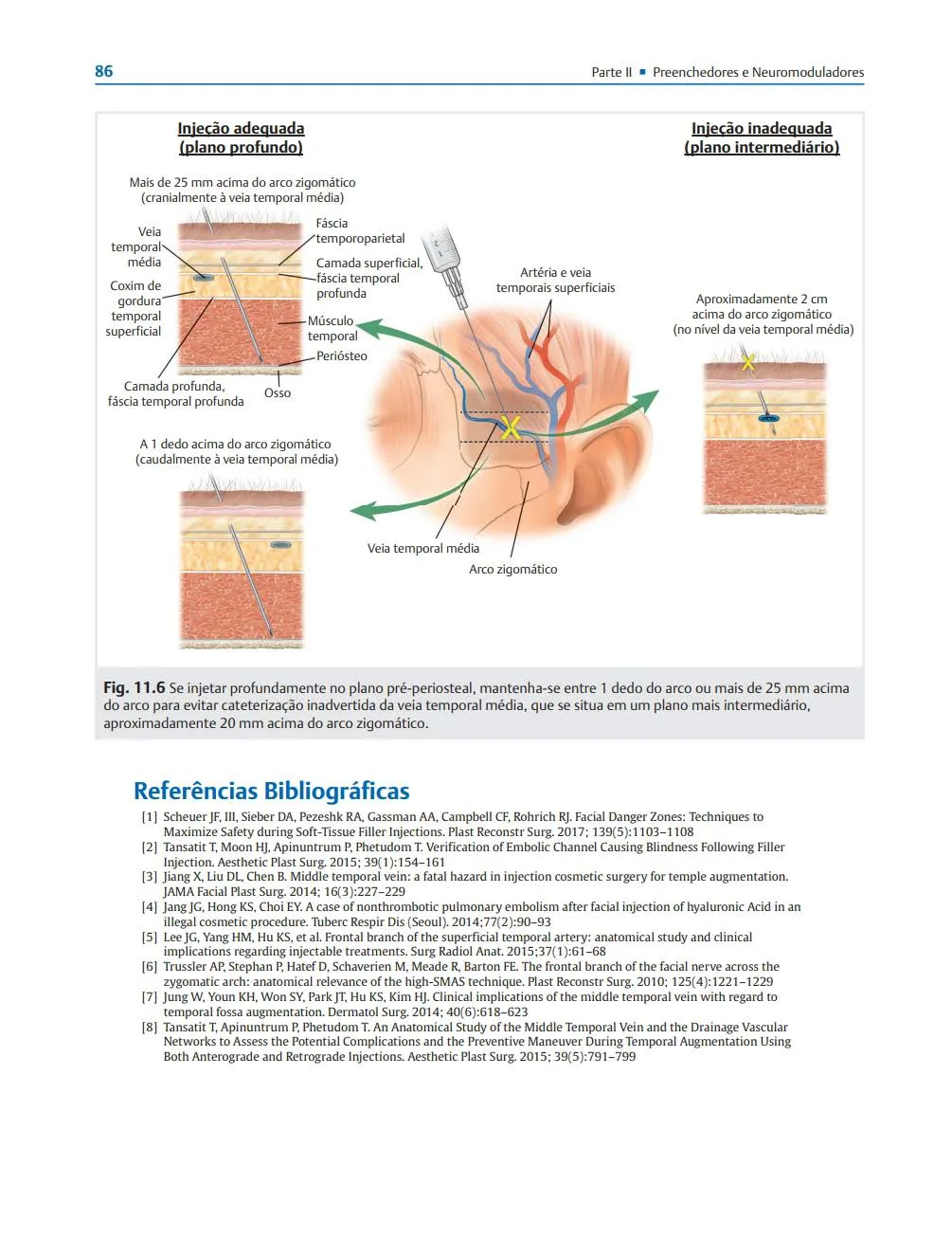 Fig 2. Why the temple is the exception that proves the rule: two safe planes (deep pre-periosteal cranial or caudal to the middle temporal vein, and superficial subdermal) bracket a lethal middle ground. "No bone under the tip" in the temple means the needle is in the venous danger layer, not deep. — (Rohrich, Zonas Faciais de Perigo, 2020, p.86).
> Fuentes: [12].
Fig 2. Why the temple is the exception that proves the rule: two safe planes (deep pre-periosteal cranial or caudal to the middle temporal vein, and superficial subdermal) bracket a lethal middle ground. "No bone under the tip" in the temple means the needle is in the venous danger layer, not deep. — (Rohrich, Zonas Faciais de Perigo, 2020, p.86).
> Fuentes: [12].
The principle, stated once. Facial soft tissue is layered — skin, superficial fat, SMAS, deep fat, periosteum — and the deep fat compartments deflate while the superficial ones descend [1][2][3]. Rebuilding the deep fundament first lets the overlying tissue reposition and move naturally, and it is generally the safer deposit because the named arteries travel in the more superficial planes [4]; Fig 1 shows the layer relationship — the deep fat compartment (layer 4) as the fundament built before the SMAS and superficial fat. That single fact makes "supraperiosteal / deep-fat, on bone, medial to the line of ligaments" the answer in the midface (D6, D7), the chin (D11), the gonial angle and prejowl (D12), and the structural support behind the brow (D2) and the nasolabial fold (D9). Where the region column reads "supraperiosteal," it is the same decision repeated, and the [n] traces to the same small set of anatomy sources [1][2][5][6][7].
The line of ligaments orders the midface (D6/D7). Medial to the line — over the anterior maxilla — a deep bolus projects the overlying cheek; lateral to it, the same plane repositions / lifts with less volume [6][7]. This is why D7's "default plane" is not one plane but one plane read against one landmark, and why the anteromedial cheek and the submalar hollow, identical from the front, take opposite answers: bone under the finger → supraperiosteal bolus; soft tissue over the masseter → a subcutaneous lamina, never a "deep" bolus into a space with no floor [6].
The five exceptions are the reason this grid exists. - Nose (D8): the deep midline is the only corridor, not merely the preferred one. The vessels are superficial and lateral on the fibromuscular layer, so strict-midline supraperiosteal/supraperichondrial deposition is the low-risk route — but the territory is terminal under a tight envelope, so the "safe plane" is safe only at low volume and low pressure [11][19]. The previously operated nose is a separate risk category and often not a candidate at all [28]. - Nasolabial fold (D9): there is no reliably safe plane in the sulcus. The facial artery ascends beside the fold and its depth is variable from subcutaneous to sub-periosteal, so the region's rule is to treat the cause (pyriform and deep-cheek support on bone) and put only a small residual subdermal pass in the fold itself [7][25][26]. - Temple (D3): two safe planes (deep pre-periosteal on bone, and superficial subdermal) bracket a lethal intermediate plane where the sentinel and middle temporal veins run. The plane discipline is binary — feel bone or stay subdermal; the middle is the venous-embolism layer [12][40] (Fig 2). - Lip (D10): safety is superficial and paramedian, the inverse of the facial default. The labial artery is submucosal in 78.1 % of subjects, so the deep and midline planes are the danger and the subcutaneous paramedian plane is the target [24]. - Neck (D13): there is no volumising plane to choose. Volume is a liability; the only sites where adding product helps are the chin and mandibular border, which rebuild the frame. The plane that matters is the one to avoid — deep or high platysma below the thyroid cartilage, where the toxin reaches the swallowing muscles [1][43][44].
Consensus: every region defaults to the deepest plane that achieves the goal, because it is generally the safest and the most structural [1][4][9]. Discrepancy that changes the gesture: deep volumising bolus (refill the compartment) versus superficial lifting/lamina (reposition or soften) is decided by the goal, not averaged — a hollow fossa or a flat maxilla takes the deep bolus, a lateral-laxity or a submalar shadow takes the superficial pass, and a mid-depth compromise is the one plane that is wrong in both the temple and the cheek [6][12].
Trampa clásica (plane): injecting a "deep" bolus where there is no bone under the needle — the submalar hollow (D7), the temple loose-areolar layer (D3), the neck. The word "deep" is only safe when the tip is on periosteum; without bone it means the intermediate soft-tissue plane where the veins and the motor nerves live. The bedside check is the same in every region: press to bone before you call the plane deep.
D0.2 · Which product / rheology per region
The grid. Brands are class examples, not doses. "First choice" is the structural/deep product; "soft option" is the superficial/mobile product; "reversible" flags whether hyaluronidase can undo it.
| # | Region | First choice (deep / structural) | Soft / superficial option | Reversible | Do NOT use here | Src |
|---|---|---|---|---|---|---|
| D1 | Forehead & glabella | temple: HA (med–high G′) · hyperdilute CaHA · PLLA | forehead: low-hydrophilicity HA, minimal | HA yes; CaHA/PLLA no | any bolus product in the glabella | [9][14][22] |
| D2 | Brow & tail | high-G′ / high-cohesivity HA deep (temple, supra-brow) | soft low-G′ HA subdermal; CaHA ± hyperdilute | HA yes | direct brow filler of any class | [9][14] |
| D3 | Temple | HA med–high G′ deep, low–med subdermal | hyperdilute CaHA / booster for skin quality | HA yes; CaHA/PLLA no | PMMA / permanent | [13][14][15] |
| D4 | Upper periorbital & eyelid | low-hydrophilicity HA, low–med G′ (water uptake first, G′ second) | higher G′ reserved for deep rim / ROOF | HA yes | hydrophilic gel that swells at 3 wk; permanent anywhere in the lid | [3][16] |
| D5 | Tear trough | low-G′ HA (Volbella/Belotero/Redensity II/RHA 2) or firm NASHA (Restylane Eyelight) | blended low-viscosity gel superficial | HA yes | hydrophilic HA (Juvéderm Ultra) · CaHA/PLLA/PMMA in the trough | [16] |
| D6 | Malar / zygoma | high-G′ / high-cohesivity HA (VYC-20L class) | med-G′ low-hydrophilia deep-cheek HA; CaHA 1:1 durable | HA yes; CaHA no | superficial hydrophilic deposit over SOOF | [1][9][14] |
| D7 | Anteromedial cheek & submalar | high-G′ cohesive HA, low-hydrophilia (keep the malar area dry) | soft low-G′ HA for the submalar lamina | HA yes | firm structural gel in the submalar subcutaneous plane | [6][9][13] |
| D8 | Nose | HA only — it is the sole reversible filler | (same; no second class) | HA yes — the whole point | CaHA, PLLA, PCL, PMMA, silicone — any permanent/semipermanent | [11][19] |
| D9 | Nasolabial fold | high-G′ cohesive HA (or CaHA) to rebuild the pyriform platform | soft low-G′ / resilient HA to soften the mobile fold | HA yes; CaHA no | permanent fillers (PMMA, polyacrylamide) — late biofilm/granuloma | [7][9] |
| D10 | Lip & perioral | firmer high-G′ HA for the white roll / vertical columns | soft cohesive low–mid-G′ HA for the red-lip body (Kysse/RHA class) | HA yes | permanent product; a firm gel driven deep and midline | [24] |
| D11 | Chin | high-G′ "bone-mimic" HA (Volux VYC-25L, 25 mg/mL) or Voluma VYC-20L; CaHA | soft HA / cannula blend for the crease | HA yes; CaHA no | CaHA/PMMA when a dissolver may be needed | [17][18] |
| D12 | Jawline & angle | high-G′ HA (Volux/VYC-25L) or CaHA on bone | high-G′ HA or CaHA subdermal on the body | HA yes; CaHA no | soft low-G′ gel spread to "build" a border | [18][23] |
| D13 | Neck & cervicomandibular | no volumiser — toxin (bands/Nefertiti) and DCA (fat) do the work | hyperdilute CaHA (1:2–1:4) / PLLA / PN / light HA for skin quality only | HA yes; CaHA/PLLA no | PLLA in thin neck skin (nodule); HA in skin of colour (PIH); a structural filler in the neck | [22][43][44] |
The rheology ladder, built once and reused everywhere. Two gel properties decide the product: G′ (elastic modulus — firmness, lift capacity) and cohesivity (how strongly the gel holds together and resists migration); hydrophilicity (water uptake) is the tie-breaker in thin-skinned regions [9][13]. The cross-region rule is monotone: the deeper and more structural the target, the higher the G′ and cohesivity; the more superficial and mobile the target, the softer and lower the G′ [13].
Target layer                         →  Product property                 →  Regions
supraperiosteal / on bone (project)  →  high G′, high cohesivity          →  D6 D7 D9 D11 D12 (chin/angle "bone-mimic")
deep fat (restore, integrate)        →  medium G′, low hydrophilicity     →  D6 D7 (deep medial cheek)
subcutaneous / mobile (soften)       →  soft, low G′                      →  D9 fold · D10 red lip · D3 subdermal
periorbital / thinnest skin          →  low hydrophilicity FIRST, then G′ →  D4 D5 (Tyndall + delayed swelling)
skin quality only (no projection)    →  hyperdilute CaHA / PLLA / booster →  D3 D6 D13 necklace lines
Why the periorbital regions invert the usual order (D4, D5). In the lid and the trough the skin is the thinnest of the body, so the first filter is water uptake, not G′: a hydrophilic gel that looks perfect at 24 h swells the lid at three weeks and can seed months of malar edema [3][16]. This is the one place where the region chapter explicitly ranks hydrophilicity above lift capacity, and why a firm low-water NASHA (Restylane Eyelight, FDA-approved 2023 for infraorbital hollows) reads as a legitimate exception to the "soft-and-low-G′-superficial" rule [16].
The irreversibility rule is a single cross-region constant. HA is the defensible default in almost every region because hyaluronidase can undo it [29][31]. CaHA, PLLA, PMMA, PCL and fat are volumising or biostimulating but not HA-reversible, so they are acceptable only where the plane is deep, the vessel risk is mapped, and the patient accepts permanence [22]. The nose takes this to its logical end — HA only, no exception — because hyaluronidase is the sole antidote to a nasal occlusion and a permanent product in a terminal territory has no rescue [11]. The tear trough and the thin neck repeat the caution for a different reason: an irreversible nodule or a Tyndall over skin < 1 mm thick is visible and hard to fix [16].
Consensus: high-G′/cohesive deep, soft/low-G′ superficial, HA-reversible by default [9][13]. Discrepancy that changes the gesture: the low-G′ "school" (Volbella/Belotero for the trough) versus the firm-NASHA option (Restylane Eyelight) for the same region — decide by skin thickness and shadow type, do not average the two into a mid-viscosity guess [16].
Trampa clásica (product): putting the firm structural gel in the superficial plane or the soft gel in the deep plane — the reverse of what each layer needs. A high-G′ gel in the mobile lip or submalar subcutaneous plane is palpable and ages badly; a soft low-G′ gel dumped deep to "build" a jawline border spreads and adds weight without defining. The product follows the plane, and the plane follows the missing layer [13][18].
D0.3 · Which instrument per region
The grid. "Needle" is the deep, precise, bone-contact tool; "cannula" is the blunt, single-entry tool for layering and large areas. The gauge column reprints the region's stated calibre.
| # | Region | Needle (deep / precise) | Cannula (layer / area) | Gauge · length | Src |
|---|---|---|---|---|---|
| D1 | Forehead & glabella | 30G, intradermal glabella; toxin 30G IM | from a lateral entry for the forehead | 30G needle · cannula lateral | [9][33] |
| D2 | Brow & tail | supraperiosteal micro-bolus to bone | temple / lateral support | needle deep + cannula | [9][33] |
| D3 | Temple | 27G sharp to bone (deep) | 22–25G blunt, lateral entry (subdermal) | 27G / 22–25G | [12][13] |
| D4 | Upper periorbital & eyelid | 30G bolus (deep sulcus); 30/32G toxin | 25/27G (ROOF, blending) | 30G / 25–27G | [9][16] |
| D5 | Tear trough | 30G vertical serial puncture on rim | 25–27G, single lateral port (fewer entries) | 25–27G cannula / 30G | [16][33] |
| D6 | Malar / zygoma | 27G to bone, aspirate (deep bolus) | 25G blunt (lateral / superficial blend) | 27G / 25G | [1][9] |
| D7 | Anteromedial cheek & submalar | 25–27G to bone (deep bolus) | 22–25G blunt (deep or the submalar lamina) | 25–27G / 22–25G | [6][13] |
| D8 | Nose | 25–30G sharp, deep midline, glabellar compression | 25G blunt, single infratip entry; 21–23G rigid preferred to hold the plane | 25–30G / 21–25G | [11][13] |
| D9 | Nasolabial fold | pyriform bolus to bone | cannula preferred; larger-barrel syringe for lower thumb pressure | cannula default | [9][33] |
| D10 | Lip & perioral | 27–30G (white-roll precision) | 25–27G, single port lateral to the commissure | 27–30G / 25–27G | [24][33] |
| D11 | Chin | needle to bone, midline (projection) | cannula (contour, labiomental crease, jawline blend) | needle / cannula | [9][17] |
| D12 | Jawline & angle | 25–27G, bone contact (angle / prejowl) | 25G 50 mm or 27G (the body / border) | 25–27G / 25G·50 mm | [9][18] |
| D13 | Neck & cervicomandibular | toxin needle, superficial; DCA 30G on a 1-cm grid | (not a cannula region for the injectables that work) | 30G grid (DCA) | [43][44] |
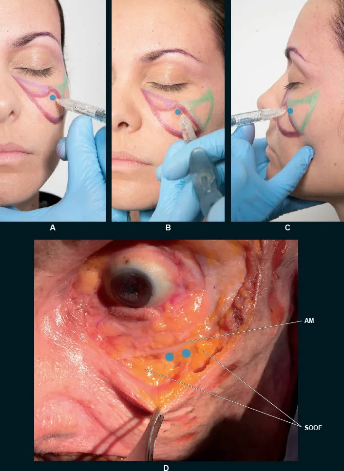 Fig 3. The needle-to-bone deep bolus, panel by panel: the tool that puts a controlled deposit on periosteum where a cannula would flex out of plane. Panel D shows why the deposit stays lateral — the medial approach nears the infraorbital foramen. — (Vieira Braz, Atlas de Anatomia e Preenchimento Global da Face, 2017, p.328).
> Fuentes: [13].
Fig 3. The needle-to-bone deep bolus, panel by panel: the tool that puts a controlled deposit on periosteum where a cannula would flex out of plane. Panel D shows why the deposit stays lateral — the medial approach nears the infraorbital foramen. — (Vieira Braz, Atlas de Anatomia e Preenchimento Global da Face, 2017, p.328).
> Fuentes: [13].
The instrument follows the plane, not the operator's habit. A needle is rigid, so it places a bolus exactly on periosteum — the reason it is the tool for every "deep supraperiosteal to bone" cell above (D3, D6, D7, D11, D12 angle, D8 midline); Fig 3 documents the needle-to-bone malar deposit panel by panel, including the cadaver correlate that shows why the deposit stays lateral to the infraorbital foramen. A cannula is blunt and flexible, so it covers a large area or a mobile plane from one entry with fewer vessel hits and less bruising — the reason it is preferred for the tear trough (D5), the nasolabial fold (D9), the submalar lamina (D7) and lip layering (D10). The flexibility is also its failure mode: a cannula can bend out of the intended plane, which is exactly why it is not the default for a precise on-bone deposit [13].
Controversy — cannula versus needle safety (the cross-region technique row). The old teaching that a blunt cannula is categorically safer than a needle does not survive the data, and the two schools disagree in a way that changes the gauge you pick:
Consensus: vascular occlusion happens with both tools; a 27G cannula behaves much like a needle; and no instrument makes any region on this page "safe" [11][32][34]. Discrepancy: cannula-preferring (a blunt tip displaces rather than pierces a vessel, lowering the occlusion signal in vessel-dense regions) versus anti-small-cannula — Zhou's series found 25 of 28 severe HA emboli (nine with blindness) were injected with a cannula of 22–27G, not a needle, and the CMAC consensus is to avoid cannulas smaller than 25G [32]. Decide by gauge and region, not by ideology: a rigid ≥ 25G cannula in a vessel-dense mobile plane (tear trough, NLF, lip) exploits the blunt-tip advantage; a small flexible cannula (< 25G) forfeits it and is where the severe complications cluster; and a precise deep bolus on bone is a needle's job because a cannula cannot be trusted to hold the supraperiosteal plane [32][33][34]. Never read "cannula" as a licence to inject faster or more carelessly — the preconception of safety is itself a documented risk factor.
The gauge is not cosmetic. A larger-diameter tool is less likely to enter and sit inside a small vessel; a very fine tool is more likely to cannulate one [11][32]. This is why the nose specifies a rigid 21–23G to reach and hold the deep midline (a long flexible microcannula drifts into the subcutaneous plane and threatens the dorsal nasal artery), and why the trough and NLF specify ≥ 25G cannulas rather than the finest available [11]. The length matters too: the jawline body uses a 50 mm cannula to run the border from one distal port.
Trampa clásica (instrument): reaching for a small flexible cannula "for safety" in a deep region and losing the plane. Below the zygomatic body, in the temple loose-areolar layer, or at the nasal dorsum, a bent cannula tip lands in soft tissue or the subcutaneous plane instead of on bone — the false-security failure Zhou describes. If the target is a precise deep bolus, the tool is a needle to bone with aspiration and motion; if the target is a diffuse mobile plane, the tool is a rigid ≥ 25G cannula from a distal entry [32][33].
D0.4 · Ceiling volume per region
The grid. Three numbers per region: the micro-aliquot per point, the per-side start, and the ceiling — per session or the point past which the region over-projects. Filler volumes per side unless stated. Reprinted from each region; where the corpus does not standardise a maximum, the cell says so rather than inventing one.
| # | Region | Per point / aliquot | Per side · start | Ceiling (session / before overfill) | Src |
|---|---|---|---|---|---|
| D1 | Forehead & glabella | glabella microdroplets (default: avoid) | forehead small, temple-first | reassess volume need at 2 weeks; never forehead + glabella same session | [9] |
| D2 | Brow & tail | supra-brow ≤ 0.1 mL micro-bolus | temple 0.5–1.0 mL/side | tail lift is millimetric (~1–3 mm); temple, not brow, holds the volume | [9][14] |
| D3 | Temple | small single deep bolus per point | titrate to contour, build across sessions | per-side maximum [MATERIAL GAP] (dose facet thin); never import cadaver multi-cc fill volumes |
[14][15] |
| D4 | Upper periorbital & eyelid | 0.05–0.1 mL micro-aliquots | sulcus 0.1–0.3 mL/side, deliberate under-correction; ROOF 0.2–0.4 mL/side | the hollow, not the lid; hydrophilic swelling is the hidden ceiling | [9][16] |
| D5 | Tear trough | deep ≤ 0.05–0.1 mL/point ("balls up" if larger) | start 0.5 mL/side | hard ceiling ~1 mL/side (Wong deep-fat 0.5–0.8) | [16] |
| D6 | Malar / zygoma | Ck1 0.1–0.3 · Ck2 0.2–0.4 · Ck3 0.1–0.3 mL; boluses < 0.3 mL | per-point, staged | ~4 mL of HA per midface session; more across sessions, not one sitting | [9] |
| D7 | Anteromedial cheek & submalar | DMCF small deep bolus, commonly ~0.2–0.5 mL | per side, staged | per-point maximum [MATERIAL GAP]; the ceiling is the overfilled face, reached a little too much many times |
[6][9] |
| D8 | Nose | microboluses ≤ 0.05 mL; radix 0.05–0.15 · supratip 0.025 · tip 0.05–0.15 · columella ≤ 0.30 mL | — | R4P mean 0.65 ± 0.17 mL total; Jung dual-plane 0.4–1.2 mL | [19][20] |
| D9 | Nasolabial fold | deep pyriform bolus 0.2–0.3 mL/point | subdermal fold 0.4–0.8 mL/side; deep fat linear ~0.4 mL | never a bolus in the fold; attenuate, never erase | [7][9] |
| D10 | Lip & perioral | per point 0.01–0.02 mL microbolus (never stationary) | 0.5–1.0 mL/lip, conservative start | ceiling is proportion (lower > upper, ~1:1.6), not a number | [24] |
| D11 | Chin | MD Codes 0.3 mL (needle) / 0.5 mL (cannula) per point | asymmetry 0.2–0.3 · mild 0.6–0.8 mL | moderate-severe 1.3–1.5 mL total; over-projection blunts the labiomental angle | [9][17] |
| D12 | Jawline & angle | angle 0.4–0.8 · body 0.4–0.8 · prejowl 0.2–0.5 mL/side | per-segment start | ceilings/side: angle ~1.2 · body ~1.0 · prejowl ~0.6 mL; VYC-25L pivotal ~3–4 mL total both sides; Voluma-type ≤ 2 mL/area | [18] |
| D13 | Neck & cervicomandibular | bands 2 U/point; DCA 0.2 mL/point on a 1-cm grid | bands ≤ 15–20 U/band; DCA ≤ 10 mL (100 mg)/session | toxin ≤ 30–40 U ona/session; DCA ≤ 6 sessions ≥ 4 wk apart; no filler volume — the neck is not filled | [43][44] |
The overfilled face is a rate, not a dose. The single cross-region volume lesson is that the ceiling is reached by adding a little too much, many times, not once [6][9]. Every region that lists a per-session maximum (midface ~4 mL, jawline ~3–4 mL, chin ≤ 1.5 mL) frames it as a stop for this sitting with more built across visits, because the tissue keeps the previous deposit and a fresh full dose on top of an incompletely-settled one is how the "pillow" cheek, the heavy jaw and the sausage lip are manufactured. The recurring instruction — reassess at 2 weeks, stage the rest — is the same brake in thirteen regions.
Undercorrection is the rule where the skin is thin. The periorbital regions (D4, D5) and the lip (D10) specify deliberate under-correction: 0.1–0.3 mL per side in the sulcus, a ~1 mL/side hard ceiling in the trough, micro-aliquots of 0.05–0.1 mL, and microboluses of 0.01–0.02 mL in the vermilion [16][24]. The reason is mechanical, not aesthetic timidity — a larger aliquot in the medial trough "balls up," and a hydrophilic gel that looks correct on the table swells into malar edema at three weeks [16].
Two regions honestly refuse to give a number. The temple (D3) and the anteromedial cheek/submalar (D7) both carry a [MATERIAL GAP]: the corpus does not standardise a per-side or per-point maximum, the dose-parameters facet is chronically thin, and RCT-grade volumes exist only for specific products (temple HA VYC-20L, temple PLLA) that are not a licence to import blind [14][15]. The grid prints the gap rather than a fabricated ceiling — the explicit refusal is the honest answer, and the region chapter proves the silence with its retrieval.
The nose is the region with the smallest total and the most exact breakdown. Where the cheek tolerates 4 mL, the whole nose sits at a mean of 0.65 ± 0.17 mL and is deposited in microboluses of ≤ 0.05 mL with a stop-and-look between each — radix 0.05–0.15, supratip 0.025, tip 0.05–0.15, columella ≤ 0.30 mL [19][20]. The contrast is the table's point: pressure that dissipates in malar fat is transmitted in a terminal territory under a tight envelope, so the same volume that is innocuous in the cheek embolises in the nose.
Consensus: small aliquots, staged sessions, reassess at 2 weeks, and a per-session stop [9]. Discrepancy that changes the gesture: where a hard millilitre ceiling exists (trough ~1 mL/side, jawline segments), use it; where the corpus refuses one (temple, submalar), titrate to contour and never transplant another region's or a cadaver study's volume [14][15]. The one number never to average is a per-point dose across two regions with different tissue floors.
Trampa clásica (volume): chasing a fixed millilitre count instead of a contour. The chin that takes 1.5 mL in one face over-projects in another; the trough that accepts 0.5 mL "balls up" at 0.8; the temple has no published per-side maximum at all. The volume is titrated to the endpoint (contour, symmetry at 2 weeks), and the ceiling is the session stop, not a target to reach [6][16].
D0.5 · Dangerous vessel per region
The grid. The vessel that occludes, the depth or landmark that finds it, and where a retrograde column drains — because the destination (ophthalmic = blindness; valveless vein = lung) is what makes a bad injection catastrophic rather than a bruise.
| # | Region | Killer vessel(s) | Depth / landmark | Drains to → consequence | Src |
|---|---|---|---|---|---|
| D1 | Forehead & glabella | supratrochlear (STr) + supraorbital (SOA) arteries | STr ~1.7–2.2 cm lateral of midline; SOA ~1 cm lateral at the notch; both deep then subcutaneous 15–20 mm above the rim | terminal ophthalmic → central retinal artery → blindness | [26][39] |
| D2 | Brow & tail | STr + SOA (arterial); temple MTV (venous) | supra-brow bolus; ≥ 1 cm above rim | ophthalmic → blindness; MTV → lung | [26][27] |
| D3 | Temple | middle temporal vein (valveless, ~2 cm above + parallel to arch); frontal branch STA; deep temporal aa. | MTV in fat between the deep-temporal-fascia laminae; STA trunk ~10 mm anterior to tragus | MTV → non-thrombotic pulmonary embolism; STA → retrograde blindness + scalp necrosis/alopecia | [12][40] |
| D4 | Upper periorbital & eyelid | STr + SOA | brow / glabella / deep sulcus | ophthalmic → blindness (retina tolerates ~90 min ischaemia) | [26][27] |
| D5 | Tear trough | angular + infraorbital arteries | on rim, medial trough | ophthalmic → blindness; behind septum → recurrent swelling | [26][27] |
| D6 | Malar / zygoma | angular / facial; zygomaticofacial; infraorbital | infraorbital foramen ~1 cm (0.6–1.2) below the rim at the medial-iris line | angular → ophthalmic → blindness; V2 dysaesthesia | [1][26] |
| D7 | Anteromedial cheek & submalar | angular + infraorbital; submalar: parotid duct, facial vessels | pyriform / nasojugal / deep medial cheek; infraorbital nerve ~8–10 mm below rim | angular/infraorbital → dorsal nasal → ophthalmic → blindness; sialocele | [6][25] |
| D8 | Nose | dorsal nasal, lateral nasal, columellar → angular | lateral & superficial on the fibromuscular layer | ophthalmic → blindness; pooled NSR VO 0.35 %, vision loss 0.09 %, necrosis 0.08 % (n=8604) | [21][28] |
| D9 | Nasolabial fold | facial artery → angular artery | no reliably safe plane; depth variable subcutaneous→sub-periosteal; upper (perialar) fold worst | angular → ophthalmic → blindness; skin necrosis | [25][26] |
| D10 | Lip & perioral | superior + inferior labial arteries (complete ring) | deep ≥ 4 mm, enter the lip ~1 cm from the commissure; submucosal in 78.1 % | inferior labial → facial → ophthalmic → blindness reported after chin/lower-lip injection | [24][26] |
| D11 | Chin | mental artery (lateral, below the 2nd premolar) | stay midline for needle-to-bone | chin/lower-lip necrosis; no blindness route | [8][25] |
| D12 | Jawline & angle | facial artery at the antegonial (premasseteric) notch (palpable); facial vein | on the lower border anterior to the masseter, deep fat; marginal mandibular nerve below the border in 19–32 % | chin/lip/floor-of-mouth embolism; asymmetric smile (nerve) | [25] |
| D13 | Neck & cervicomandibular | not a vessel — the killer is dysphagia | below the thyroid cartilage, no toxin at any dose; DCA no-zone 1–1.5 cm below the mandibular border | deep/high platysma → swallowing muscles → dysphagia / aspiration; marginal mandibular (DCA) | [43][44] |
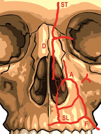 Fig 4. One picture for the whole "blindness" column. The external-carotid facial system (F, SL, A) and the internal-carotid ophthalmic system (ST, D) are a single continuous network across the midline, so a retrograde intra-arterial column from the fold, nose, glabella, cheek or lip can reach the retina. This is why every region except the chin, jaw and neck lists the ophthalmic artery as its terminal destination. — (Jones, Injectable Fillers, 2019, p.215).
> Fuentes: [26].
Fig 4. One picture for the whole "blindness" column. The external-carotid facial system (F, SL, A) and the internal-carotid ophthalmic system (ST, D) are a single continuous network across the midline, so a retrograde intra-arterial column from the fold, nose, glabella, cheek or lip can reach the retina. This is why every region except the chin, jaw and neck lists the ophthalmic artery as its terminal destination. — (Jones, Injectable Fillers, 2019, p.215).
> Fuentes: [26].
One anastomosis explains eleven of the thirteen rows. The external-carotid (facial) and internal-carotid (ophthalmic) territories connect through the angular–dorsal nasal–supratrochlear anastomosis, so a retrograde column of filler driven into any facial-artery tributary can reach the central retinal artery [26][27] (Fig 4). That is why "→ ophthalmic → blindness" is the destination for the forehead, brow, periorbital, trough, cheek, submalar, nose, NLF and lip. The retina infarcts after roughly 90 minutes of ischaemia, which is the clock behind every "any visual symptom is a time-critical emergency" line [26].
The two rows that break the pattern are the two that make the table useful. - Temple (D3) adds a venous mechanism the arterial regions do not have: the middle temporal vein is large, thin-walled and valveless, sitting ~2 cm above and parallel to the zygomatic arch in the fat between the deep-temporal-fascia laminae, so a mid-plane deposit can embolise to the pulmonary circulation as well as, via the STA anastomosis, to the eye [12][40]. It is the classic venous-embolism site in the face — hence the plane discipline in D0.1. - Chin, jaw and neck (D11, D12, D13) have no blindness route. The chin's risk is the mental artery (lateral, below the second premolar) and the answer is to stay midline; the jaw's is the facial artery at the palpable antegonial notch plus the marginal mandibular nerve that runs below the border in up to a third of sides; the neck's is not a vessel at all but the depth of the platysma over the swallowing muscles [8][25][43]. This is why the lower face is where a beginner is sent and the periorbital/nasal midface is not.
Depth is a number worth memorising per region. The forehead vessels are deep in their first 1.5 cm then subcutaneous 15–20 mm above the rim, so injections near the rim stay superficial and injections high on the forehead go supraperiosteal [26]. The infraorbital foramen sits ~1 cm below the rim on the medial-iris line and the infraorbital nerve ~8–10 mm below it, so the cheek is approached from lateral, never speared from below [1][6]. The labial arteries run deep (≥ 4 mm) and submucosal in most subjects and enter ~1 cm from the commissure, so the lip is threaded superficial and paramedian and the commissure is never bolused [24].
Emergency card, cross-region. For any HA vascular sign (blanching, disproportionate pain, dusky livedo, delayed capillary refill): stop, high-dose pulsed hyaluronidase, flood the ischaemic territory and repeat hourly until colour and capillary refill return, aiming to finish within 72 h; per-area dosing scales with the territory (≈ 500 IU/area, more for the nose) [29][31]. For any visual symptom, ocular pain or ophthalmoplegia: ocular emergency, do not "wait and see," involve ophthalmology, follow the UK vision-loss consensus [30]. For cough/breathlessness during a temple injection: suspect venous embolism, stop, oxygen [12]. Ultrasound (Doppler) mapping before, and guidance during, is the fastest-moving safety layer and turns a statistical vessel into this patient's vessel [40][41].
Trampa clásica (vessel): treating the depth landmark as generic. "Deep is safe" holds for the cheek and chin but is exactly wrong in the temple (deep-fat plane = the veins), the nose (the deep midline is safe only at micro-volume) and the lip (deep = the arterial ring). The vessel, its depth and its drainage are region-specific, and the destination — retina, lung, or a nerve — is what the injector must know before the needle moves [12][24][26].
D0.6 · Toxin per region
The grid. Doses in onabotulinumtoxinA (ona) units; abo (Dysport/Azzalure) is roughly 1:2.5–3 [38][42] and never interchangeable unit-for-unit. "Target muscle" is the aesthetic indication in that region; several regions have no primary toxin target and the cell says so.
| # | Region | Target muscle(s) | Units per point · per side | Antagonist to spare / red line | Src |
|---|---|---|---|---|---|
| D1 | Forehead & glabella | glabellar complex (corrugator + procerus + depressor supercilii); frontalis | glabella 20 U total, 5 points × 4 U; frontalis 10–20 U, 4–6 points × 2–4 U; lateral orbicularis tail 1–2 U/side | frontalis (sole brow elevator); lateral corrugator point ≥ 1 cm above the bony rim (below → lid ptosis) | [38][9] |
| D2 | Brow & tail | lateral orbicularis oculi tail; glabellar depressors | brow-tail 2–5 U, 1 point/side, ≥ 1 cm outside the rim; medial-brow lift = glabella 20 U | do not weaken frontalis (drops the brow); net tail lift ~1–3 mm | [38] |
| D3 | Temple | none aesthetic (temporalis toxin = bruxism/masseteric hypertrophy, cross-ref) | — | not a cosmetic temple target | [—] |
| D4 | Upper periorbital & eyelid | orbicularis oculi (crow's feet); glabella; lateral tail | crow's feet 4 U/point × 3 = 12 U/side (24 U total); brow-tail 1–2 U/point (2–4 U/side) | stay ≥ 1–1.5 cm lateral of the canthus (lateral rectus → diplopia); off the zygomatic arch (lip droop) | [38] |
| D5 | Tear trough | pretarsal orbicularis oculi (only role) | 1–2 U/side (max 3–4), midpupillary, 2–3 mm below margin | only with a good snap test; over-dose → lid malposition | [38] |
| D6 | Malar / zygoma | no primary IM target; microbotox intradermal for pore/sheen only | intradermal microdroplets | filler must not paralyse the elevators the toxin needle would reach | [44] |
| D7 | Anteromedial cheek & submalar | none owned (nasalis / LLSAN are borders, cross-referenced) | — | not a primary toxin region | [—] |
| D8 | Nose | nasalis (bunny lines); depressor septi nasi (droopy tip); dilator naris (alar flare) | bunny lines 1.25–5 U/side; tip 2–4 U at columella base; alar 2–5 U/side | spare levator labii superioris (lip ptosis) | [38] |
| D9 | Nasolabial fold | not a fold tool; LLSAN only for a gummy smile | LLSAN 3 U/side (~6 U total) | over-weakening LLSAN drops the upper lip; toxin "to relax the fold" = worst benefit-risk | [38] |
| D10 | Lip & perioral | orbicularis oris (lip flip); LLSAN (gummy smile); DAO | lip flip 2–4 U total; gummy smile 1–2 U/side (Yonsei); DAO 2–5 U/side | tiny orbicularis-oris dose (oral competence); effect 6–8 wk | [38] |
| D11 | Chin | mentalis; depressor anguli oris (DAO) | mentalis 2–8 U (12 U abo), 1–2 deep low-midline points; DAO 2–5 U/side | mentalis too high hits orbicularis oris; DAO too medial hits depressor labii → asymmetric smile | [9] |
| D12 | Jawline & angle | masseter (width); platysma (Nefertiti); DAO; mentalis | masseter 20–30 U ona × 3 points; platysma Nefertiti ~15–20 U; DAO 4–8 U; mentalis 4–8 U | masseter ≥ 1 cm above the border, anterior to the parotid; MMN below border in 19–32 % | [42][43] |
| D13 | Neck & cervicomandibular | the region's core: platysma bands; Nefertiti; microbotox | bands 2 U/point, 3–6 points/band, ≤ 30–40 U ona/session (75–100 abo); Nefertiti 2–3 U/point, ≤ 20–25 U/side; microbotox 28 U/mL/side | ⚠ never below the thyroid cartilage, never deep (dysphagia) | [43][44] |
 Fig 5. The one dose every injector knows, as the reference the toxin grid is built around: glabella ≈ 20 U in five points, the lateral correctors kept ≥ 1 cm above the orbital rim so the toxin does not diffuse through the septum to the levator. Every region's toxin cell is a variation on placing units against a target muscle while sparing its antagonist. — (Lipham, Cosmetic and Clinical Applications of Botox and Dermal Fillers, 2015, p.95).
> Fuentes: [38].
Fig 5. The one dose every injector knows, as the reference the toxin grid is built around: glabella ≈ 20 U in five points, the lateral correctors kept ≥ 1 cm above the orbital rim so the toxin does not diffuse through the septum to the levator. Every region's toxin cell is a variation on placing units against a target muscle while sparing its antagonist. — (Lipham, Cosmetic and Clinical Applications of Botox and Dermal Fillers, 2015, p.95).
> Fuentes: [38].
Distances keep toxin safe, not doses. The cross-region rule is that the complications of aesthetic toxin are almost never "too many units" — they are the wrong muscle or the wrong distance: the lateral corrugator point below 1 cm above the rim diffuses to the levator (ptosis); crow's-feet toxin < 1 cm lateral of the canthus reaches the lateral rectus (diplopia); masseter injected too high or too anterior weakens the risorius (an asymmetric smile) [38][42]. The glabella at 20 U in five points is the fixed reference (Fig 5), and every other region is the same logic — units against a target, a measured distance from the antagonist.
The elevator/depressor balance is the brow's whole story (D1, D2, D4). Toxin weakens, it does not fill. The frontalis is the sole brow elevator and thins laterally; weaken a depressor and the brow rises locally, weaken the elevator and it drops. This is why the brow-tail lift is a 2–5 U depressor dose, not a frontalis dose, and why the "frontalis crutch" patient — using the forehead to hold a ptotic lid open — is a relative contraindication to forehead toxin [38].
Four regions have no primary toxin target, and saying so is the point. The temple (D3), malar (D6), anteromedial cheek/submalar (D7) and the nasolabial fold as a fold (D9) are filler or energy regions; their only neuromodulator roles are at their borders (bunny lines, a gummy smile) or a non-projective skin treatment (microbotox), which are cross-referenced rather than owned [44]. A hub that listed a toxin dose for the cheek would be inventing an indication the region does not have.
The neck inverts the emphasis (D13). It is the one region where toxin is the core tool and filler is the liability, and it carries the single most dangerous aesthetic-toxin complication of the whole body: dysphagia from a platysma injection that is too deep or too high-dose. The dose is capped at ≤ 30–40 U ona per session, the injection is shallow and pinched, and the absolute red line — no toxin below the thyroid cartilage at any dose — is the same rule the plane grid states from the other side [43][44].
Consensus: target the muscle, spare its antagonist, keep the measured distance, review at 2 weeks [38]. Discrepancy that changes the gesture: unit totals shift with sex and muscle mass (men often need more in the glabella and masseter), so the grid gives ranges, not fixed doses, and the abo conversion (~1:2.5–3) is applied per product, never averaged into a single "unit" [38][42]. Trampa clásica (toxin): treating a muscle whose action the patient needs — the compensating frontalis, the LLSAN "to relax the fold," an over-dosed pretarsal orbicularis — buying an unhappy, heavier or asymmetric result for weeks. Test the muscle's role before the first unit [38].
D0.7 · Classic trap per region
The grid. Each region's single most common error, the sign that betrays it, and the fix. These are not generic prudence — they are the specific mistake each region punishes, and the signature the follow-up patient shows.
| # | Region | The classic trap | Clinical signature | The fix | Src |
|---|---|---|---|---|---|
| D1 | Forehead & glabella | treating the constantly-raised resting forehead | one deep low crease + a heavy/tired gaze after toxin; visual field narrows | screen the frontalis compensator (hidden lid ptosis) before the first unit | [38] |
| D2 | Brow & tail | erasing forehead lines while the patient depends on the frontalis | smooth forehead, dropped brow (and often lid) at follow-up | test frontalis dependence first; treat depressors, support the temple | [38] |
| D3 | Temple | free-hand "deep" bolus at mid-depth without bone contact | cough/breathlessness (MTV) or vision loss during injection | feel bone or use ultrasound; no bone under the tip = venous danger layer | [12][40] |
| D4 | Upper periorbital & eyelid | filling a droopy lid that is skin/levator/brow, not volume | hood worsens; the syringe "did nothing" or made it heavier | measure MRD1, lift the brow manually; refer dermatochalasis/true ptosis | [3] |
| D5 | Tear trough | treating every dark circle as a hollow | trough fills, shadow remains; months of malar edema from a hydrophilic gel | classify shadow vs pigment vs vessel vs bag before the first syringe | [16] |
| D6 | Malar / zygoma | anchoring the whole cheek with one big supraperiosteal bolus | over-projected point, "sausage" fold on the smile, pillow cheek | Ck1/Ck2/Ck3 are three targets and three vectors, not one bolus | [1][9] |
| D7 | Anteromedial cheek & submalar | a supraperiosteal bolus aimed at the submalar hollow | the malar eminence projects above a hollow that is still there → worse | press to bone: bone = anteromedial deep bolus; soft tissue = submalar lamina | [6] |
| D8 | Nose | treating the nose like the cheek (same volume/speed) | blanching, pain, vision loss — a terminal territory under a tight envelope | HA only, microboluses ≤ 0.05 mL, low pressure, stop-and-look | [11][28] |
| D9 | Nasolabial fold | starting the treatment in the fold | fold recurs; treating the symptom at the point of maximum vascular risk | treat the cause first — deep cheek + pyriform — reassess at 2–4 weeks | [7][26] |
| D10 | Lip & perioral | chasing the volume the patient asks for, ignoring proportion | upper ≥ lower, flat Cupid's bow "sausage," product migrated over the border | keep lower the larger (~1:1.6), thread the border, judge at 2 weeks | [24] |
| D11 | Chin | treating a "double chin" with lipolysis | submental fat unchanged; the real deficit was an under-projected chin | examine the chin first — projecting the mentum opens the cervicomental angle | [8] |
| D12 | Jawline & angle | starting at the gonial angle "for character" | the face reads wider, not sharper — manufactured square young face | build the continuous border first; angle last, only if the ramus is short | [18] |
| D13 | Neck & cervicomandibular | running one protocol on every neck | a full-price session with zero change, or DCA that unmasks bands | classify the four phenotypes (fat / band / skin / skeleton) first | [44] |
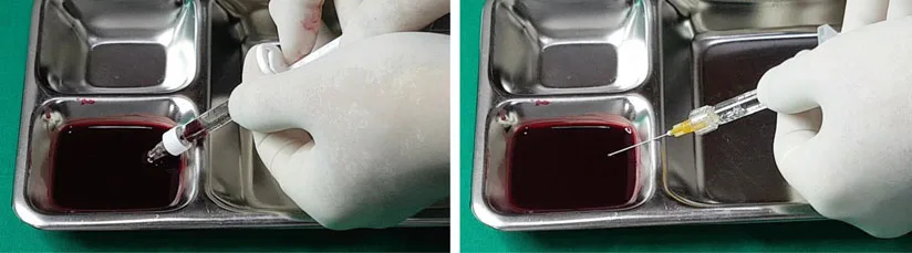 Fig 6. Why "I aspirated and it was clear" is not a safety claim. The blunt cannula and the viscous gel column make a false-negative common, so a negative aspirate does not prove the tip is extravascular — the safety layer is movement, low pressure, micro-aliquots and reversibility, not the plunger pull. — (Hong, Art and Science of Filler Injection, 2020, p.199).
> Fuentes: [13].
Fig 6. Why "I aspirated and it was clear" is not a safety claim. The blunt cannula and the viscous gel column make a false-negative common, so a negative aspirate does not prove the tip is extravascular — the safety layer is movement, low pressure, micro-aliquots and reversibility, not the plunger pull. — (Hong, Art and Science of Filler Injection, 2020, p.199).
> Fuentes: [13].
The traps share one root: solving the wrong problem for the region. Nine of the thirteen are a diagnostic error, not a technical one — a compensating frontalis mistaken for a wrinkle (D1, D2), a pigmented circle mistaken for a hollow (D5), a submalar shadow mistaken for a cheek deficit (D7), an under-projected chin mistaken for submental fat (D11), a lax border mistaken for a volume deficit (D12), and a four-phenotype neck run as one (D13). The fix in every case is the same: classify before you inject, and know which of the region's problems is actually in front of you [3][8].
Controversy — aspiration before injection (the second cross-region technique row). Whether to pull back on the plunger before depositing is unsettled, and the disagreement is worth stating because it changes the ritual at every deep bolus:
Consensus: a negative aspirate is not proof of an extravascular tip. Sensitivity is poor — flashback appears in only ~33 % within one second, and Casabona's series put it near 53 %; a viscous gel column, a fine lumen, a blunt cannula and vessel-wall adherence all produce false negatives [35][37] (Fig 6). It gives a false sense of security, and it is never a substitute for the real safety layer: small aliquots, low pressure, retrograde motion, reversibility and hyaluronidase in the room [36]. Discrepancy: against routine aspiration (Goodman/Van Loghem: unreliable, and pausing to aspirate conflicts with the moving-needle, low-pressure principle that actually reduces intravascular volume) versus for aspiration as an adjunct (de Maio's alert areas, deep static boluses on bone: a positive aspirate is still true-positive and worth heeding, so aspirate, wait, and re-enter elsewhere if blood returns — regardless of false negatives) [35][36]. Decide by injection type: for a deep static bolus near bone (chin, angle, deep cheek) aspiration is a cheap adjunct with a real true-positive yield; for a moving retrograde thread in a mobile plane it adds little and interrupts the technique that matters more. Neither school claims aspiration makes the region safe [35][36][37].
The nasolabial fold makes the aspiration debate concrete. The region chapter marks a [MATERIAL GAP]: no retrieved source proves aspiration prevents an NLF occlusion, and the facial artery's unpredictable depth there means a negative aspirate is worth nothing [25]. The fold's rule — cannula default, larger-barrel syringe for lower pressure, cause-first — is the safety layer that survives the aspiration argument.
Trampa clásica (the meta-trap): trusting a ritual (a negative aspirate, "I used a cannula," "I stayed deep") in place of the region-specific discipline. Each of those has a documented failure mode — false-negative aspiration, blunt-cannula misplacement, deep-plane venous embolism in the temple — and the table exists so the injector reaches for the right discipline for the region in front of them, not the reassuring habit [12][32][35].
The seven grids, transposed — one governing line per region. Read column-wise the tables answer "which plane / which vessel across all regions"; read row-wise they compress each region to its single defining fact, the way a clinician holds it:
- D1 Forehead & glabella — toxin first (glabella 20 U), filler almost never; the glabella is the max-blindness zone [26][38].
- D2 Brow & tail — the volume that lifts the tail lives at the temple, not the brow; depressor toxin before any filler [14][38].
- D3 Temple — two safe planes, a lethal valveless-vein middle; feel bone or use ultrasound [12][40].
- D4 Upper periorbital & eyelid — a droopy lid is skin/levator/brow before it is volume; measure MRD1 first [3].
- D5 Tear trough — classify shadow vs pigment vs vessel vs bag before the syringe; on bone, 3–4 mm below the rim, ~1 mL/side ceiling [16].
- D6 Malar / zygoma — three targets (Ck1/Ck2/Ck3), three vectors, not one bolus; ~4 mL/session ceiling [1][9].
- D7 Anteromedial cheek & submalar — press to bone: bone → deep supraperiosteal bolus, soft tissue → subcutaneous lamina [6].
- D8 Nose — HA only, deep midline, microbolus ≤ 0.05 mL, mean 0.65 mL total; a terminal territory [11][19].
- D9 Nasolabial fold — no safe plane; treat the cause (cheek + pyriform) first, the fold last [7][25].
- D10 Lip & perioral — superficial paramedian, keep lower > upper, thread the border, never bolus the commissure [24].
- D11 Chin — bite test first (microgenia → filler; Class II → refer); needle to bone, midline, no blindness route [8][17].
- D12 Jawline & angle — continuous border before the angle; palpate the antegonial pulse; subtract (masseter) in the wide face [18][42].
- D13 Neck — classify four phenotypes; toxin is the core tool; never below the thyroid cartilage [43][44].
Where a region's governing line contradicts the cross-region default (nose, NLF, temple, lip, neck), the contradiction is the reason that region has its own chapter — and the reason this hub prints the exceptions instead of smoothing them into an average.
Coverage vs UPO
UPO teaches injection region by region, module by module; it never lays the regions side by side. That side-by-side comparison is exactly what this hub adds, and it is where the discrepancies between modules become visible.
| UPO master topic (module) | Status in this chapter | What the atlas adds |
|---|---|---|
| Per-region injection technique (M2 facial modules: glabella, cheek, lips, chin…) | Covered as the source of every grid cell | The cross-region comparison — plane/product/instrument/volume/vessel/toxin/trap in one row per region, so two modules that disagree are caught |
| Complications & acute ischaemia (M2 T10, Tejero) | Covered in D0.5 + D0.7 | Corroborated externally: the UPO slide's Zhou citation and cannula-vs-needle claim are re-anchored to [32]; hyaluronidase to the HDPH protocol [29] and 2026 consensus [30][31] |
| Cannula vs needle safety (UPO complications slides) | Covered as the D0.3 controversy | The slide is [D] never_sufficient_alone; the atlas keeps the CMAC "avoid < 25G" rule but attaches the primary series [32] and the gauge-by-region decision |
| Toxin dosing by facial zone (M1 toxina módulos) | Covered in D0.6 | The antagonist-and-distance framing across regions, and the four regions with no primary toxin target stated explicitly |
| Dilutions / mesobotox / microbotox (UPO slides) | Referenced in D0.6 (D13) | Flagged as the fastest-ageing lane; microbotox anchored to [44], never cited from the slide alone |
| Not in UPO: a single comparative table of all regions | New — this chapter is that table | The whole artifact |
Not in UPO: explicit [MATERIAL GAP] where a per-region maximum is unstandardised |
New (D3, D7 volume) | Declares the silence with its retrieval instead of printing a fabricated ceiling |
| Not in UPO: the "deep = safe except here" exception map | New (D0.1) | Names the five regions where the deep-first default is wrong, and why |
UPO is the lane that ages fastest, and every value that traces only to a UPO slide is treated as never_sufficient_alone: the corpus slides (Tejero on acute ischaemia, the dilution/mesobotox decks) set the shape of the clinical discussion, but the numbers in the grids are anchored to external, identifier-bearing sources [29][32][35]. No grid cell in this chapter rests on a UPO slide alone.
Self-assessment
Ten active-recall questions built only from facts already stated above. Answers folded.
1. What is the cross-region default plane, and the anatomical reason for it?
Answer
Supraperiosteal / deep-fat first, on bone, medial to the line of ligaments — because the named facial arteries run mainly in the superficial and subcutaneous planes, so the deep plane is generally both the structural fundament and the low-risk corridor [4].2. Name the five regions where the deep-first default breaks, and how.
Answer
Nose (deep **midline** is the only corridor), nasolabial fold (**no reliably safe plane**), temple (two safe planes bracket a **lethal middle**), lip (safety is **superficial paramedian**), neck (the danger is **dysphagia**, not a vessel) [11][12][24][25][43].3. What is the per-session HA ceiling for the midface, and what is the general rule about ceilings?
Answer
~4 mL of HA per midface session, more built across sessions — because the overfilled face is reached by a little too much many times, not once [9].4. What is the mean total filler volume for a nonsurgical rhinoplasty, and how is it delivered?
Answer
R4P mean **0.65 ± 0.17 mL** total, in microboluses ≤ 0.05 mL with a stop-and-look between deposits [19][20].5. Which vessel makes the temple uniquely dangerous, and by what mechanism distinct from the other regions?
Answer
The **middle temporal vein** — large, thin-walled, **valveless**, ~2 cm above and parallel to the zygomatic arch — giving a **venous** route to non-thrombotic pulmonary embolism that the purely arterial regions do not have [12].6. What did Zhou (2020) find about cannula versus needle, and what is the resulting gauge rule?
Answer
25 of 28 severe HA emboli (nine with blindness) were injected with a cannula of 22–27G, not a needle; the CMAC consensus is to **avoid cannulas smaller than 25G** [32].7. State the glabella toxin dose and the corrugator safety distance.
Answer
**20 U** ona in five points (5 × 4 U); the lateral corrugator point stays **≥ 1 cm above the bony orbital rim** to avoid diffusion to the levator (lid ptosis) [38].8. Which three regions have no blindness route, and what is the risk instead?
Answer
Chin (mental artery, stay midline), jawline (facial artery at the antegonial notch + marginal mandibular nerve), and neck (dysphagia) — the risk is a nerve or the swallowing muscles, not the ophthalmic artery [8][25][43].9. Why is a negative aspirate unreliable, and what is the real safety layer?
Answer
Flashback appears in only ~33 % (Van Loghem) to ~53 % (Casabona) within a second, and a viscous gel or blunt cannula produces false negatives; the safety layer is small aliquots, low pressure, retrograde motion and reversibility [35][37].10. In the tear trough, how far below the rim and how medial may the deep deposit go, and what is the per-side ceiling?
Answer
On bone, **3–4 mm below the palpable rim edge**, **no more medial than the inner limbus**; start 0.5 mL/side with a hard ceiling of ~1 mL/side [16].What's new and trends (2023–2026)
Dated changes that move a cell in the grids. This section synthesises sources already cited; it introduces no new claim.
| Year | Change | Which grid it moves | Maturity | Ref |
|---|---|---|---|---|
| 2023 | Restylane Eyelight (firm low-water NASHA) FDA-approved for infraorbital hollows | D0.2 product (D5): a firm-gel option legitimises an exception to "soft-superficial" in the trough | clinically actionable now | [16] |
| 2024 | Doppler / high-frequency ultrasound best-practice guidance for filler mapping and guidance | D0.5 vessel: pre-procedure vascular mapping becomes the fastest-moving safety layer, region-agnostic | promising but not validated | [40][41] |
| 2024 | VYC-25L jawline RCT; chin RCT data mature | D0.2/D0.4 (D11, D12): "bone-mimic" high-G′ HA and session ceilings (~3–4 mL jawline) firm up | clinically actionable now | [17][18] |
| 2025 | VYC-20L temple RCT | D0.4 (D3): a controlled temple volume exists, though the per-side maximum stays [MATERIAL GAP] |
clinically actionable now | [14] |
| 2025–26 | The blanket "a cannula is categorically safer than a needle" claim, refuted; gauge-by-region rule instead | D0.3 instrument controversy: "avoid < 25G" reaffirmed against the marketing generalisation | unsupported commercial claim | [32][33][34] |
| 2025–26 | Doppler/AI-assisted vessel mapping toward automated per-region occlusion-risk scoring | D0.5 vessel: mapping exists; an automated risk score does not yet | preclinical/speculative | [40][41] |
| 2026 | PLLA temporal-augmentation RCT | D0.2 (D3): a staged irreversible biostimulator gains RCT support at the temple | promising but not validated | [15] |
| 2026 | UK consensus on filler-induced vision loss; hyaluronidase VO management guide | D0.5 emergency card: the vision-loss pathway and HDPH dosing are consensus-anchored | clinically actionable now | [30][31] |
What did NOT change, and why the older references are still the state of the art. The backbone of this hub is anatomy, and anatomy is stable: the facial fat compartments (Rohrich & Pessa 2007; Schenck 2018; Surek 2016–2019; Cotofana 2016–2019) and the facial-skeleton ageing model (Mendelson & Wong 2012) still define the planes and the deep-first principle [1][2][5][6][8]. The ophthalmic anastomosis that makes eleven regions potentially blinding is a fixed anatomical fact, described for over a decade and only re-confirmed by newer imaging [26][27]. DeLorenzi's high-dose pulsed hyaluronidase (2017) remains the rescue protocol the 2026 consensus documents build on, not replace [29][31]. de Maio's MD Codes volumes and the glabella 20 U five-point scheme are unchanged references [9][38]. The trend is additive — ultrasound guidance, region-specific RCTs, updated consensus wording — layered on an anatomy and a rescue pharmacology that have not moved. A chapter that chased only the 2026 papers would lose the 2007–2019 anatomy that the whole table stands on.
Unexplored directions (AI speculation)
> [IA-ESPEC] The following are model-generated research directions, not clinical recommendations. Each is tagged [IA-ESPEC], states the cited anchor it grows from, the proposal, and what would settle it. None contains a dose, a product or a protocol a reader could act on. They are hypotheses for investigators, never point-of-care guidance.
-
[IA-ESPEC] A registry-derived per-region volume ceiling for the two gap regions. Anchor: the temple (D3) and submalar (D7) both carry a
[MATERIAL GAP]— the corpus does not standardise a per-side maximum [14][15]. Proposal: the side-by-side grid exposes that every other region has a stated ceiling while these two do not; a prospective multicentre registry could derive an evidence-based per-side maximum stratified by product G′. Expected effect: a stated per-side ceiling for the temple and submalar reduces overfill and nodule events in the two regions that currently lack one. Confounder: product G′ and individual tissue thickness vary, so a single ceiling may not transfer across gels or faces and must be reported per product. What would settle it: a dose-finding study reporting contour outcome and complication rate against per-side volume, the way the jawline VYC-25L pivotal already does [18]. -
[IA-ESPEC] A composite per-region occlusion-risk index (plane × vessel × gauge). Anchor: D0.3 shows severe emboli cluster with < 25G cannulas [32] and D0.5 shows the vessel and its drainage differ by region. Proposal: the three grids together contain the variables (default plane, killer vessel depth, instrument gauge) that a single stratified risk score could combine, turning thirteen separate danger discussions into one comparable number. Expected effect: a single stratified index predicts severe-occlusion risk more usefully than any one variable (plane, vessel or gauge) alone. Confounder: injector experience and regional case-mix differ across registries and would confound the region and gauge effects unless adjusted for. What would settle it: a prospective adverse-event registry that records region, plane, gauge and outcome, letting the index be fitted and validated rather than asserted.
-
[IA-ESPEC] Ultrasound as a routine versus selective pre-injection standard, decided by region. Anchor: Doppler/HFUS mapping is the fastest-moving safety layer [40][41] and the temple, NLF and nose are the regions with the most unpredictable vessel depth (D0.5). Proposal: rather than "ultrasound for everyone" or "for no one," the grid suggests the regions where mapping changes the plane decision most, so a selective standard could be tested where the yield is highest. Expected effect: selective ultrasound in the high-variability regions (temple, NLF, nose) lowers the vascular-event rate without the cost of imaging every patient in every region. Confounder: operator ultrasound skill and equipment access vary widely and would confound the guidance effect unless standardised. What would settle it: an RCT comparing landmark versus ultrasound-guided injection, powered on vascular-event rate, stratified by the high-variability regions.
-
[IA-ESPEC] Aspiration value stratified by injection kinematics, not by region. Anchor: the D0.7 controversy splits on injection type — a deep static bolus versus a moving retrograde thread [35][36]. Proposal: the disagreement may dissolve if aspiration is evaluated separately for static and dynamic techniques, since its only defensible yield (a true-positive) is available only when the tip is momentarily still on bone. Expected effect: aspiration retains a measurable true-positive value for a deep static bolus but not for a moving retrograde thread, resolving the apparent contradiction between the two schools. Confounder: gel viscosity and needle gauge affect flashback probability independently of injection kinematics and would have to be controlled. What would settle it: a bench-plus-clinical study reporting true-positive and false-negative rates separately for stationary-bolus and moving-thread techniques.
§Safety
Three safety constants hold across all thirteen regions, and they are the take-home of the whole hub.
-
Hyaluronidase is in the room for every HA injection, in every region on this page. It is the reason HA is the defensible default and the argument against any permanent product in these regions; the rescue is high-dose pulsed hyaluronidase, flooding the ischaemic territory and repeating hourly toward completion within 72 h, with the dose scaled to the territory [29][31]. If hyaluronidase is not in the room, the region is not treated that day.
-
Assume the ophthalmic anastomosis until the region proves otherwise. Eleven of thirteen regions drain, through the angular–dorsal nasal–supratrochlear network, to the central retinal artery, so every deep facial injection is done with small aliquots, low pressure, retrograde motion and — increasingly — ultrasound mapping, on a 90-minute clock for the retina [26][27]. Any visual symptom is an ocular emergency, not routine bruising, and follows the vision-loss consensus pathway [30]. Only the chin, jaw and neck are outside this network, and each has its own killer — the mental and marginal mandibular nerves, and dysphagia from a deep or high platysma injection below the thyroid cartilage [8][25][43].
-
Classify before you inject — the commonest region-specific trap is a diagnostic error, not a technical one. A droopy lid is skin, levator or brow before it is volume (D4); a dark circle is shadow, pigment, vessel or bag (D5); a "cheek" deficit is anteromedial (bone) or submalar (soft tissue) (D7); a "double chin" may be an under-projected mentum (D11); a "neck" is one of four phenotypes (D13). Dermatochalasis, true ptosis, skeletal Class II, festoons, gland ptosis and skin excess are surgical or energy problems, and no dose fixes them [3][8].
Sequence — which region first, when several are treated in one plan. The grids answer what to do in each region; the order across regions follows one rule the individual chapters share: structural and lateral first, mobile and medial second, superficial and skin last. In a full-face plan the temple and lateral cheek scaffold (D3, D6) are treated early, because the lateral structural support reduces the medial volume the midface then needs; the anteromedial cheek and deep pyriform (D7, D9) follow, unloading the nasolabial fold and the tear trough so they often need less or nothing; the tear trough and lip (D5, D10) are treated after their support is in place; and toxin is generally placed before filler where both are planned in the upper face, since relaxing the depressors settles brow position and lowers the volume the region needs [9]. The cross-reference for the whole sequence is L2 — Combination Sequencing and Treatment Planning; the hub's contribution is only to show that "lateral before medial, deep before superficial, toxin before filler" is the same order in region after region, not a per-chapter idiosyncrasy. Treating the medial midface or the fold first — before its lateral scaffold exists — is the sequencing version of the D0.7 traps: the deficit is chased at the wrong point and over-corrected.
One session or several? The grids also expose where a single sitting is wrong on principle: never forehead and glabella in the same session (D1); toxin in its own session when brow compensation is in doubt (D1, D2); staged volume rather than one large deposit in the midface, chin and jawline (D6, D11, D12); and the neck's four phenotypes are sequenced by which tool the finding actually needs, not run as one protocol (D13). The recurring "reassess at 2 weeks" line is the same brake in thirteen regions — the settled result, not the on-table result, is what the next deposit is judged against.
Region-specific absolute red lines, restated as a checklist: glabella/supra-brow — highest-blindness zone, avoid or treat with extreme restraint (D1, D2); temple — feel bone or use ultrasound, never mid-depth (D3); tear trough — on bone, 3–4 mm below the rim, never behind the septum (D5); nose — HA only, microbolus, never the operated nose blindly (D8); NLF — no safe plane, treat the cause first (D9); lip — superficial paramedian, never bolus the commissure (D10); jaw — palpate the antegonial pulse before any bolus (D12); neck — never toxin below the thyroid cartilage, never DCA between the mandibular border and the submental crease (D13) [12][16][24][28][43].
Brand and off-label note. Product names appear as class examples for a private clinical reference, not endorsements; several indications named here (microbotox, Nefertiti platysma, hyperdilute CaHA, many region-specific filler placements) are off-label, and the numbers are reprinted from the region chapters, which hold the evidence tier and the consent language. (P) No unit, volume or product in this chapter is a substitute for the region chapter's full technique, contraindication and consent set.
References
[B]Cotofana S, et al. Anatomy of the Facial Fat Compartments and Their Relevance in Aesthetic Surgery. J Dtsch Dermatol Ges. 2019. DOI 10.1111/ddg.13737[B]Schenck TL, et al. Functional Anatomy of the Facial Superficial Fat Compartments. Plast Reconstr Surg. 2018;141(6). DOI 10.1097/PRS.0000000000004364[C]Cotofana S, et al. The Anatomy of the Aging Face: A Review. Facial Plast Surg. 2016;32:253-260. DOI 10.1055/s-0036-1582234[B]Freytag L, Alfertshofer M, Frank K, ... Cotofana S. Understanding Facial Aging Through Facial Biomechanics. Facial Plast Surg Clin North Am. 2022. DOI 10.1016/j.fsc.2022.01.001[B]Rohrich RJ, Pessa JE. The Fat Compartments of the Face: Anatomy and Clinical Implications. Plast Reconstr Surg. 2007;119(7):2219-2227. DOI 10.1097/01.prs.0000265403.66886.54[B]Surek CC, et al. The Facial Fat Compartments Revisited: Clinical Relevance. Plast Reconstr Surg. 2019. DOI 10.1097/PRS.0000000000006181[B]Surek CC, et al. Deep Pyriform Space: Anatomical Clarifications and Clinical Implications. Plast Reconstr Surg. 2016. DOI 10.1097/PRS.0000000000002262[C]Mendelson B, Wong CH. Changes in the Facial Skeleton With Aging: Implications and Clinical Applications. Aesthetic Plast Surg. 2012. DOI 10.1007/s00266-012-9904-3[A]de Maio M. MD Codes: A Methodological Approach to Facial Aesthetic Treatment With Injectable Hyaluronic Acid Fillers. Aesthetic Plast Surg. 2021. DOI 10.1007/s00266-020-01762-7[B]de Maio M. Periorbital Filling With the MD Codes Algorithm: A Narrative Review and Practical Guide. J Cosmet Dermatol. 2021. PMID 34623020[C]Facial Fillers: Relevant Anatomy, Injection Techniques, and Complications. 2023. PMID 37780674[B]Rohrich RJ, et al. Facial Danger Zones (Zonas Faciais de Perigo). Thieme/DiLivros; 2020. ISBN 9786555720044[B]Hong K. Art and Science of Filler Injection. Springer; 2020. ISBN 9789811306105[A]Improvement in Temple Hollowing With VYC-20L Hyaluronic Acid Filler: A Randomized Study. Plast Reconstr Surg. 2025. DOI 10.1097/PRS.0000000000011957[A]Efficacy and Safety of a Poly-L-Lactic Acid Filler for Temporal Augmentation: A Randomized, No-treatment Control, Evaluator-blinded, Multicenter Study. 2026. PMID 41637104[A]Biesman BS, et al. A Multicenter, Randomized, Evaluator-Blinded Study to Examine the Safety and Effectiveness of a NASHA Gel (Restylane Eyelight) for Infraorbital Hollows. 2024. PMID 38573527[A]Beer K, Kaufman-Janette J, Bank D, et al. Safe and Effective Chin Augmentation With the Hyaluronic Acid Injectable Filler VYC-20L. Dermatol Surg. 2019. DOI 10.1097/DSS.0000000000002795[A]Safe and Effective Restoration of Jawline Definition With Hyaluronic Acid Injectable Gel VYC-25L: A Randomized Controlled Trial. Aesthet Surg J. 2024. DOI 10.1093/asj/sjae147[A]Jalali A. Nonsurgical Rhinoplasty Using the Hyaluronic Acid Filler VYC-25L: Safety and Patient Satisfaction. J Cosmet Dermatol. 2023. DOI 10.1111/jocd.15997[A]Silikovich F, Kroumpouzos G. Nonsurgical Rhinoplasty: Results From a Retrospective Study of the Rino-4-Puntos (R4P) Technique. Aesthetic Plast Surg. 2024. DOI 10.1007/s00266-024-04263-z[A]DeVictor S, Ong AA, Sherris DA. Complications Secondary to Nonsurgical Rhinoplasty: A Systematic Review and Meta-analysis. Otolaryngol Head Neck Surg. 2021. DOI 10.1177/0194599820987827[A]Consensus Recommendations for the Use of Hyperdiluted Calcium Hydroxyapatite (Radiesse) as a Face and Body Biostimulator. 2019. PMID 31044123[A]Calcium Hydroxylapatite for Jawline Rejuvenation: Consensus Recommendations. 2014. PMID 24641600[B]Cotofana S, et al. Distribution Pattern of the Superior and Inferior Labial Arteries: Impact for Safe Upper and Lower Lip Injection. Plast Reconstr Surg. 2017. DOI 10.1097/PRS.0000000000003244[B]Yang HM, Lee JG, Hu KS, et al. New Anatomical Insights on the Course and Branching Patterns of the Facial Artery: Clinical Implications. 2014. PMID 24445874[A]Beleznay K, Carruthers JDA, Humphrey S, Jones D. Avoiding and Treating Blindness From Fillers: A Review of the World Literature. Dermatol Surg. 2015. DOI 10.1097/DSS.0000000000000486[A]Beleznay K, et al. Update on Avoiding and Treating Blindness From Fillers: A Recent Review of the World Literature. Aesthet Surg J. 2019. DOI 10.1093/asj/sjz053[B]Yang Q, et al. Fatal Cerebral Infarction and Ophthalmic Artery Occlusion After Nasal Augmentation With Hyaluronic Acid. Aesthetic Plast Surg. 2019. DOI 10.1007/s00266-019-01589-x[A]DeLorenzi C. New High-Dose Pulsed Hyaluronidase Protocol for Hyaluronic Acid Filler Vascular Adverse Events. Aesthet Surg J. 2017. DOI 10.1093/asj/sjw251[A]Consensus Guidelines for the Management of Tissue Filler-induced Vision Loss in the United Kingdom. 2026. PMID 41490283[A]Guide for Managing Vascular Occlusion Caused by Fillers With Hyaluronidase. 2026. PMID 40770496[B]Zhou SB, Chiang CA, Liu K. False Sense of Safety: Blunt Cannulas Cause the Majority of Severe Vascular Complications in Hyaluronic Acid Injection. Plast Reconstr Surg. 2020. PMID 33759817[C]Needles Versus Cannulas: Advantages, Disadvantages, and Selection. 2026. PMID 41457906[C]Cannula Is Safer Than Needle in Filler Injection? 2025. PMID 41541903[B]Van Loghem JAJ, Fouché JJ, Thuis J. Sensitivity of Aspiration as a Safety Test Before Injection of Soft Tissue Fillers. J Cosmet Dermatol. 2016. DOI 10.1111/jocd.12437[A]Goodman GJ, Magnusson MR, Callan P, et al. Neither Positive Nor Negative Aspiration Before Filler Injection Should Be Relied Upon as a Safety Maneuver. Aesthet Surg J. 2020. DOI 10.1093/asj/sjaa215[B]Casabona G. Blood Aspiration Test for Cosmetic Fillers to Prevent Accidental Intravascular Injection in the Face. 2015. PMID 26079591[A]SAMCEP Society Consensus on the Treatment of Upper Facial Lines With Botulinum Neurotoxin Type A: A Tailored Approach. 2023. PMID 37408173[B]Evaluation of Supratrochlear Artery Depth and Course Variations by Doppler Ultrasonography Along the Glabellar Frown Lines for Safer Filler Injections. 2022. PMID 36066329[B]Sigrist R, Desyatnikova S, Chammas MC, Vasconcelos-Berg R. Best Practices for the Use of High-Frequency Ultrasound to Guide Aesthetic Filler Injections. 2024. PMID 39202206[B]Lee W, et al. Doppler Ultrasound for Vessel Mapping in Hyaluronic Acid Filler Injection. 2023. PMID 37564711[D]Hong JY, Jeong GJ, Kwon TR, et al. Efficacy and Safety of a Novel Botulinum Toxin A for Masseter Reduction: A Randomized Trial. Dermatol Surg. 2019. DOI 10.1097/DSS.0000000000002475[B]Levy PM. The 'Nefertiti Lift': A New Technique for Specific Re-contouring of the Jawline. J Cosmet Laser Ther. 2007. DOI 10.1080/14764170701545657[B]Wu WTL. Microbotox of the Lower Face and Neck: Evolution of a Personal Technique and Review of the Literature. Plast Reconstr Surg. 2015. DOI 10.1097/PRS.0000000000001827
Verificación: Hub chapter (template HUB, 7 grids), authored in English, artifact_class: CHEATSHEET_CANDIDATE. Built entirely from data already published in the region chapters D1–D13 (zero new claims): each grid cell reprints its region's stated value and the [n] of the source that region cites, re-numbered into this chapter's Vancouver list of 44 references (42 carrying a DOI or PubMed URL, checked through the RM refverify lane — Crossref existence plus Retraction-Watch — alongside 2 textbook ISBNs). No prior version existed on disk (no D0.*.es.md); closed with --no-prior. Kept the 7 HUB blocks (D0.1–D0.7) — no extra subchapter added, because the hub's job is comparison across the fixed seven decisions, not new content; the two scope CONTROVERSY rows (cannula-vs-needle, aspiration) were folded into D0.3 and D0.7 as cross-region technique rows rather than new blocks. Two [MATERIAL GAP] declarations retained (temple and submalar per-side volume maxima) rather than fabricating a ceiling. Retrieval lane: 5 medlib-MCP facet searches (evaluation/runs/D0.rag.jsonl, top scores 0.73–0.82) plus the pre-existing scope contract (scouts/D0/D0.scope.jsonl). 6 figures sourced through the medlib figure lane, each opened with Read before captioning, bundled to _images/D0/: midface vasculature (Jones), deep-first plane (Freytag/Cotofana), temple plane/MTV (Rohrich), supraperiosteal needle (Vieira Braz), aspiration test (Hong), glabella toxin points (Lipham). UPO slide material treated as never_sufficient_alone and externally re-anchored. (P) The comparative grid surfaces cross-region discrepancies but resolves none by averaging — where two regions differ, the instruction is to check the region, per the hub's contract.