D2 · Brow and Brow Tail
> Currency and provenance — 41 references · median 2022, range 2016-2026, 44 % from 2022 on · provenance: verified external 63 % (26) · MEDLIB corpus 37 % (15, of which 2 from the UPO master's).
Domain: D — Region-by-Region - Face & Head · Third: upper · Pass 3 (regions by third: WHERE it is injected).
Subchapters
- [ ] D2.1 — In 30 seconds: doses, volumes, planes and red lines of this region
- [ ] D2.2 — Layered anatomy, skin to bone (6-layer stack; ROOF; the temple's 10 layers)
- [ ] D2.3 — Vessels, nerves and danger zone: course, depth, variants, what occludes → J2
- [ ] D2.4 — Ageing of the region, in order (bone → deep fat/ligament → superficial fat → skin)
- [ ] D2.5 — Assessment: measured, photographed, tested dynamically, scanned
- [ ] D2.6 — Goal and patient selection: who benefits, who does not, what is referred
- [ ] D2.7 — Technique, the full grid (product, instrument, plane, movement, school, volume)
- [ ] D2.8 — Toxin of the region: muscle, units/point, points, safety distances, antagonist spared
- [ ] D2.9 — Combination and sequence: what before, what after, at what interval → L2
- [ ] D2.10 — Region-specific complications (only the ones that happen here) → J1-J8
> Tags: [A] label / guideline / dated consensus · [B] primary literature with PMID/DOI · [C] monograph · [D] slide or opinion, never sufficient alone · [MEDLIB] own corpus · [MODELO] structure only, never a figure · [IA-ESPEC] AI speculation, never actionable · (P) model reasoning, never a dose · ⚠ disputed/stale number.
Voices/schools: the brow has two operators who disagree at the chair, and the chapter keeps both. Toxin repositions the brow by unbalancing depressors against the frontalis; filler/thread/energy/surgery support or reposition the descended tail. The region is not filled: it is balanced and supported. Cross-links: technique fundamentals in C1 — Injection Planes, Tools & Technique Fundamentals, toxin map in C3 — Botulinum Toxin - Full Technique Map, upper-face region in D1 — Upper Face (Forehead, Glabella, Temple, Brow), vascular emergency in J2 — Vascular Occlusion & Emergency Response, hyaluronidase in J3 — Hyaluronidase - Pharmacology & Clinical Protocols, combination logic in L2 — Combination & Sequencing.
D2.1 · In 30 seconds
The region in one screen. Doses in onabotulinumtoxinA (ona) units unless stated; verify per-side vs total before copying any figure.
| Axis | Number to leave the room with | Source |
|---|---|---|
| Chemical brow-tail lift | 2-5 U ona into the lateral orbicularis oculi tail, 1 point/side, ≥1 cm outside the orbital rim | [A][36][6] |
| Medial brow lift | glabellar depressor release: 20 U ona in 5 points of 4 U (2/corrugator + 1 procerus), each ≥1 cm above the rim | [A][7] |
| Antagonist NOT paralysed | frontalis (especially lower and lateral fibres): treat it and the brow drops | [6][24] |
| Expected brow displacement | lateral brow edge rises, medial/central brow falls after frontalis dosing; net tail lift is millimetric (~1-3 mm) | [6][7] |
| Temple support (lifts the tail indirectly) | 0.5-1.0 mL/side; supraperiosteal bolus on bone OR interfascial cannula, US-confirmed | [25][10][2] |
| Supra-brow micro-support | ≤0.1 mL supraperiosteal micro-bolus, lateral to the supraorbital notch | [2][25] |
| Product | high-G′/high-cohesivity HA deep (temple, supra-brow); soft low-G′ HA subdermal temple; CaHA ± hyperdilute for temple biostimulation; reversibility on HA is the safety argument here | [2][25][26] |
| Reversal ready | hyaluronidase in the room for every HA in this region, no exception | [19][8] |
Red lines of the brow/tail (the region's specific killers, not generic prudence):
> ⚠ Supratrochlear artery emerges ~1.7-2.2 cm lateral to the glabellar midline; supraorbital artery ~1 cm lateral to it at the notch/foramen. Both are terminal branches of the ophthalmic artery. A supra-brow or glabellar filler bolus can embolise retrograde to the retina (blindness) or forehead skin (necrosis). The glabella/supra-brow and the temple are among the highest-risk facial zones for filler blindness [8][12][17][18].
> ⚠ Temple carries a second, venous mechanism: the middle temporal vein and the sentinel (medial zygomaticotemporal) vein are large, thin-walled and valveless; a temporal bolus can embolise to the pulmonary circulation, and the ophthalmic anastomosis adds a blindness/stroke route [2][25][17].
The four problems that must be separated before touching anything (confusing them produces the region's worst results): 1. Dynamic depressor overactivity (lateral orbicularis tail, glabellar complex pulling the brow down) → toxin. 2. Loss of lateral support (temple/lateral cheek deflation) → the tail descends although the muscle is intact → volume/support at the temple, not filler in the brow. 3. True descent + skin excess (dermatochalasis, heavy brow ptosis) → energy or surgery, not an injectable. 4. Frontalis-dependent compensation (a hidden eyelid ptosis held up by the forehead) → a relative contraindication to forehead/brow toxin: paralysing the crutch unmasks the ptosis.
Decision ladder (descended brow tail): 1) toxin to depressors → 2) lateral support at the temple/supra-brow (± thread on a temporal→tail vector) → 3) energy for true laxity → 4) thread lift, with its complication rate → 5) surgery [C3.1 salvage][11][16].
Trampa clásica (block-level): treating the frontalis to erase forehead lines while the patient depends on it to open the eye. The forehead smooths and the brow (and often the lid) drops; the follow-up is an unhappy patient with a heavier, tired gaze. Test frontalis dependence before the first unit [6][24].
The brow in one page (D2.1, exhaustive layer under the glance box)
Why the brow is a decision chapter, not a gesture chapter. The doses and planes already live in the toxin and filler technique maps; what changes region by region is what the brow loses and in what order, and therefore which tool is first line. The brow is the one facial unit with the most extreme benefit-risk ratio in both directions at once: toxin here is the highest-evidence, highest-satisfaction, lowest-risk aesthetic act in the specialty, and filler in the immediately adjacent glabella/supra-brow/temple concentrates, with the nose, the highest published incidence of injectable blindness [8][17][18][23]. The rule that organises the chapter follows from that asymmetry.
> In the brow region the toxin is first line almost always, and filler is a deliberate, justified decision, never the natural extension of the treatment. When the correct answer is volume, it is usually volume at the temple, not in the brow itself [C3.1 salvage][25].
The brow has no bony anchor, and that single fact explains the whole region. Unlike the malar or chin soft tissue, the eyebrow is not tethered to bone by a strong ligament. It is suspended by the frontalis-galea sheet (the sole elevator) and tethered laterally by the temporal ligamentous adhesion and the superior temporal septum, and inferomedially by the orbital retaining ligament [2][1]. There is no elevator lateral to the frontalis, and the frontalis fibres thin out laterally. The unsupported lateral tail is therefore the first part to descend with ageing and the hardest to hold up, which is why this chapter exists as its own region and why "brow tail" is in its title. Splitting it across a toxin chapter and a temple chapter would reproduce exactly the modality dispersion the region restructure removed.
The elevator/depressor balance, stated once. Toxin does not fill; it weakens. Every brow toxin decision is a tug-of-war between one elevator (frontalis) and four depressors (orbicularis oculi, corrugator supercilii, depressor supercilii, procerus). Weaken a depressor and the elevator wins locally (lift); weaken the elevator and the depressors win (drop). The bad toxin brow is almost never "too much dose": it is the wrong muscle or the broken balance [6][24][C3.1 salvage].
Two layers, never averaged. Above: the glance box and the ladder. Below, in each block: the full grid of products, planes, movements, schools, volumes and the discrepancies between schools kept side by side with their clinical decider. A brow closed on one technique when five exist is a bug, exactly like a dropped contraindication.
What this chapter deliberately does NOT do: it does not print an "ideal brow position" as a target (the ideal is a shifting aesthetic convention and differs by sex and population [20]); it does not fill the glabella routinely (rejected on the benefit-risk of the highest-blindness zone in the face [8][17]); and it does not present the fox-eye / cat-eye barbed-thread brow lift as a durable evidence-based procedure, because the retrieved evidence is uncontrolled and explicitly temporary [13][15] and the durable-RCT claim was flagged and excluded at scope.
Schools of the region (the two operators, and where each fails):
| School | Thesis | Where it fails |
|---|---|---|
| Toxin-first, almost always ✅ | the brow is muscle territory; balance the depressors against the frontalis | the atlas position; leaves out the patient whose tail dropped from true temple deflation |
| Volume-first | ageing is volume loss; restore it | false for the dynamic depressor pull; exposes the patient to the region's blindness risk for a small gain |
| Temple-support / lateral-first ✅ | the tail is suspended laterally; support the temple and it lifts | correct for volumetric descent; not a substitute for releasing the depressors |
| Chemical brow lift as a shaping tool ✅ | small depressor doses reshape the arch reversibly | modest in millimetres; oversold as a "lift" it disappoints |
| Direct brow filler | fill the brow to raise it | ⚠ rejected here: adds weight to an unanchored structure and drops the tail |
| Thread-first | an immediate mechanical lift is what patients want | temporary, uncontrolled evidence; not durable, and misused as a substitute for surgery |
| Surgery-first | true descent needs a lift | correct past the injectable threshold; over-applied to mild cases that toxin would fix reversibly |
The decision tree, in one pass: is the brow low? → is it dynamic (depressor pull, lifts on request) → toxin to depressors; is it volumetric (temple hollow, intact muscle) → temple support; is it skin/bone (dermatochalasis, resorbed rim, heavy ptosis) → energy or surgery; is the frontalis compensating a hidden lid ptosis → do not treat the forehead; refer/treat the lid first. Every downstream block elaborates one branch of this tree.
The benefit-risk asymmetry, quantified in kind. Upper-face toxin has among the best evidence, highest satisfaction and lowest risk in aesthetics (a Cochrane-level evidence base for facial wrinkles) [23]; peri-brow/temple filler carries one of the highest published incidences of injectable blindness [8][17][18]. The two live centimetres apart. That asymmetry is why the region's default is toxin, and why every filler here is a justified exception with hyaluronidase in the room.
Map of the ten blocks: anatomy by layers (D2.2) → vessels/nerves/danger zone (D2.3) → what ages and in what order (D2.4) → assessment (D2.5) → goal and selection (D2.6) → the full technique grid (D2.7) → toxin of the region (D2.8) → combination and sequence (D2.9) → region-specific complications (D2.10). Everything downstream depends on getting D2.2-D2.4 right, because the plane you choose and the tool you choose are dictated by the layer the deficit lives in and the vessel that lives in that plane.
D2.2 · Layered anatomy, skin to bone (this region only)
| Layer | At the brow | What lives there / matters |
|---|---|---|
| 1 skin | thick, sebaceous, hair-bearing | - |
| 2 superficial fat | sparse; brow fat pad laterally | subdermal soft-HA target for flattening |
| 3 SMAS / muscle | orbicularis oculi (depressor) meets frontalis-galea (elevator) | the elevator/depressor balance = the whole game |
| 4 deep fat / glide | ROOF + sub-brow glide plane | supra-brow supraperiosteal micro-bolus target |
| 5 periosteum | frontal bone / supraorbital rim | STr/SO arteries run deep here near the rim |
The six-layer stack over the brow (fixed order), with what actually occupies each layer at the eyebrow and how it differs from the generic face:
                          BROW  (over the supraorbital rim)        TEMPLE (over the tail, 10 layers)
  1  SKIN                 thick, sebaceous, hair-bearing            thin, mobile
  2  SUPERFICIAL FAT      sparse; brow fat pad laterally            subcutaneous fat (STA runs deep to it in L3)
  3  SMAS / MUSCLE        orbicularis oculi (depressor) meets       superficial temporal fascia (= SMAS cont.)
     (musculo-aponeurotic) frontalis-galea (the ONLY elevator)      → carries anterior + posterior STA
  4  DEEP FAT / GLIDE     ROOF (retro-orbicularis oculi fat) +      upper temporal compartment (avascular)
     (loose areolar)       sub-brow glide plane                     lower temporal compartment (facial n.,
                                                                     sentinel vein)  → deep temporal fascia
  5  PERIOSTEUM /         periosteum of frontal bone / supra-       deep temporal fascia; then superficial
     DEEP FASCIA           orbital rim                              temporal fat pad, temporalis, periosteum
  --------------------------------------------------------------------------------------------------------
  KEY REGIONAL FACT: the brow has NO direct bony attachment. It hangs from frontalis-galea (L3) and is
  tethered by the orbital retaining ligament (inferomedial) and the temporal ligamentous adhesion /
  superior temporal septum (lateral). Descent begins at the unsupported tail.
Consensus (what every school agrees on): the face is a five-layer stack, and the acronym is SCALP - Skin, Connective tissue (subcutaneous fat), Aponeurosis (musculo-aponeurotic / SMAS), Loose areolar (deep fat), Periosteum/deep fascia [1][2]. The stack is continuous from neck to scalp but the layer count is regional: three layers in the tear trough, ten in the temple [2]. The brow sits at the transition where the galea/frontalis (L3) becomes the elevator and the orbicularis oculi (also L3) becomes the depressor, both in the same musculo-aponeurotic plane.
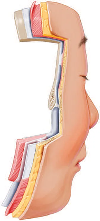 Fig 1. The five-layer stack folded over the brow and orbital rim; the brow soft tissue is suspended from the aponeurotic layer, not anchored to bone - (Azizzadeh, 2018, p.23).
> Sources: Azizzadeh 2018 [35]; layered concept per Cotofana 2016/2019 [1][2].
Fig 1. The five-layer stack folded over the brow and orbital rim; the brow soft tissue is suspended from the aponeurotic layer, not anchored to bone - (Azizzadeh, 2018, p.23).
> Sources: Azizzadeh 2018 [35]; layered concept per Cotofana 2016/2019 [1][2].
The layer that defines the brow: the ROOF (retro-orbicularis oculi fat, layer 4). Deep to the orbicularis oculi and superficial to the periosteum of the frontal bone, bounded superiorly by the inferior frontal septum and inferiorly by the orbicularis retaining ligament, and sitting lateral to the emergence of the supraorbital neurovascular bundle [2]. It is continuous with the lower temporal compartment through the superior interval, which is the anatomical bridge that makes temple support translate into brow-tail support [2][25]. First described in 1909 (Charpy) [2]. The ROOF is the deep fat that a supra-brow supraperiosteal micro-bolus augments; the sub-brow glide plane over it is why a heavy or superficial deposit here reads immediately as brow heaviness.
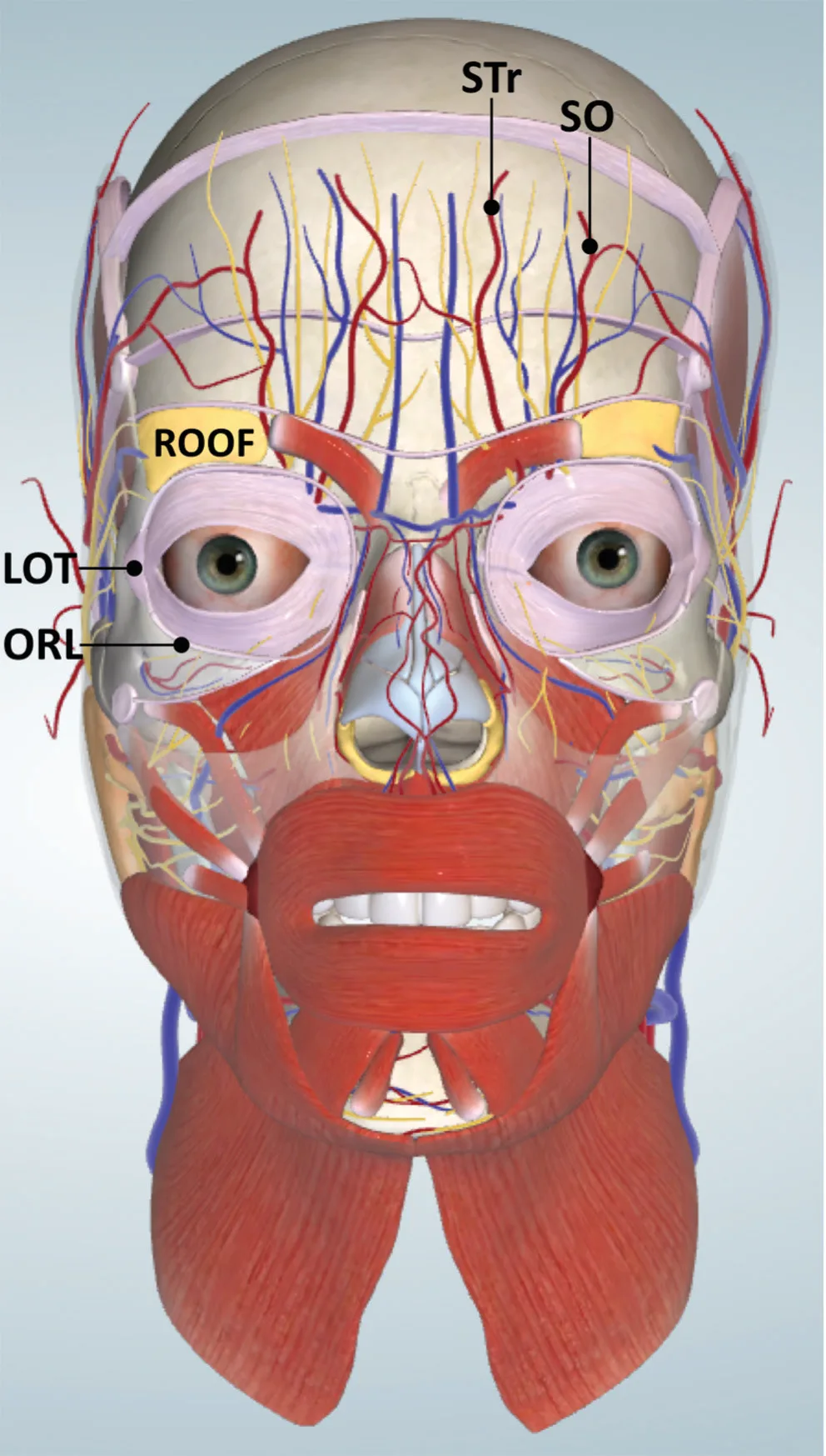 Fig 2. The ROOF and its relations: the two forehead arteries ascend medial to it, the ORL and LOT bound it below - (Cotofana & Lachman, 2019, p.7).
> Sources: Cotofana & Lachman 2019 [2].
Fig 2. The ROOF and its relations: the two forehead arteries ascend medial to it, the ORL and LOT bound it below - (Cotofana & Lachman, 2019, p.7).
> Sources: Cotofana & Lachman 2019 [2].
The forehead compartments above the brow (where a supra-brow filler actually goes). Three superficial forehead compartments sit in layer 2 between skin and frontalis (first delineated 2007, updated 2017); three deep forehead compartments sit deep to the frontalis and its fascia, bounded by the middle frontal septum (below) and superior frontal septum (above) [2]. Between the frontalis and its underlying fascia is the subfrontal fat, and inside that subfrontal fat run the supraorbital and supratrochlear vessels as they ascend from their foramina, crossing the inferior and middle frontal septa from deep to superficial [2]. This is the single most important plane fact for D2.3: over the low forehead the arteries are deep, and they become subcutaneous as they climb.
The temple stack over the tail (the 10 layers, because the tail is supported from here). The SCALP layers continue into the temple but change names past the superior temporal septum: galea becomes superficial temporal fascia (STF), periosteum becomes deep temporal fascia (DTF) [2]. The anterior and posterior branches of the superficial temporal artery run inside the STF (layer 3), so a subcutaneous temple deposit is superficial to them. Two compartments sit between STF and DTF in layer 4: the upper temporal compartment (no relevant neurovascular structures) and the lower temporal compartment (frontal branch of the facial nerve, zygomaticotemporal sensory branch, temporal part of the sentinel vein) [2]. Between 2 and 5 cm above the zygomatic arch the DTF splits into a superficial and a deep lamina enclosing the superficial temporal fat pad and the proximal sentinel vein (medial zygomaticotemporal vein); deeper still lie the deep temporal fat pad, then the temporalis muscle (layer 9) fed by the anterior and posterior deep temporal arteries running on the periosteum (layer 10) [2].
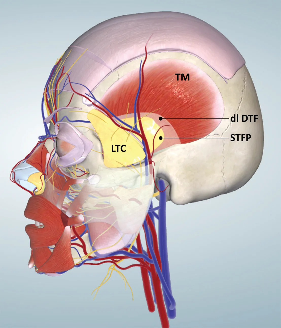 Fig 3. The temple is a 10-layer region; the two safe deep targets are the supraperiosteal plane on bone near the temporal crest and the interfascial space, both away from the STA in the STF - (Cotofana & Lachman, 2019, p.8).
> Sources: Cotofana & Lachman 2019 [2].
Fig 3. The temple is a 10-layer region; the two safe deep targets are the supraperiosteal plane on bone near the temporal crest and the interfascial space, both away from the STA in the STF - (Cotofana & Lachman, 2019, p.8).
> Sources: Cotofana & Lachman 2019 [2].
On cadaver, the same stack looks like this, and the point of showing it is that the fascial planes are real, thin and separable, which is what makes an interfascial deposit a genuine compartment and not a wish.
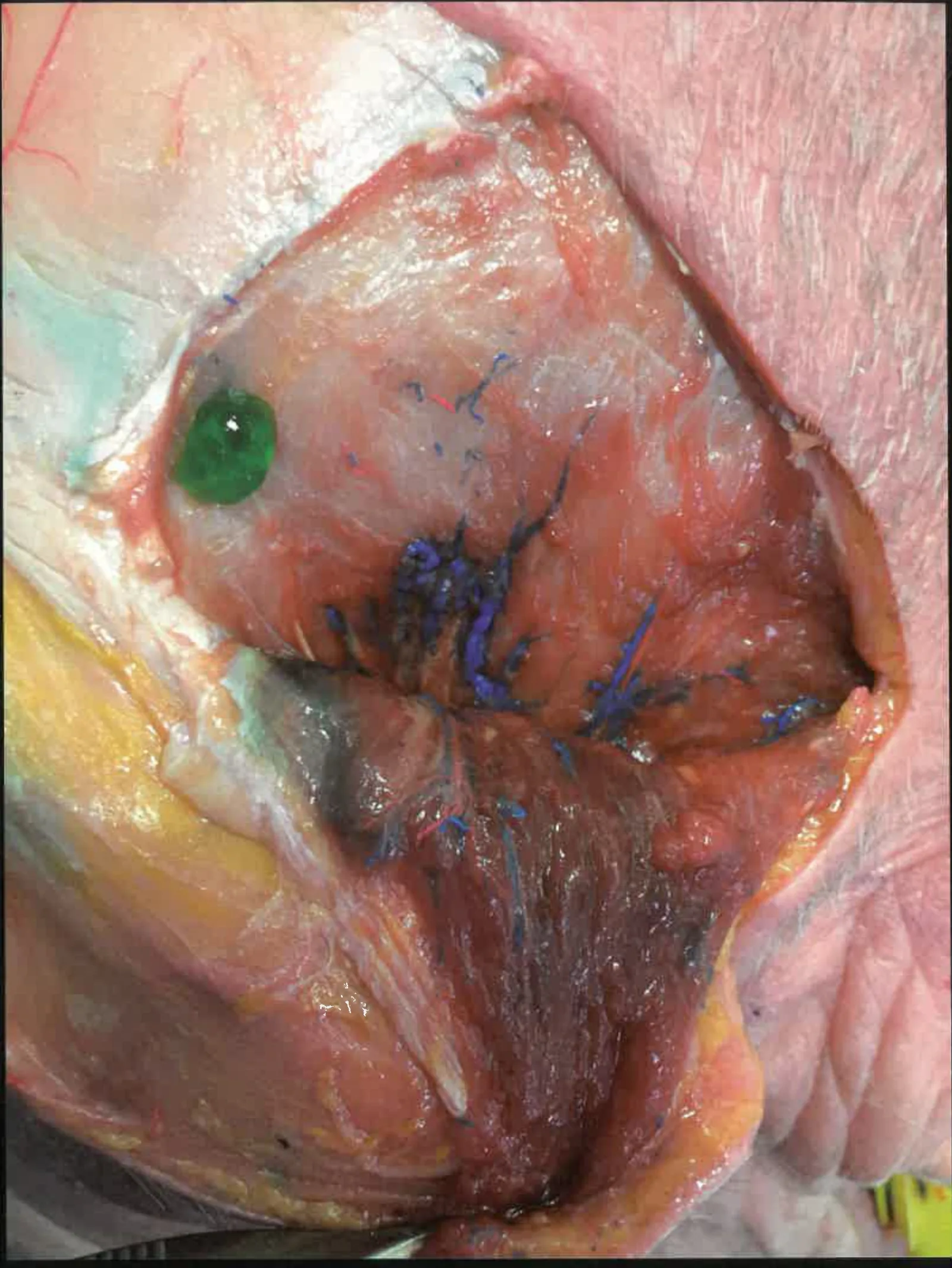 Fig 4. The temporal layers on dissection; the marked venous channels are the sentinel/middle temporal territory that makes the temple a venous danger zone as well as an arterial one - (Atlas de Anatomía y Relleno de la Cara, corpus, p.170).
> Sources: Gobla, Atlas de Anatomía y Relleno de la Cara [37]; layer scheme per Cotofana 2019 [2].
Fig 4. The temporal layers on dissection; the marked venous channels are the sentinel/middle temporal territory that makes the temple a venous danger zone as well as an arterial one - (Atlas de Anatomía y Relleno de la Cara, corpus, p.170).
> Sources: Gobla, Atlas de Anatomía y Relleno de la Cara [37]; layer scheme per Cotofana 2019 [2].
Line of ligaments (why medial fills project and lateral fills lift). All major facial ligaments align in a single line immediately lateral to the lateral orbital rim, from the temporal crest to the mandible: temporal ligamentous adhesion, lateral orbital thickening (LOT), zygomatic ligament, mandibular ligament [2]. Injections medial to this line project the overlying tissue; injections lateral to it lift more inferior regions [2]. The brow tail sits right on this line, which is exactly why supporting the temple/lateral zone (lateral to the line) repositions the tail upward instead of merely projecting it forward.
The muscles of the brow, by part (they are all in layer 3, and their balance is the whole game). The orbicularis oculi has an orbital part (the outer ring, the brow depressor whose lateral fibres pull the tail down) and palpebral/pretarsal parts (the lid); the corrugator supercilii runs obliquely from the medial supraorbital rim to the mid-brow skin (medial depressor, glabellar lines); the depressor supercilii is a small medial depressor; the procerus pulls the medial brow down over the nasal root; and the frontalis (continuous with the galea aponeurotica, with no bony origin) is the sole elevator [2][1]. Four depressors, one elevator, all in the same plane: that is why the region is dosed as a balance, not as isolated points.
The brow fat pad and the ROOF, distinguished. The brow fat pad is a superficial-layer fat lateral to the brow that gives the tail its youthful convexity; the ROOF is the deep-layer (layer 4) retro-orbicularis fat over the superior orbit. They deflate/descend differently: brow-fat-pad deflation flattens the tail, ROOF descent drops it. A supra-brow micro-bolus targets the ROOF plane; a subdermal soft deposit addresses brow-fat-pad flattening. Confusing the two is a plane error [2][3].
The three superficial forehead compartments (where a supra-brow filler that is not on bone ends up). First delineated 2007, updated 2017: a central superficial forehead compartment flanked by two lateral ones, all in layer 2 between skin and frontalis [2]. A too-superficial supra-brow deposit lands here and reads as a visible ridge; the intended plane for medial-brow support is deeper, on or just above periosteum, lateral to the notch. The subfrontal fat (between frontalis and its fascia) is the danger plane because it carries the ascending supraorbital/supratrochlear vessels [2].
Why the temple is the lever and not the brow (restated anatomically). The tail sits on the line of ligaments; the temple lies lateral to it. Because the superficial temporal compartments increase projection rather than displace when filled [3], and because the ROOF connects to the lower temporal compartment through the superior interval [2], a temple deposit transmits support to the tail along the fascial chain instead of merely bulging forward. This is the anatomical justification for "support the tail from the temple," not a stylistic preference.
Trampa clásica: treating the brow as if "deep on bone is safe everywhere". Over the low forehead the supraorbital/supratrochlear arteries are deep on bone in the subfrontal fat, so a periosteal supra-brow deposit near the rim is closer to them, not safer; over the temple the safe deep planes (supraperiosteal at the crest, interfascial) exist precisely because they avoid the arteries in the STF and the deep temporal arteries on the periosteum. The plane is only "safe" relative to the specific vessel of that sub-region [2][12].
D2.3 · Vessels, nerves and danger zone → J2 — Vascular Occlusion & Emergency Response
The vessels of the brow/tail, with course, depth, plane-change and what fails if you hit them:
| Vessel | Origin | Where it is | Depth / plane behaviour | If occluded / injured |
|---|---|---|---|---|
| Supratrochlear a. | terminal branch of ophthalmic a. | emerges ~1.7-2.2 cm lateral to glabellar midline, at the medial brow | deep on periosteum near the rim (in subfrontal fat), ascends and becomes subcutaneous over the mid/high forehead | retrograde embolus → ophthalmic → central retinal a. = blindness; antegrade → forehead/glabella skin necrosis [8][12][17] |
| Supraorbital a. | terminal branch of ophthalmic a. | supraorbital notch/foramen, ~1 cm lateral to the supratrochlear | same deep-to-superficial ascent through the frontal septa | same ophthalmic route → blindness; scalp/forehead necrosis [12][17] |
| "Avascular corridor" | - | the strip between supratrochlear and supraorbital, above the central brow | relatively vessel-poor but NOT vessel-free | the corridor is a lower-risk lane, never a licence to bolus [12] |
| Superficial temporal a. (ant + post branch) | external carotid | in the superficial temporal fascia (L3) of the temple | superficial; a subcutaneous temple deposit is superficial to it, a supraperiosteal one is deep to it | temple skin necrosis; retrograde to ophthalmic anastomosis → blindness/stroke [9][2][17] |
| Deep temporal aa. (ant + post) | maxillary a. | on the periosteum (L10), deep to temporalis | deepest temple plane | reached only by a periosteal deposit far from the temporal crest; bleeding/haematoma [2] |
| Middle temporal v. / sentinel (medial zygomaticotemporal) v. | to pterygoid plexus / ophthalmic | lower temporal compartment (L4) and interlaminar (L6) | large, thin-walled, valveless | venous embolus → non-thrombotic pulmonary embolism; the temple's own complication mechanism [2][17] |
| Angular v. | facial v. | ~4.2 ± 0.7 mm inferior to the inferior orbital rim | deep to orbicularis in the nasojugal groove | relevant to the infra-brow/tear-trough border, a venous route to the ophthalmic system [2] |
Sensory nerves (spare them; a numb brow is a complication too): the supraorbital nerve (through notch/foramen with its artery) and the supratrochlear nerve (medial) carry forehead/scalp sensation; the zygomaticotemporal nerve gives temple sensation and shares the lower temporal compartment with the sentinel vein [2]. Motor: the frontal (temporal) branch of the facial nerve runs in the lower temporal compartment and over the zygomatic arch; a deposit that catches it gives a transient frontalis palsy that mimics, and is worse than, an over-treated frontalis.
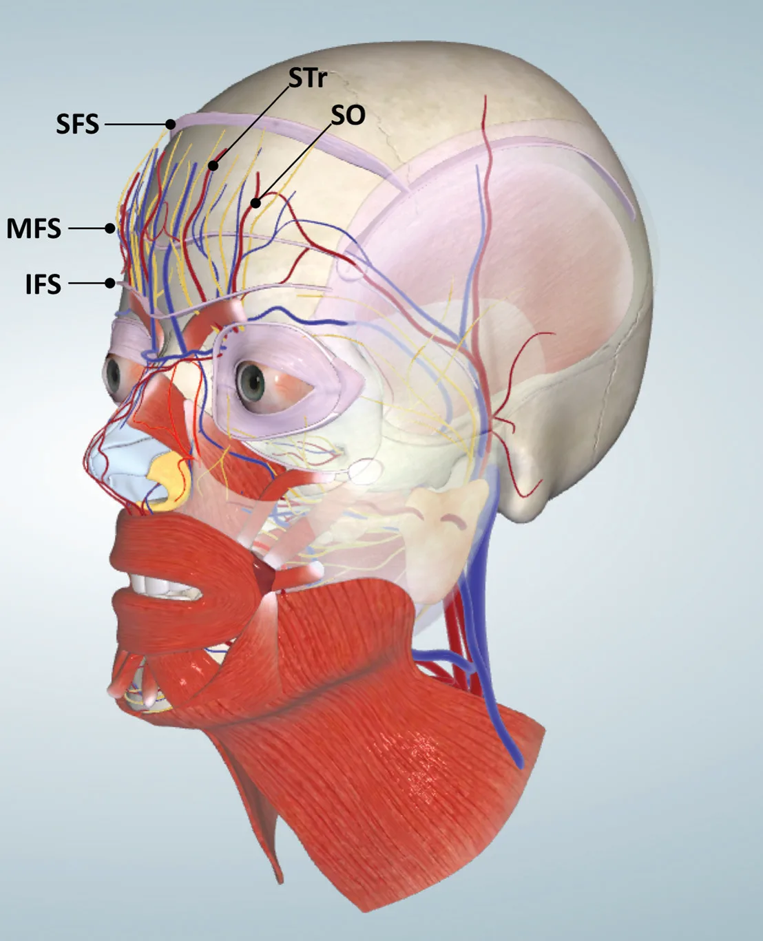 Fig 5. Supratrochlear (STr) and supraorbital (SO) arteries change plane as they climb, so no single depth is safe over the whole forehead - (Cotofana & Lachman, 2019, p.6).
> Sources: Cotofana & Lachman 2019 [2]; forehead arterial pattern per Kliniec 2024 [12].
Fig 5. Supratrochlear (STr) and supraorbital (SO) arteries change plane as they climb, so no single depth is safe over the whole forehead - (Cotofana & Lachman, 2019, p.6).
> Sources: Cotofana & Lachman 2019 [2]; forehead arterial pattern per Kliniec 2024 [12].
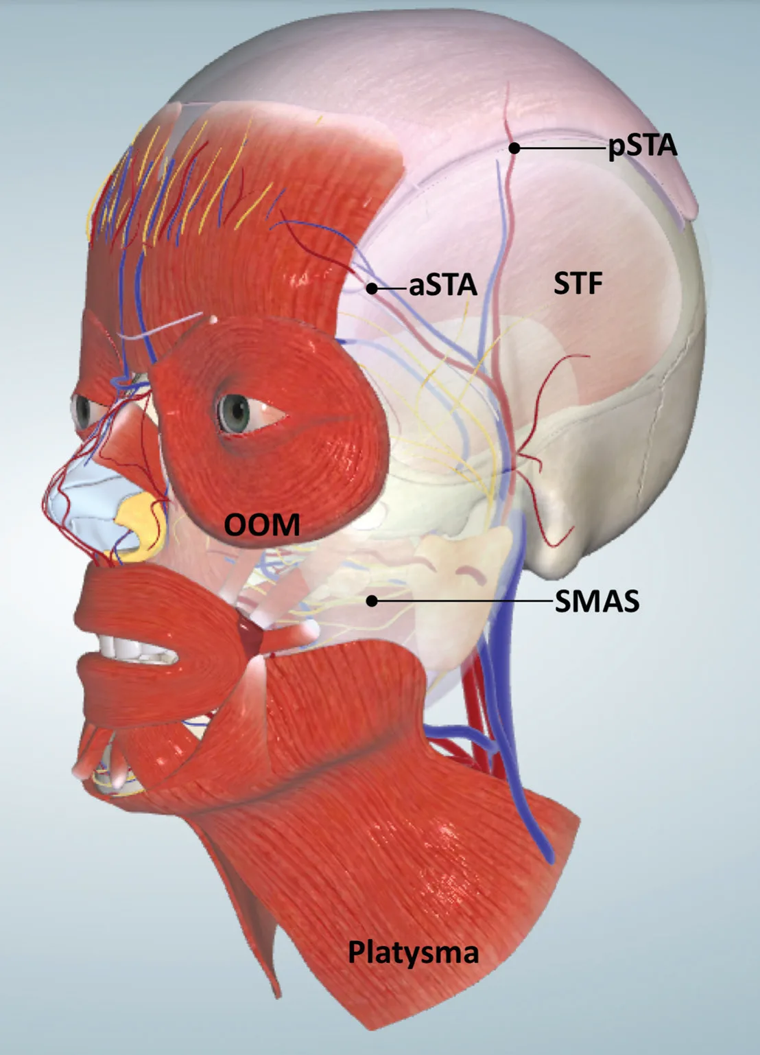 Fig 6. The STA sits in the STF of the temple; the two vascular-safe deep planes are deliberately deep (supraperiosteal) or interfascial to this - (Cotofana & Lachman, 2019, p.7).
> Sources: Cotofana & Lachman 2019 [2]; STA anatomy per Daskalopoulou 2022 [9].
Fig 6. The STA sits in the STF of the temple; the two vascular-safe deep planes are deliberately deep (supraperiosteal) or interfascial to this - (Cotofana & Lachman, 2019, p.7).
> Sources: Cotofana & Lachman 2019 [2]; STA anatomy per Daskalopoulou 2022 [9].
Variants, and why frequency matters at the chair. The supraorbital neurovascular bundle exits through a notch in most people and through a true foramen in a substantial minority; a foramen sits higher and more lateral and cannot be palpated, so the "1 cm lateral to supratrochlear" rule locates a moving target [12]. The forehead arterial pattern (dominant supratrochlear vs dominant supraorbital, and the height at which each turns subcutaneous) varies between individuals, which is why 3D and Doppler mapping outperform surface landmarks [12][10]. Arterial branching in general is variable enough that the corpus complication teaching classifies it into named types.
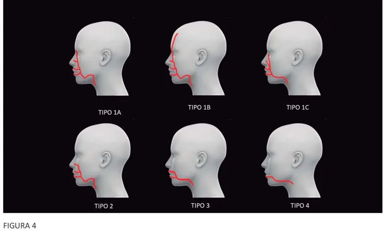 Fig 7. Arterial course is a variable, not a constant; the safe injector treats every landmark as probabilistic and keeps a reversal agent ready - (UPO Sorted, Tejero, 2024, slide 96).
> Sources: UPO Sorted, Tejero 2024 [30] (
Fig 7. Arterial course is a variable, not a constant; the safe injector treats every landmark as probabilistic and keeps a reversal agent ready - (UPO Sorted, Tejero, 2024, slide 96).
> Sources: UPO Sorted, Tejero 2024 [30] ([D], never sufficient alone); vascular variability corroborated by Kliniec 2024 [12].
The danger-zone map, region-specific. The brow region touches two of the face's top-tier danger zones. The glabella/supra-brow zone (supratrochlear + supraorbital, both terminal ophthalmic branches, short retrograde path to the retina) and the temporal fossa (STA superficial, deep temporal arteries deep, sentinel/middle temporal veins valveless, ophthalmic anastomosis). Between them, the medial-canthal/angular territory adds a venous route.
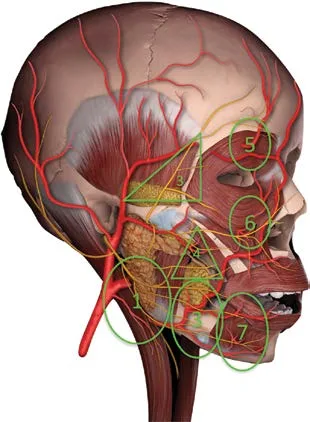 Fig 8. The brow tail sits in the temporal danger triangle; the medial brow sits under the supraorbital/glabellar circle - both are arterial-and-venous, not one or the other - (Pirayesh, 2020, p.126).
> Sources: Pirayesh 2020 [32]; danger-zone concept per Rohrich [33].
Fig 8. The brow tail sits in the temporal danger triangle; the medial brow sits under the supraorbital/glabellar circle - both are arterial-and-venous, not one or the other - (Pirayesh, 2020, p.126).
> Sources: Pirayesh 2020 [32]; danger-zone concept per Rohrich [33].
The safe-injection rule set for this region, each rule tied to its vessel. (1) Low injection pressure so a bolus cannot be driven retrograde past the ophthalmic origin [8][17]. (2) Small aliquots so the volume in any vessel, if entered, is sub-embolic. (3) Moving needle/cannula so the tip is never stationary in one vessel while pressure builds. (4) Aspirate where it means something (a static needle in a viscous gel gives false negatives, but a positive is informative). (5) Cannula for the temple planes where feasible, accepting it does not abolish venous entry. (6) Ultrasound to see the STA and sentinel vein before depositing [10][25]. (7) Reversible product as default and hyaluronidase in the room. (8) Never bolus on the notch, the glabellar midline, or an intermediate temple plane. Each rule exists because of a specific vessel in a specific plane, not as generic caution [2][12][19].
The aspiration debate, stated fairly. Aspiration before injection can detect an intravascular needle tip, but a negative aspiration does not guarantee an extravascular position (viscous gels and small-gauge needles give false negatives), so it is one layer of safety, not a licence. The stronger layers are plane choice, low pressure/small aliquots/movement, and direct ultrasound visualisation; aspiration supplements them and does not replace them [17][10].
Why hyaluronidase does not fully rescue the eye. Once filler has embolised the central retinal artery, the retina is ischaemic within minutes and enzymatic dissolution reaches the intraluminal gel unreliably; the protocol (retrobulbar and territory flooding) is attempted because the downside of not trying is total, but the honest expectation is that established retinal embolism often does not recover even with correct, immediate treatment [8][17][18]. This is the anatomical reason the region's whole strategy is prevention over rescue, and why filler here is a justified exception rather than a default.
What actually reaches the eye, mechanistically. A filler bolus injected under enough pressure into a branch of the ophthalmic system (supratrochlear, supraorbital, dorsal nasal, or via the STA/angular anastomoses) can be pushed retrograde past the ophthalmic origin; when injection pressure falls, antegrade flow carries it into the central retinal artery and its branches, producing sudden, usually painful, monocular vision loss that hyaluronidase does not reliably reverse once the retina is embolised [8][17][18]. The corollary rules are anatomical, not decorative: low injection pressure, small aliquots, moving needle or cannula, aspirate where it means something, and hyaluronidase in the room [8][19]. Detailed emergency algorithm in J2 — Vascular Occlusion & Emergency Response and reversal pharmacology in J3 — Hyaluronidase - Pharmacology & Clinical Protocols.
Depth, quantified where the corpus gives a number. The angular vein runs ~4.2 ± 0.7 mm inferior to the inferior orbital rim, deep to the orbital part of orbicularis, an oblique inferolateral-to-superomedial course that is a venous route into the ophthalmic system from the infra-brow border [2]. The supratrochlear and supraorbital arteries sit on the periosteum in the subfrontal fat near the rim and turn subcutaneous as they cross the inferior and middle frontal septa; there is no single "safe depth" over the whole forehead, which is the reason the region is dosed by sub-zone, not by a global rule [2][12].
Sensory territory and the block that spares it. The supraorbital nerve (with its artery, through notch/foramen) supplies forehead and anterior scalp; the supratrochlear nerve (medial) supplies the lower medial forehead and glabella; the zygomaticotemporal nerve supplies the temple and shares the lower temporal compartment with the sentinel vein [2]. A supraorbital/supratrochlear nerve block at the notch anaesthetises the field for a thread or a resurfacing pass without adding volume near the arteries, but the needle for the block is in the same territory as the vessels, so it is placed with the same care.
Variant frequency, stated honestly. The supraorbital exit is a notch in the majority and a true foramen in a substantial minority; a foramen sits higher and more lateral and is not palpable, so the landmark rule "1 cm lateral to supratrochlear" points at a distribution, not a fixed spot [12]. Forehead arterial dominance (supratrochlear-dominant vs supraorbital-dominant) and the height of the deep-to-subcutaneous transition also vary between individuals; the corpus complication teaching classifies arterial branching into named types precisely because the variance is large enough to matter at the chair [30][12]. This is the anatomical case for Doppler mapping over surface landmarks in any filler candidate here [10][39].
The avascular corridor, and its limit. Between the supratrochlear and supraorbital arteries, over the central brow, lies a relatively vessel-poor strip; it is a lower-risk lane for a superficial deposit, but it is not vessel-free, and it narrows or shifts with the arterial variance above. It is a reason to prefer one lane, never a licence to bolus with confidence [12].
Trampa clásica: placing a "safe deep bolus" on the supraorbital rim to support the medial brow. Near the rim the supraorbital and supratrochlear arteries are deep on bone, so the periosteal plane is the vessel's plane, not an escape from it; the safe medial-brow support is a small, superficial-to-periosteum micro-aliquot lateral to the notch, or no filler at all [2][12].
D2.4 · Ageing of the region, in order
The order matters because it dictates the tool. Treating a bone/ligament problem with superficial filler, or a descent problem with more toxin, is the commonest strategic error in this region.
| # | Structure | What it loses at the brow | Clinical sign | Correct lever |
|---|---|---|---|---|
| 1 | Bone (first) | supero-medial + infero-lateral orbital-rim resorption; orbital aperture widens; glabellar/orbital/maxillary/pyriform angles decrease; supraorbital ridge remodels | loss of support for the medial brow head, apparent hollowing, skeletonised superior orbit | structural support (deep/supraperiosteal), or accept and treat soft tissue; bone is not reversible with filler [1][21][22] |
| 2 | Deep fat + retaining ligaments | ROOF descent; orbital retaining ligament loses its horizontal set and inclines inferiorly, destabilising the orbicularis; lateral tail unsupported | lateral brow-tail descent - the earliest visible sign and the target of this chapter | toxin to depressors first; then temple/supra-brow support [1][2] |
| 3 | Superficial fat | deflation of the sparse brow/temporal superficial fat | flattening, loss of the youthful convexity above the tail | soft superficial volume or biostimulation at the temple, not the brow [2][3] |
| 4 | Skin / dermis | thinning, elastosis, static rhytids, actinic change | fixed forehead/glabellar/lateral-canthal lines | skin-quality tools (resurfacing, microneedling, boosters), toxin for the dynamic component [23] |
| ↳ | Muscle response | frontalis over-recruits to hold the brow up; resting tone rises, dynamic lines become static | forehead lines at rest, a raised resting brow that masks incipient eyelid ptosis | this is the compensation that makes forehead toxin risky (D2.6) [1][6] |
Consensus (the sequence, stated once): ageing is multi-tissue and starts deep. Bone resorbs first and widens the orbital aperture, removing the platform the medial brow head rested on; the retaining ligaments then fatigue and the deep fat (ROOF) descends, which is felt earliest and worst at the unsupported lateral tail; superficial fat deflates; skin thins and grabs static lines last [1][2][21][22]. The frontalis then over-recruits to keep the eye field open, which both writes forehead lines and hides a developing eyelid ptosis until toxin unmasks it.
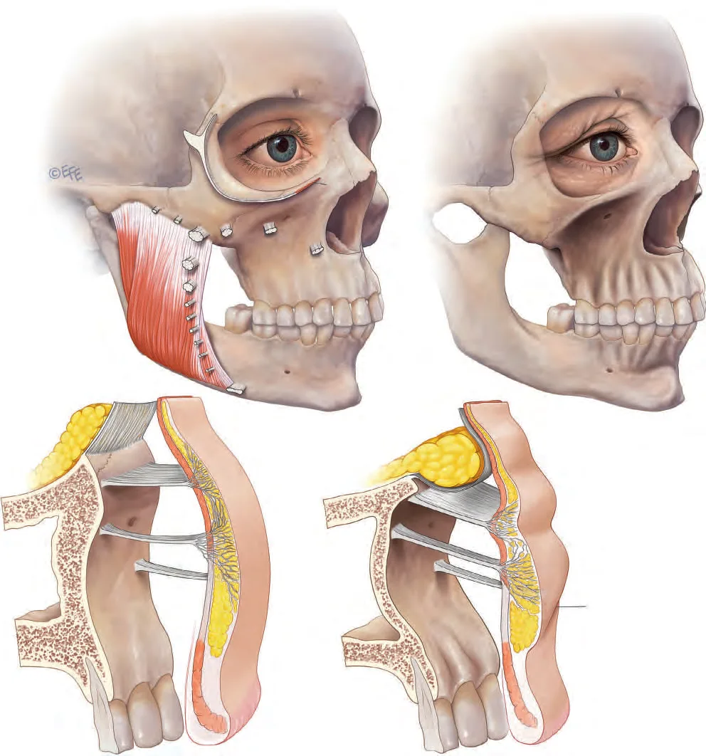 Fig 9. Bone first: the aged orbit is larger and its rim resorbed, so the medial brow loses its bony platform before any soft-tissue change is visible - (Standring, Gray's Anatomy, 2016, p.960).
> Sources: Standring, Gray's Anatomy 2016 [34]; skeletal ageing per Walczak 2023 [21], Karunanayake 2017 [22].
Fig 9. Bone first: the aged orbit is larger and its rim resorbed, so the medial brow loses its bony platform before any soft-tissue change is visible - (Standring, Gray's Anatomy, 2016, p.960).
> Sources: Standring, Gray's Anatomy 2016 [34]; skeletal ageing per Walczak 2023 [21], Karunanayake 2017 [22].
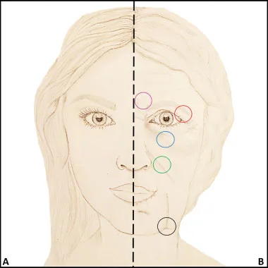 Fig 10. The superior-orbit circle is the brow story: ROOF laxity plus orbicularis/ORL fatigue plus bone change drop the tail - (Cotofana, 2016, p.5).
> Sources: Cotofana 2016 [1].
Fig 10. The superior-orbit circle is the brow story: ROOF laxity plus orbicularis/ORL fatigue plus bone change drop the tail - (Cotofana, 2016, p.5).
> Sources: Cotofana 2016 [1].
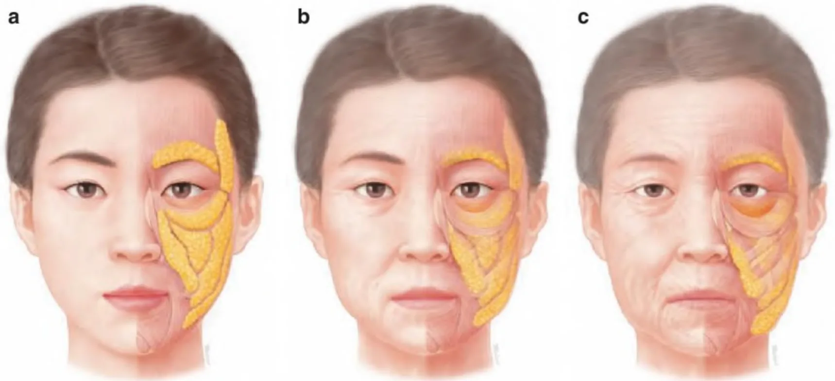 Fig 11. Superficial-fat deflation and descent is step 3, visible after the deep changes have already dropped the tail - (Anatomía Clínica de la Cara, corpus, p.59).
> Sources: Anatomía Clínica de la Cara para Relleno y Toxina Botulínica [38]; compartment behaviour per Schenck 2018 [3].
Fig 11. Superficial-fat deflation and descent is step 3, visible after the deep changes have already dropped the tail - (Anatomía Clínica de la Cara, corpus, p.59).
> Sources: Anatomía Clínica de la Cara para Relleno y Toxina Botulínica [38]; compartment behaviour per Schenck 2018 [3].
The ligaments, ranked (why the tail goes first). Biomechanically the zygomatic ligament is the stiffest, then the orbital retaining ligament, then the mandibular [1]. The lateral brow tail is held only by the temporal ligamentous adhesion and the superior temporal septum, with no strong bony ligament of its own; as the orbital retaining ligament loses its horizontal position the orbicularis destabilises and the ROOF above it sags [1][2]. That is the mechanical reason the tail descends before the head, and why lateral support (temple, on the lateral side of the line of ligaments) lifts it while a medial bolus only projects.
Muscle ageing, quantified in behaviour not dose. Facial muscles undergo sarcopenia: they lengthen, raise resting tone toward maximum-contracture tone, and shorten their amplitude, converting dynamic lines to static ones [1]. Clinically this means an ageing brow both depresses more at rest (orbicularis/glabellar tone up) and is held up more by the frontalis (compensation), a double change that a single-muscle toxin plan will get wrong.
What this does NOT change (still state-of-the-art): the deep-first sequence and the ROOF/orbital-retaining-ligament mechanism come from 2016-2019 cadaveric and imaging work [1][2][3] and are unchanged by newer skeletal-ageing imaging [21], which reinforces rather than revises them. The corpus's own 2016 review remains the reference frame.
The sequence over time (schematic, not a fixed clock, because onset varies by individual and ethnicity): the deep changes (bone resorption, ligament attenuation, ROOF descent) begin earlier than they are visible and accumulate; the tail descent is typically the first change a patient notices, often as a "tired/heavy" look before any line is fixed; superficial-fat deflation follows; static skin lines and actinic change come last, on top of the already-descended frame [1][2][21]. Because the deep changes precede the surface ones, a patient can have a real structural brow problem while the skin still looks good, which is exactly the patient who benefits most from toxin plus lateral support and least from waiting for the skin to demand attention.
The three physical signs that place the patient in the sequence: a flatter medial brow (lost bony shelf, step 1); a lower, less-supported tail with a hollow temple (ligament/deep-fat + temple deflation, step 2); and static forehead/lateral-canthal lines with thinned skin (step 4), usually with a raised resting brow from frontalis compensation layered on top. Reading which signs dominate tells you which lever leads: a flat medial brow with good skin is a support/structural question; a low tail with a hollow temple is a temple question; static lines with a compensating forehead are a toxin-plus-skin question with a ptosis screen first [1][6].
Deflation vs descent (attenuation), and why it is not an idle debate here. One school reads facial ageing as volume loss (deflation), another as ligamentous attenuation and descent; in the brow the honest answer is both, in sequence: the deep fat deflates and the retaining ligaments attenuate over a resorbing rim, so the tail loses volume and drops [1][2]. The clinical consequence is that neither "just fill it" nor "just lift it" is complete; the tail needs the depressors released (toxin), the platform restored where volume is truly lost (temple), and, past a threshold, surgical repositioning. Averaging the two models into "add filler" is exactly the error the region punishes.
The bony detail that removes the medial-brow platform. Orbital-aperture widening is not uniform: resorption is greatest supero-medially and infero-laterally, so the superomedial rim under the medial brow head recedes while the inferolateral rim recedes under the lateral canthus [1][21][22]. The medial brow loses its bony shelf first; the lateral canthus and tail lose their inferolateral support second. This is why an aged brow looks both flatter medially (lost shelf) and lower laterally (lost lateral support plus soft-tissue descent).
The compensation cascade, spelled out. As the brow drops and the visual field narrows, the frontalis is recruited at rest to hold the brow (and the lid) up; sustained recruitment writes forehead lines and, critically, masks an incipient eyelid ptosis [1][6]. The clinical trap is that this patient looks like a forehead-line candidate and is in fact a hidden-ptosis patient: toxin to the frontalis erases the lines and unmasks the ptosis, closing the visual field. The ageing sequence therefore ends not at the skin but at a behaviour (frontalis over-recruitment) that the assessment (D2.5) must detect before treatment.
Muscle ageing, the number that is behaviour not dose. Facial muscles shorten their amplitude and raise resting tone toward maximal-contracture tone, so dynamic lines become static and the depressors pull harder at rest [1]. An older brow is therefore simultaneously more depressed at rest and more frontalis-dependent; a single-muscle plan gets one of the two wrong. The lever remains toxin-to-depressors first, then support, then surgery, tracking the tissue that actually failed.
Trampa clásica: reading a descended tail as a volume deficit and filling the brow. The tail dropped because the deep fat and ligaments failed over a resorbing rim; adding superficial volume to the brow makes it heavier and lower, not higher. The lever for descent is toxin (depressors) plus lateral support, never brow filler [1][2][C3.1 salvage].
D2.5 · Assessment: what is measured, photographed, tested dynamically, scanned
The assessment answers four questions in order: is the brow low, is it asymmetric, is the descent muscular / volumetric / skin / bony, and is the frontalis compensating a hidden lid problem. Getting the last one wrong is how the region produces its worst outcomes.
| What | How | Why it changes the plan |
|---|---|---|
| Brow height / position | brow-to-mid-pupil and brow-to-lid-margin distances at rest; a brow-positioning grade | quantifies descent and gives a pre/post baseline you can defend [6][20] |
| Brow shape / apex | mark the apex; describe the arc | the apex sits near the lateral limbus-to-lateral canthus in most aesthetic conventions, higher/more medial in women, flatter in men [20] |
| Symmetry | photograph and measure both sides; document baseline asymmetry before, not after | baseline asymmetry is the rule, not the exception; undocumented, it becomes "your" complication |
| MRD1 / MRD2 | margin-reflex distances | separates true eyelid ptosis (low MRD1) from brow ptosis and from dermatochalasis |
| Lid laxity / snap test | pull the lower lid, release; canthal tone | governs any peri-orbital toxin and flags dry-eye risk |
| Skin quality | pinch, actinic damage, static lines | decides whether the residual line needs skin tools, not more toxin [23] |
| Vessel / layer map | high-frequency Doppler ultrasound before filler | locates the STA, sentinel/middle temporal vein and the target plane in real time; the temple is where this pays for itself [10][39][25] |
The three photographs, non-negotiable: frontal at rest, at maximum frown (glabellar complex + corrugator/orbicularis depressor action), and at maximum voluntary elevation (frontalis recruitment). Without the elevation photo you cannot see frontalis compensation, and without the frown photo you cannot dose the depressors. Standardise distance, head position and lighting so the follow-up photo is comparable [6].
The dynamic exam separates four look-alikes that need opposite treatments: - True brow ptosis - the brow itself is low; frontalis lifts it when asked → candidate for depressor toxin ± lateral support. - Dermatochalasis - redundant upper-lid skin overhangs the lash line; the brow may be normal → surgical, not injectable. - Levator-based eyelid ptosis - low MRD1, the lid margin is down; the brow is often held high by compensation → refer; forehead toxin is hazardous here. - Frontalis compensation - the patient unconsciously recruits the frontalis at rest to keep the visual field open. The relax-forehead test: ask the patient to close the eyes, relax the forehead, then gently open the eyes without raising the brows; watch the lid margin drop and the true resting brow position appear. Observe the forehead when the patient thinks they are unobserved [6][24].
Why ultrasound is now part of a serious brow/temple work-up. The temple is a 10-layer region with a superficial artery, a valveless venous system and a bone-safe deep plane; real-time high-frequency ultrasound identifies the STA and sentinel vein, confirms the interfascial or supraperiosteal target, and lets the operator watch the deposit rather than infer it [10][25]. In a region where the failure mode is blindness or pulmonary embolism, "I aspirated and it was landmark-based" is a weaker standard than "I saw the vessel and I saw the plane" [10][39]. Ultrasound is not yet the standard of care everywhere, and its access and learning curve are real; the honest position is that it converts the temple from a landmark procedure to a visualised one where available.
Sex and population, measured not assumed. The male brow sits lower (at or near the rim), is flatter and thicker, with a less lateral apex; anthropometric analysis confirms these are distinct, not variations of one female template [20]. Elevating and arching a male brow feminises the gaze, which is one of the most frequent and least-discussed unwanted outcomes of upper-face treatment in men [20][C3.1 salvage]. The apex target and the acceptable lift are therefore a decision, documented with the patient, not a default.
How to measure the brow so the follow-up is defensible. Use a fixed facial landmark (mid-pupil, medial canthus) and measure brow-to-mid-pupil and brow-to-lid-margin at the head, mid and tail, both sides, on a standardised frontal photo; record the apex position relative to the lateral limbus/lateral canthus; and repeat the identical measures at the two-week review [6][20]. A millimetric brow lift is only demonstrable against a measured baseline; "it looks higher" is not a record. Baseline asymmetry is documented before treatment, because an asymmetry discovered after is read as a complication [6].
The dynamic exam, in sequence. (1) Rest - the starting brow and lid position. (2) Maximum frown - isolates the glabellar depressors and the corrugator/orbicularis pull; this photo sets the depressor dose. (3) Maximum voluntary elevation - shows frontalis strength and pattern; a weak or asymmetric elevation warns of a poor lift response. (4) Relax-forehead test - close eyes, relax forehead, open without brow recruitment; watch the lid margin drop and the true resting brow appear, exposing frontalis compensation [6][24]. (5) Snap/lid-laxity - governs any peri-orbital toxin and flags dry-eye risk. Skipping (3) and (4) is how the region's worst outcome (unmasked lid ptosis) gets missed.
Ultrasound protocol for the temple, where the safety payoff is largest. With a high-frequency linear probe: identify the superficial temporal artery in the superficial temporal fascia, the middle temporal / sentinel vein in the lower temporal compartment, and the target plane (interfascial or supraperiosteal); then deposit under real-time visualisation rather than by landmark [10][39][25]. In a region whose failure mode is blindness or pulmonary embolism, seeing the vessel and the plane is a materially higher standard than "landmark plus aspiration," where ultrasound is available and the operator is trained [10]. Its access and learning curve are real limits, stated honestly, not reasons to omit it where it exists.
Grading the brow, so the plan is graded too. A workable brow-position grade separates mild (tail sits at or just below the ideal, good skin, lifts well on request → chemical brow lift ± temple), moderate (visible tail descent, some skin change, temple hollow contributing → toxin + temple support, consider thread), and severe (heavy static descent, skin excess, poor lift on request → surgical). The grade is recorded with the measurements so the tool follows the grade, not the request [6][20]. The apex is graded separately (over the lateral limbus/lateral canthus in most conventions; flatter and lower for men) because a shaping request is a different goal from a descent problem [20].
What ultrasound actually shows here (so the scan is read, not just done): the superficial temporal artery as a pulsatile channel in the superficial temporal fascia; the middle temporal / sentinel vein as a compressible, valveless channel in the lower temporal compartment; the fascial leaves that define the interfascial space; and the layer depth from skin to bone, which confirms the target plane and the safe entry angle [10][39][25]. A scan that is performed but not interpreted adds time without safety; the point is to see the vessel and the plane before the needle moves.
What goes in the record (so a result is reproducible and a complication is traceable): the four photos, the brow measurements both sides, the compensation-test result, the product/lot/dilution, and the units or millilitres per point with plane and tool. Without the dilution and the pre-treatment photo, neither the operator nor anyone else can reproduce the result or investigate a failure [C3.1 salvage].
Trampa clásica: dosing the depressors off the rest photo and skipping the elevation photo. The frontalis compensation is invisible at rest; the first sign is a post-toxin heavy brow or an unmasked lid ptosis at the two-week review, by which point it is a three-month problem. The elevation photo and the relax-forehead test are five seconds that prevent it [6][24].
D2.6 · Goal and patient selection: who benefits, who does not, what is referred
The goal is to reposition and support the tail, not to fill the brow. Every good candidate maps to a lever; every poor candidate is a referral or a different region.
| Candidate | Finding | First-line lever | Note |
|---|---|---|---|
| Ideal injectable candidate | mild-moderate lateral tail descent, good skin quality, brow rises on request | toxin to depressors ± temple support | best benefit-risk of the whole region [C3.1 salvage][7] |
| Young shaping request | wants a defined arch / small lift, no descent | low-dose depressor toxin (chemical brow lift) | reversible, cheap, low risk; manage the "how much" expectation [6] |
| Volumetric temple deficit | hollow temple pulling the tail down, intact muscle | temple volume (support), then reassess brow | the tail is a temple problem here, not a brow one [25][2] |
| Frontalis-compensated lid ptosis | brow held high at rest, drops on relaxing forehead, low MRD1 | relative contraindication to forehead/brow toxin | toxin unmasks the ptosis; refer or treat the lid first [6][24] |
| True heavy brow ptosis | significant static descent, brow will not lift much | surgery (brow/forehead lift) | injectables underdeliver and erode trust [11][16] |
| Dermatochalasis / skin excess | redundant upper-lid skin over the lashes | surgery (blepharoplasty ± browpexy) | no injectable removes skin [16] |
| Structural bony deficiency | skeletonised superior orbit, resorbed rim beyond volume correction | structural support ± surgery | manage expectations; filler alone will not rebuild bone [21][22] |
Consensus: the injectable sweet spot is mild-to-moderate tail descent with good skin in a patient whose frontalis is not doing hidden work. Outside that, the correct answer is often not an injectable, and saying so is part of the treatment. The two referral triggers that must be spoken aloud are heavy brow ptosis / marked skin excess (surgical) and a frontalis-compensated eyelid ptosis (unmask risk) [11][16][6][24].
Who does NOT benefit (and why offering treatment harms): - The patient using the forehead as a crutch: toxin closes their visual field; they leave worse [6][24]. - The dermatochalasis patient: skin hanging over the lashes is not a muscle or volume problem; an injectable produces a disappointing result that reads as a failure [16]. - The overfilled-brow chaser: adding brow volume to "lift" a descended tail makes it heavier and lower; this is the commonest self-inflicted iatrogenic brow [2][C3.1 salvage]. - The male patient handed a female template: an arched, elevated brow feminises the gaze and is often regretted [20].
Expectation setting, in numbers the patient can hold. A chemical brow lift moves the tail millimetres, not centimetres; the perceptual change (a more open, rested, less angry gaze) is larger than the metric change [6][7]. Threads give an immediate mechanical lift that is temporary (months, not years) [13][15]. Surgery is the durable answer for true descent and skin excess [11][16]. A patient who wants a surgical result from a syringe is a patient who will be unhappy with a technically perfect injection.
Psychology and the "angry/tired" complaint. The brow encodes emotion: a low medial brow reads as anger, a low tail as tiredness or sadness. Patients describe the emotion, not the anatomy; translating "I look angry/tired" into "medial depressor overactivity / lateral tail descent" is the selection step that decides the tool. Reframing the goal as changing a gesture, not erasing a line, improves both selection and satisfaction (see B2 — Patient Psychology & Selection).
The surgical ladder, so the referral is specific and not a shrug. When descent or skin excess crosses the injectable threshold, the options are graded: transblepharoplasty browpexy (a small internal brow anchor added to an upper blepharoplasty, for mild tail support with skin excess); temporal (lateral) brow lift (a lateral vector for isolated tail descent); endoscopic / gliding forehead lift (minimal-incision elevation of the whole brow); direct and deep-plane direct brow lift (a supra-brow excision with, in the deep-plane version, orbicularis transection/suspension and brow-fat suspension for a stronger, more durable lift) [11][16]; and extended suprabrow lift with fat grafting for the skeletonised, deflated brow. The decision rule that sends the patient to any of these is significant/heavy ptosis or marked skin excess, where an injectable underdelivers and erodes trust [11][16].
Energy vs thread vs surgery, decided by the tissue. Mild true laxity with good skin: energy-based tightening (radiofrequency, microfocused ultrasound) is a reasonable non-surgical option. Moderate tail descent wanting an immediate visual lift, accepting temporary results: a temporal→tail thread. Heavy descent or skin excess: surgery. Filler and toxin sit before this ladder, not on it, and are the wrong tool once true descent dominates [11][16][13].
Consent that matches the region's risk. For any filler here, the consent names the blindness risk explicitly and records that hyaluronidase is available; for toxin, it records the possibility of transient asymmetry, brow/lid heaviness and the two-week review; for a male patient, it records that a lift/arch is a deliberate, discussed change because it feminises the gaze [20][C3.1 salvage]. A generic aesthetic-consent clause does not meet the standard for a zone that can blind.
Expanded "not a candidate" list (with the harm of proceeding): the frontalis-crutch patient (toxin closes the visual field); the dermatochalasis patient (an injectable cannot remove skin, so the result disappoints and reads as failure); the true-eyelid-ptosis patient (needs a lid procedure, not a brow one); the overfilled-brow chaser (more brow volume drops the tail further); and the male patient handed a female template (a feminised, regretted result) [6][16][20][2]. Naming why each is excluded is part of the treatment, not a hedge.
Trampa clásica: accepting the patient's own diagnosis. They point at the brow and ask for filler; the problem is a resorbing rim, a deflating temple, or a compensating frontalis. Treat the mechanism you found on exam (D2.5), not the request, and refer the surgical ones without apology [11][16].
D2.7 · Technique - the full grid
The region is not closed with one technique; it is closed with the whole grid of ways to support or reposition the tail, each row with its school, plane, tool, volume and evidence. In the brow the filler almost never goes into the brow: it goes into the temple (to reposition the lateral face and lift the tail) or as a supra-brow micro-bolus (to support the medial brow head over a resorbed rim). Direct brow filler is the exception, and usually the wrong one.
Product grid (class → plane → reversibility → when NOT to use it):
| Product class | Typical plane here | Reversible? | Use / avoid in this region |
|---|---|---|---|
| High-G′ / high-cohesivity HA | supraperiosteal temple; supra-brow micro-bolus | yes (hyaluronidase) | first choice for lift/projection where it resists compression [25][2] |
| Soft low-G′ HA | subdermal temple | yes | diffuse temple smoothing (Cotofana/Freytag subdermal school); avoid deep where projection is needed [25] |
| CaHA (normal) | supraperiosteal temple | no | biostimulation + volume; irreversibility is a strong argument against it in a blindness zone [26] |
| CaHA hyperdilute | subdermal temple | no | skin-quality/biostimulation over a skeletonised temple; dilution figures vary widely and are not averaged here [26] |
| PLLA | supraperiosteal / subdermal temple | no | gradual temple volume/skin quality over sessions; not for acute correction |
| PCL | supraperiosteal temple | no | durable biostimulatory volume; irreversible |
| PMMA | - | no (permanent) | avoid in this vascular, mobile region; permanence + blindness risk is the wrong trade |
| Autologous fat | supraperiosteal temple / brow fat | partial | durable temple/brow volume in the right hands; embolic risk is real, technique-dependent |
| Polynucleotides / skin booster | intradermal peri-orbital | n/a | skin quality, not lift; adjunct not a repositioner |
| Toxin | see D2.8 | n/a (wears off) | the actual first-line "tool" of the region |
Instrument grid: needle by gauge/length (e.g. 27-30 G, 13 mm for supra-brow micro-aliquots; single-point supraperiosteal temple bolus on bone); cannula by gauge/length (22-25 G, 40-50 mm for the interfascial temple approach from an infra-zygomatic entry); with vs without ultrasound guidance (US-confirmed is the safer temple standard where available) [25][10][39]. Cannula reduces but does not abolish intravascular risk; the temple's valveless veins are wide enough to admit a cannula tip.
Plane grid (deep to superficial): supraperiosteal (on bone, near the temporal crest to avoid the deep temporal arteries) · interfascial (between superficial and deep temporal fascia, US-guided) · deep fat (ROOF, via supra-brow) · SMAS/STF (avoid: the STA lives here) · superficial (subdermal) fat · subdermal · intradermal (boosters only). Movement grid: single bolus (bone) · microbolus · retrograde linear · fan · cross-hatch · serial puncture · tower (avoid tower in the temple).
Point-by-point technique (region support):
| Target | Plane / entry | Direction | Tool | Vol / point | Vol / side | Movement | Touch-up |
|---|---|---|---|---|---|---|---|
| Temple, deep (bone) | supraperiosteal, needle perpendicular to firm bone contact near temporal crest | straight down to bone, withdraw 0.5 mm | 27 G needle | 0.2-0.5 mL slow | 0.5-1.0 mL | single slow bolus, low pressure | reassess at 2 wk; top up on bone only |
| Temple, interfascial | between superficial/deep temporal fascia, infra-zygomatic entry | advance under US to the interfascial space | 22-25 G cannula, US-guided | retrograde threads | 0.5-1.0 mL | retrograde linear/fan in-plane | US-confirm plane before each pass [25][10] |
| Temple, subdermal | subcutaneous, tangential from lateral entry | tangential, superficial to STA | 25 G cannula | small aliquots | 0.3-0.8 mL | retrograde linear, diffuse | avoid over-superficial lumping |
| Supra-brow (medial head support) | supraperiosteal micro-bolus, lateral to the supraorbital notch | perpendicular, tiny | 30 G needle | ≤0.1 mL | ≤0.1-0.2 mL | microbolus | rarely needed; do not bolus on the notch |
| Thread (temporal→tail vector) | subcutaneous, temporal hairline anchor to tail | vector from anchor to tail/outer canthus | barbed PDO cannula | n/a | n/a | linear traction vector | see non-injectable row [13][15] |
Ceilings before overfill: temple ~1.0 mL/side is a working ceiling for a first session; beyond it the temple reads heavy and the lateral brow can be pushed into an unnatural shelf. Supra-brow filler above ~0.2 mL starts to read as brow heaviness. When in doubt, under-fill and reassess at two weeks; the region punishes over-correction more than under-correction [25][C3.1 salvage].
School grid + the discrepancy that changes the gesture (never averaged):
Consensus: support the tail from the temple/lateral zone (lateral to the line of ligaments, so it lifts rather than projects), use the smallest effective volume, keep hyaluronidase ready, and prefer a reversible product in this blindness zone [2][25][26].
Discrepancy - which temple plane to use (the schools genuinely do the opposite, decide by the variable, do not average): - Interfascial cannula, US-guided (Bravo; Desyatnikova/Tejero school): 22-25 G cannula between the fascial leaves, infra-zygomatic entry, deposit watched on ultrasound. Argument: safest around the STA and sentinel vein because the deposit and the vessels are both seen. Decider: you have ultrasound and the skill; the temple is deep/hollow [25][10]. - Supraperiosteal single-point needle bolus on bone (classic MD Codes temple, deep code): one deep bolus with firm bone contact near the temporal crest, deep to all vessels. Argument: simple, reproducible, deep to the arterial and venous planes. Decider: no ultrasound, experienced hand, bone-safe target; cross-ref C2 — MD Codes Systematic Approach for the exact code nomenclature (temple T-points) [25][C3.1 salvage]. - Subdermal high-viscoelastic repositioning (Cotofana/Freytag temporal-lift): soft/robust product placed subdermally to reposition the lateral face and lift the tail, exploiting that the superficial temporal compartments increase projection rather than displace when filled [3][25]. Argument: a lifting/repositioning effect rather than a hollow-fill. Decider: the goal is a lateral lift vector, not a deep volume deficit.
> The three schools use a different tool, entry, depth and vascular-risk profile. There is no averaged "middle plane"; picking one is picking its risk profile. The one plane to avoid is the intermediate one that loses bone contact without confirming the fascial space, which is where the sentinel vein lives [2][25].
MD Codes and named techniques (the exhaustive school column): the temple in the MD Codes system is addressed with a superficial (subdermal) and a deep (supraperiosteal) point; the exact code letters and injection sequence are catalogued in C2 — MD Codes Systematic Approach and attributed to de Maio, not reprinted here as a bare dose. Named region techniques include the Cotofana "six techniques for the temple" framework [2][25] and the temporal-lift repositioning concept; each is a specific tool+plane+volume, not a brand of the same act.
Ultrasound-guided temple deposit, step by step (the visualised standard where available). (1) Map the superficial temporal artery in the superficial temporal fascia and the middle temporal / sentinel vein in the lower temporal compartment with a high-frequency linear probe. (2) Choose the plane: interfascial (cannula) or supraperiosteal (needle on bone near the crest). (3) Enter from an infra-zygomatic point, advance under real-time view to the target plane, confirm the tip is clear of the mapped vessels. (4) Aspirate where meaningful, deposit slowly in small aliquots, watch the gel spread in-plane. (5) Re-scan after deposit to confirm placement. This converts the temple from a landmark procedure to a seen one and is the single biggest safety upgrade available in the region [10][39][25].
Filler aftercare and retouch (region-specific). Low-pressure technique, brief compression of any bleeding point, and a clear instruction set: no massage of a temple deposit that must stay in plane, watch for delayed pain/vision change and report it immediately, and a two-week review to judge the settled result before any top-up. Document product, lot, plane, tool and volume per point so a result is reproducible and a complication is traceable. Over-correction is harder to fix than under-correction here, so the default is to under-fill and reassess [25][C3.1 salvage].
When direct brow filler IS justified (the rare exception, stated so it is not a loophole). A supra-brow micro-bolus (≤0.1 mL, supraperiosteal, lateral to the supraorbital notch) can support a medial brow head over a resorbed rim when temple support alone has not restored the medial platform; and a small subdermal soft-HA deposit can address focal brow-fat-pad flattening. Both are small, superficial-to-periosteum or subdermal, reversible, and placed away from the notch and midline. Anything larger, deeper on the notch, or in the glabellar midline is the rejected direct-fill pattern [2][12][26].
Movement-by-plane cheat (which motion belongs to which depth): supraperiosteal on bone → single slow bolus; interfascial → retrograde linear/fan under ultrasound; subdermal temple → retrograde linear, diffuse; supra-brow ROOF → microbolus; skin quality → serial intradermal puncture (boosters). The tower technique (stacked column from bone to skin) is avoided in the temple, where it crosses the venous plane [2][25].
Non-injectable rows (when the correct answer is NOT an injectable): - PDO thread lift on a temporal→lateral-brow vector: immediate mechanical lift + collagen stimulation, durability months not years (temporary), evidence uncontrolled and modest [13][15]. - Energy-based tightening (radiofrequency, microfocused ultrasound): for mild true laxity where the problem is skin, not muscle or volume. - Surgery (see D2.6): direct brow lift, deep-plane direct brow lift [11], endoscopic/gliding forehead lift, temporal (lateral) brow lift, transblepharoplasty browpexy [16] - the durable answer for heavy ptosis or skin excess.
Product-by-product, for the region (what each is actually for here): - High-G′/high-cohesivity HA: the workhorse for supraperiosteal temple lift and the rare supra-brow micro-bolus; resists compression, reversible; first choice where projection/lift is the goal [2][25]. - Soft/low-G′ HA: subdermal temple smoothing and focal brow-fat-pad flattening; integrates and spreads; reversible; wrong choice deep where it gives no lift [25]. - CaHA (normal): supraperiosteal temple biostimulation + volume; irreversible; a considered choice for a thin-skinned temple, kept as an exception in a blindness zone [26]. - CaHA hyperdilute: subdermal temple skin-quality/biostimulation over a skeletonised temple; irreversible; dilution figures vary widely and are not averaged here [26][C3.1 salvage]. - PLLA: gradual temple volume and skin quality over sessions; irreversible; judged over months, not at the two-week review. - PCL: durable biostimulatory temple volume; irreversible. - PMMA: permanent; avoided in this mobile, high-risk region. - Autologous fat: durable temple/brow-adjacent volume plus skin benefit; variable take, real embolic risk, irreversible; a considered surgical-adjacent choice, not a first-line office injectable. - Polynucleotides / skin boosters: intradermal peri-orbital skin quality; adjunct, not a repositioner. - Toxin: the region's actual first-line tool (D2.8), the only one that treats the depressor cause rather than the volume symptom.
Rheology, decoded for this region (why G′ and cohesivity choose the plane). G′ (elastic modulus) is resistance to deformation: a high-G′ gel holds its shape against the compression of the temporalis and the overlying fascia, so it is the deep supraperiosteal choice where lift/projection is needed; a low-G′ gel spreads and integrates, so it is the subdermal choice for diffuse smoothing. Cohesivity is how the gel holds together: high cohesivity resists fragmentation and migration, useful in a mobile region. Calibre/particle size tracks with plane depth. The practical rule: high-G′/high-cohesivity deep, soft/low-G′ superficial, never the reverse (a soft gel deep gives no lift; a firm gel superficial gives nodules and Tyndall) [2][25][26].
Biostimulator logic without a memorised dilution table (the atlas does not average dilutions). CaHA, PLLA and PCL work by collagen induction, not volume alone; in the temple they are chosen for a skeletonised, thin-skinned hollow where skin quality matters as much as fill. The transferable logic: more dilution = more biostimulation and less immediate volume; less dilution = more volume and more nodule risk in a superficial plane [26][C3.1 salvage]. The dilution numbers vary widely between authors and indications and are not printed here as a single figure; the constraint that matters is irreversibility - none of the three is reversible, which in a blindness zone is a strong reason to keep them as the exception, not the default [26].
Cannula vs needle, evidence-graded for the temple. A cannula reduces (does not abolish) intravascular risk and is the safer default for the interfascial and subdermal temple approaches; a single-point needle bolus on bone is the classic deep approach where bone contact is the safety mechanism. The valveless temple veins are wide enough to admit a cannula tip, so "cannula = safe" is false; the safety comes from the plane and from seeing the vessel on ultrasound, not from the instrument alone [10][25][2].
Autologous fat, the durable non-HA volume option. Fat grafting gives durable temple/brow-adjacent volume and improves overlying skin, at the cost of variable take, a real embolic risk if injected under pressure into the temporal vessels, and irreversibility. It is a considered choice for the skeletonised temple in the right hands, not a first-line office injectable, and it belongs in the same "reversible-by-default" caution as the biostimulators [2][25].
Named-technique compendium for the region (school column, exhaustive): the Cotofana "six techniques for the temple" framework (each a specific plane/tool/volume) [2][25]; the MD Codes temple points (superficial + deep, catalogued and attributed to de Maio in C2 — MD Codes Systematic Approach); the Cotofana/Freytag subdermal temporal-lift repositioning [3][25]; and, for the tail specifically, the temporal→lateral-brow thread vector [13]. Each is a different act, not a rebrand of one bolus.
Trampa clásica: filling the brow itself to lift the tail. It adds weight to a structure with no bony anchor and drops it further; the correct move is toxin to the depressors plus temple support lateral to the line of ligaments. Second classic error: an intermediate temple plane that has lost bone contact but not entered the interfascial space, seeded straight into the sentinel-vein territory [2][25].
D2.8 · Toxin of the region
Toxin is the first-line tool of the brow, and it lifts by unbalancing depressors against the frontalis. Doses in onabotulinumtoxinA (ona) units; brands are not interconvertible and off-label targets are marked. The one rule that governs the block: the frontalis is the only elevator, so it is the antagonist you do NOT paralyse when the goal is a lift.
The target grid (muscle → points → units/point → plane → safety distance → antagonist spared):
| Target | Role | Points | Units/point (ona) | Plane | Safety distance | Note |
|---|---|---|---|---|---|---|
| Lateral orbicularis oculi (tail) | depressor of the tail | 1/side (the "double-dose" tail point) | 2-5 U | subcutaneous, superficial | ≥1 cm outside the orbital rim (≥1.5-2 cm if intradermal) | the actual chemical brow-tail lift point [36][4] |
| Corrugator supercilii | medial depressor (classic) | 2/side | 4 U | intramuscular, on the belly, needle up | ≥1 cm above the rim | over-low medial point diffuses to levator → lid ptosis [27][7] |
| Depressor supercilii | medial depressor | 1/side | 2-4 U | at the medial brow head | ≥1 cm above rim | part of the medial-lift release [7] |
| Procerus | central depressor | 1 | 4 U | intramuscular midline | - | completes glabellar depressor release [27] |
| Frontalis (if treated) | sole elevator - the antagonist | high points only | small, tapered | IM/superficial (thin muscle) | keep ≥2 cm above the rim; spare lower + lateral fibres | treating lower/lateral fibres drops the brow [6][24] |
Label anchor (glabella, approved) [A]: onabotulinumtoxinA 20 U total in 5 points of 4 U (2 per corrugator + 1 procerus), intramuscular, needle angled up and away from the orbit [27]. Brand equivalents are not convertible: abobotulinumtoxinA (Azzalure) 50 U Speywood in 5 points of 10 U [28]; incobotulinumtoxinA (Bocouture) 20 U in 5 points [29]; other type-A brands quote their own 5-point totals. The ona:abo ratio published spans 1:2.5 to 1:3 by scenario and is not averaged; re-dose from scratch per brand [28][C3.1 salvage].
Label anchor (frontalis, approved combined) [A]: onabotulinumtoxinA 20 U in 5 points of 4 U, and the approved indication requires treating the glabella simultaneously (20 U frontal + 20 U glabella) precisely because paralysing the sole elevator without releasing the depressors drops the brow [27][6]. The five frontalis rules (keep high, treat elevator + depressors together, less lateral dose to keep the tail, adapt the pattern to the forehead, screen frontalis compensation) are the region's core safety net [6][24][C3.1 salvage].
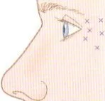 Fig 12. The tail lift is a small, superficial deposit into the lateral orbicularis, ≥1 cm outside the rim, sparing the frontalis above it - (Carruthers, Toxina Botulínica, p.52).
> Sources: Carruthers & Carruthers [36]; lateral canthal dosing per label [27] and Benedetto [41].
Fig 12. The tail lift is a small, superficial deposit into the lateral orbicularis, ≥1 cm outside the rim, sparing the frontalis above it - (Carruthers, Toxina Botulínica, p.52).
> Sources: Carruthers & Carruthers [36]; lateral canthal dosing per label [27] and Benedetto [41].
The chemical brow lift, mechanistically. Weaken the depressors selectively and the frontalis wins locally, so the brow rises: the tail rises by releasing the lateral orbicularis (the double-dose tail point), the medial brow rises by releasing the glabellar depressor complex (corrugator, depressor supercilii, procerus, medial orbicularis) [C3.1 salvage][7]. Expected displacement is millimetric and quantified: after frontalis dosing the medial and central brow fall while the lateral brow edge rises, and upper-forehead injection patterns prevent brow ptosis better than lower ones [6]. An anatomy-respecting 3-point glabellar technique (procerus + corrugator origins) achieved brow-line control without eyelid or brow ptosis in a 105-patient series [7].
Discrepancy 1 - intradermal vs intramuscular forehead toxin (decide by frontalis dependence, do not average the depth or dose): > Consensus: the depressors are released to lift; the frontalis is spared where possible. > Discrepancy: classic intramuscular dosing of the forehead relaxes lines but lowers the brow at weeks 2-4 when lower/lateral frontalis fibres are caught; intradermal / microtox (low total dose, e.g. 8 U to the forehead) preserves brow position with the same anti-wrinkle effect and the same duration, at the cost of more pain [4]. Decider: a frontalis-dependent patient or one at brow-drop risk favours the intradermal/low-dose route; a purely dynamic line with no compensation tolerates classic IM. The depth and the dose are not averaged [4][6].
Discrepancy 2 - the function of the corrugator supercilii (this changes which fibres you inject to lift vs to smooth): > Classic teaching: the corrugator is a pure medial brow depressor (draws the brows together and down, writes glabellar lines). > Reappraisal (Muñoz-Gonzalez & Fakih-Gomez 2024, 5-year split-face, 298 patients): targeting specific portions of the corrugator and depressor supercilii produces medial-brow elevation and superior lift versus traditional dosing, but is frequently associated with omega-shaped wrinkles (the depressor/omega complex) [5][14]. Decider: if the goal is a medial lift, portion-specific targeting can help but risks the omega deformity; if the goal is a smooth glabella, classic full-belly dosing is simpler. The dispute is embedded in the current upper-face teaching decks [31][5]. Kept side by side, not merged.
Mephisto / Spock brow (the region's signature toxin complication): over-elevation of the tail when the central frontalis is treated and the lateral fibres are left free, so the untreated lateral frontalis over-pulls the tail up. Correct with 1-2 U into the lateral frontalis just above the point of maximum elevation, and prevent it by distributing the frontalis pattern evenly from the start [C3.1 salvage][24].
Apraclonidine rescue for the lid, not the brow. If diffusion reaches the levator and produces an eyelid ptosis, topical apraclonidine (alpha-agonist, contracts Müller's muscle, raises the lid 1-2 mm) buys symptomatic time while the toxin wears off; it does not reverse the toxin [C3.1 salvage]. Detail in J4 — Botulinum Toxin Complications.
Onset, duration and interval. The neuromodulator effect begins at ~3-4 days and peaks at ~2 weeks, which is why the review and any retouch are at two weeks, not at day 7 [7][C3.1 salvage]. Duration in the upper face is on the order of 3-4 months, longer with repeated cycles as the depressor bulk reduces; the lift is not permanent and the plan is a course, not a single act. Re-treatment is scheduled by the return of movement, not by a fixed calendar, and dose creep is avoided by re-measuring the brow each cycle.
Dose titration philosophy (start low, add later). In a region where over-treatment drops the brow or the lid, the rule is the lowest effective dose first, add at two weeks if needed. You can always add units; you cannot remove them. This applies most to the frontalis (where a low dose protects the tail) and to the lateral-orbicularis tail point (where a small dose lifts and a large one can over-elevate into a quizzical look) [6][24][C3.1 salvage].
Microtox / intradermal for brow-adjacent skin quality (not a lift, and labelled honestly). Very dilute toxin in multiple intradermal micropapules over the forehead/temple can soften fine superficial lines and reduce oiliness without abolishing expression; the sweat/sebaceous-gland effect is the well-established part (the same mechanism as hyperhidrosis), the superficial-muscle-tone effect is plausible but less proven, and the primary literature under the name is thin. It is an adjunct to skin quality, [D]/[C] in strength, and the consent says so; it is not a substitute for a chemical brow lift or for a skin-quality programme with better evidence [23][C3.1 salvage].
The antagonist principle, applied to the tail lift specifically. To lift the tail you weaken its depressor (lateral orbicularis) while sparing the lateral frontalis that elevates it; treat both and you cancel the lift or drop the tail. The Mephisto/Spock deformity is the mirror error: sparing the lateral orbicularis is fine, but sparing the lateral frontalis while over-treating the central frontalis lets the untreated lateral fibres over-pull the tail up. The tail is a two-muscle balance, and both errors come from treating one and forgetting the other [C3.1 salvage][24].
Contraindications (region-relevant): neuromuscular disease (myasthenia, Eaton-Lambert, ALS) is an absolute contraindication; aminoglycosides potentiate the block; pregnancy/lactation, active local infection, and prior non-response/resistance are screened [23][41]. And the region-specific relative contraindication: the frontalis-compensated eyelid ptosis (D2.5/D2.6), where forehead toxin unmasks the ptosis [6][24].
Brand-equivalence table for the glabellar complex (the doses are NOT interconvertible; re-dose per brand):
| Brand (INN) | Glabella total | Points | Note |
|---|---|---|---|
| Botox/Vistabel (ona) | 20 U | 5 × 4 U | reference for every "U" in this chapter [27] |
| Azzalure/Dysport (abo) | 50 U Speywood | 5 × 10 U | Speywood units ≠ ona units; ratio 1:2.5-1:3, not averaged [28] |
| Bocouture/Xeomin (inco) | 20 U | 5 × 4 U | no complexing protein; clinically ~1:1 with ona but re-titrate [29] |
| Nuceiva/Jeuveau (prabo) | 20 U | 5 × 5 pts | quote the product's own total [C3.1 salvage] |
| Letybo (leti) | 20 U | 5 pts | as above [C3.1 salvage] |
Adjacent upper-face points that share the brow's balance (so the plan is coherent, not piecemeal):
- Lateral canthal lines (crow's feet), label [A]: 24 U total, 12 U/side in 3 points of 4 U, subcutaneous and superficial, kept outside the bony orbital rim (medial creep reaches the lateral rectus, diplopia; inferior/anterior creep reaches the zygomaticus major, lip drop) [27][36]. The most superior of these three points is functionally continuous with the lateral-orbicularis tail lift, which is why crow's-feet dosing and brow-tail shaping are decided together, not separately [36].
- Pretarsal orbicularis, off-label [C]: 1-2 U/side, only with a good snap test; opens the palpebral aperture but risks epiphora/ectropion in a lax lower lid. Not a brow point, but it changes the peri-orbital gestalt the brow sits in [C3.1 salvage].
Reconstitution, marking and aftercare (region-specific): reconstitute with preservative-free (or preserved) saline without agitation; record dilution, lot and units per point so a result, good or bad, is reproducible and a complication is traceable [C3.1 salvage]. Mark with the patient seated and animating (frown, elevate, close), because the brow's true muscle vectors are dynamic. Post-procedure: no rubbing/massage of the area for a few hours, stay upright, avoid intense heat and exercise the same day. Review and retouch at two weeks, never at day 7, when the effect is incomplete and early retouch is the classic route to over-dosing [6][C3.1 salvage].
The per-side vs total trap, stated for this region. Published brow/forehead doses are sometimes per-side and sometimes total; the glabella figure (20 U) is a total, the crow's-feet figure (24 U) is a total split 12/12, and the tail point (2-5 U) is per side [27][36][C3.1 salvage]. Before copying any figure, including these, confirm the reporting unit [27][C3.1 salvage].
Trampa clásica: dosing the frontalis low to chase the inferior forehead line. The inferior frontalis band is the part that holds the brow up; catching it drops the brow and, in a compensating patient, the lid. Stay high, treat the depressors in the same session, and taper the lateral dose to protect the tail [6][24][C3.1 salvage].
D2.9 · Combination and sequence → L2 — Combination & Sequencing
The order is fixed by physiology: relax the depressors first, let the brow find its true resting position, then decide whether any volume is still needed. Volume placed before the toxin settles is volume placed against a brow that is about to move.
| Step | Timing | What | Why this order |
|---|---|---|---|
| 1. Toxin to depressors | day 0 | lateral orbicularis tail ± glabellar complex | releases the down-pull; the brow rises and settles [C3.1 salvage][7] |
| 2. Reassess | ~2 weeks | re-photograph at rest/frown/elevation | the true residual deficit only appears after the toxin peaks; retouch here, not at day 7 [6] |
| 3. Lateral support | ≥2 weeks | temple volume (± supra-brow micro-bolus), lateral to the line of ligaments | repositions the tail; often the "deficit" is smaller than it looked [25][2] |
| 4. Thread (optional) | after toxin | temporal→tail barbed vector | toxin protects the vector by relaxing the depressors that would fight it [13][15] |
| 5. Skin / energy | any time, with toxin in place | resurfacing, boosters, RF/MFU for laxity | treats the static line / true laxity the toxin cannot [23][16] |
Consensus (sequence + interval): toxin first, wait ~2 weeks, then volume. The typical toxin→filler interval in this region is about two weeks, long enough for the depressor release to settle so the volume decision is made against the corrected brow, not the pre-treatment one [C3.1 salvage][7][25]. Treating filler and toxin in the same visit is possible for experienced hands but forfeits the reassessment that prevents over-volumising.
"Upper-face-first / lateral-face-first." Because the tail is suspended from the lateral fascial chain and sits lateral to the line of ligaments, treating the temple (and lateral midface) repositions the tail before any medial work; a medial-midface-first plan chases a symptom whose cause is lateral [2][25]. In a pan-facial plan the brow tail is addressed by the temple step, not by a dedicated brow step.
How the tools protect each other: - Toxin protects a thread: relaxing the depressors removes the down-force that would migrate or extrude a temporal→tail thread, improving its (already limited) durability [13][15]. - Toxin protects filler: a released glabella/orbicularis lets a supra-brow or temple deposit sit without being kneaded by repeated depressor contraction. - Filler/temple support protects the toxin result: where the tail descent is partly volumetric, toxin alone plateaus; temple support restores the platform the toxin lift needs [25][2]. - Skin tools finish what neither muscle nor volume can: a static forehead or lateral-canthal line engraved in a thinned dermis needs resurfacing with the toxin already in place, so the dermis is not re-folded a hundred times a day [23].
What NOT to combine in one act: high-risk filler zones in the same session (glabella + forehead), or a temple bolus plus a supra-brow bolus before reviewing the toxin, both stack risk and blur which step produced which result. Separate the high-risk vascular steps; document each product, plane and volume so a complication can be traced to a step. Detailed sequencing logic and pan-facial ordering in L2 — Combination & Sequencing and regional context in D1 — Upper Face (Forehead, Glabella, Temple, Brow); the classic anatomy underpinning the plane choices is catalogued in Radlansky's dissection atlas [40].
Session planning across a course of treatment (not one visit). A rational brow course is often two or three visits: visit 1, toxin to the depressors (± a conservative frontalis pattern), photograph; visit 2 at ~2 weeks, reassess against the measured baseline, retouch the toxin, and, if a true deficit remains, place temple support lateral to the line of ligaments; visit 3, address skin quality or a thread if the plan calls for it. Compressing all of this into one visit forfeits the reassessment that keeps volume small and the result natural [6][25][C3.1 salvage].
The interval logic, stated once. Toxin peaks at ~2 weeks; that is why the volume decision waits for it, and why retouches are made then. A biostimulator placed at the temple works over weeks to months, so it is judged over a longer horizon, not at the two-week toxin review. Threads give an immediate mechanical change that fades over months. Matching each tool's time course to the review schedule prevents both premature retouching and premature disappointment [13][15][25].
Pan-facial ordering, where the brow fits. In a full-face plan the brow tail is treated by the upper-face-first / lateral-face-first step (temple before medial midface), because the fascial chain repositions the tail from the lateral side; a plan that starts medially chases a symptom whose cause is lateral [2][25]. The brow is therefore rarely a standalone target in a pan-facial session: it is the beneficiary of the temple step plus the depressor toxin, with a dedicated brow step reserved for the residual after those settle. Detailed cross-regional ordering is in L2 — Combination & Sequencing.
What protects what, expanded into a rule set. Toxin before thread (relaxed depressors reduce migration/extrusion) [13]; toxin before or with filler (a released glabella/orbicularis lets a supra-brow or temple deposit sit) ; temple support before judging the toxin plateau (restores the platform the toxin lift needs) [25][2]; skin tools with the toxin already in place (so the dermis is not re-folded) [23]. The one anti-pattern: two high-risk vascular filler steps (glabella + forehead, or temple bolus + supra-brow bolus) in the same act, which stacks risk and blurs attribution.
Trampa clásica: retouching at day 7 because the patient says "nothing happened," then adding temple filler the same visit. The toxin is not yet at peak, so the retouch over-doses and the filler is placed against a brow that is still moving. Review and retouch at two weeks, and let the toxin settle before any volume [6][C3.1 salvage].
D2.10 · Region-specific complications → J1 — Injectable Complications Overview · J2 — Vascular Occlusion & Emergency Response
Only the complications that happen here, by mechanism. The generic set (bruising, oedema, infection, biofilm) lives in J1-J8; this block is the brow/temple's own failure modes.
| Modality | Complication | Mechanism (region-specific) | First move |
|---|---|---|---|
| Toxin | Mephisto / Spock brow | central frontalis treated, lateral fibres free → lateral over-pull of the tail | 1-2 U lateral frontalis above the peak [C3.1 salvage][24] |
| Toxin | brow ptosis | lower/lateral frontalis caught → the sole elevator weakened | wait; treat any untreated depressor to rebalance [6][24] |
| Toxin | eyelid ptosis | diffusion through the septum to the levator, or unmasking a frontalis-compensated ptosis | apraclonidine 1-2 mm symptomatic; wait [C3.1 salvage] |
| Toxin | omega-shaped wrinkles | portion-specific corrugator/DSM targeting for medial lift | anticipate with the reappraised technique; dose conservatively [5][14] |
| Toxin | brow asymmetry / quizzical look | uneven frontalis vs depressor balance between sides | small corrective touch-up at 2 weeks [24] |
| Filler | supra-brow / glabellar VO → blindness + skin necrosis | supratrochlear/supraorbital (terminal ophthalmic) retrograde embolism | STOP, hyaluronidase flood, J2 emergency path [8][17][18] |
| Filler | temple VO → blindness / stroke / pulmonary embolism | STA/ophthalmic anastomosis (arterial) or valveless middle temporal / sentinel vein (venous) | STOP, hyaluronidase, urgent referral [2][17][26] |
| Filler | brow heaviness / overfill | volume added to a structure with no bony anchor | hyaluronidase to debulk; re-plan to temple support [25][C3.1 salvage] |
| Filler | contour lump / Tyndall | superficial deposit in thin sub-brow skin | hyaluronidase; deeper plane next time [26] |
| Thread | extrusion / dimpling / migration / asymmetry | barbed thread under tension on a mobile vector | release/trim; manage expectation of temporary result [13][15] |
The two that can blind, in the order you act. A supra-brow or glabellar filler bolus that embolises the supratrochlear or supraorbital artery (both terminal branches of the ophthalmic artery) can reach the central retinal artery: sudden, usually painful, monocular vision loss, often with forehead skin mottling/necrosis in the same territory [8][17][18]. The temple adds an ophthalmic-anastomosis arterial route and a valveless venous route (middle temporal / sentinel vein) that can throw a non-thrombotic pulmonary embolism [2][17]. Hyaluronidase does not reliably reverse retinal embolism once established, but the protocol is attempted regardless, and high-dose/intra-arterial approaches are described [18][19]. The full emergency algorithm (recognition, flooding dose, retrobulbar debate, timelines, referral) is in J2 — Vascular Occlusion & Emergency Response and J3 — Hyaluronidase - Pharmacology & Clinical Protocols.
Prevention that is specific to this region (not generic prudence): - Reversible product only as the default in this blindness zone; if an irreversible biostimulator is chosen for the temple, it is a justified exception, not a default [26][C3.1 salvage]. - Low pressure, small aliquots, moving cannula/needle, and hyaluronidase in the room for every HA [8][19]. - Ultrasound where available to see the STA and sentinel vein before the temple deposit [10][25]. - Never bolus on the supraorbital notch or the glabellar midline; the safe medial-brow support is a tiny superficial-to-periosteum aliquot lateral to the notch, or none [2][12]. - Separate the high-risk vascular steps across visits; do not stack glabella + forehead + temple in one act. - Screen frontalis compensation before any forehead/brow toxin to avoid unmasking an eyelid ptosis [6][24].
Region emergency-readiness checklist (before any filler in the brow/temple): - Hyaluronidase in the room, in adequate quantity, in date, with the dilution and the J2/J3 flooding protocol posted. - Written VO pathway: recognise (blanch/pain/vision), stop, flood, warm compress/massage, escalate vision loss to emergency ophthalmology, timelines documented. - Ophthalmology referral route identified in advance (a vision-loss window is minutes, not hours). - Aspirin/vasodilator and monitoring per the J2 protocol; ultrasound available for the temple where possible. - Consent naming blindness explicitly, signed. - Reversible product loaded by default; any irreversible biostimulator justified and documented. - Documentation of product/lot/plane/tool/volume per point, so a complication is traceable to a step.
This checklist is what an institutional SOP for this region contains; the corpus institutional protocols are a structural template for its shape (activation criteria, roles, escalation path), while the clinical content is corroborated against the primary vascular literature [30][8][17].
Generic vs region-specific, drawn explicitly. Bruising, oedema, transient headache, injection-site infection and biofilm are generic injectable complications handled in J1 — Injectable Complications Overview and J5 — Asepsis, Biofilm & Sterilization; this block covers only what the brow/temple adds on top: the terminal-ophthalmic blindness route, the valveless-temple venous route, the frontalis-balance toxin deformities, and the brow-heaviness-from-overfill failure. Keeping the two lists separate prevents a generic checklist from masquerading as region-specific safety.
The corpus complications teaching ([D], never sufficient alone) classifies the filler adverse-effect set and the arterial variants that drive it; it is a structural template for the clinic protocol, corroborated against the primary vascular literature above [30][31].
Differential of the "my brow dropped after treatment" complaint (each has a different fix): brow ptosis (frontalis over-weakened, brow lower on the treated side, wait/rebalance); eyelid ptosis (levator diffusion or unmasked compensation, lid margin down, apraclonidine + wait); pseudo-ptosis from over-fill (brow heavier and lower after a brow/temple bolus, hyaluronidase to debulk); and Mephisto over-elevation on the other side making a normal side look dropped by contrast (treat the over-elevated lateral frontalis). Naming which one is present prevents the reflex of "add more toxin," which makes brow ptosis worse [24][C3.1 salvage].
Recognising the two vascular catastrophes in the seconds that matter. Skin VO (supra-brow/forehead): immediate blanching in the supratrochlear/supraorbital territory, disproportionate pain, then dusky reticulate mottling; the window to flood with hyaluronidase is short and the response is graded to territory and dose, per J2 — Vascular Occlusion & Emergency Response and J3 — Hyaluronidase - Pharmacology & Clinical Protocols (the flooding dose is protocolised there, not quoted here from memory). Retinal/ophthalmic VO: sudden, usually painful, monocular vision loss, sometimes ophthalmoplegia or ptosis, occasionally with forehead skin signs in the same act; this is an emergency with a very short window and an uncertain response even to correct treatment [8][17][18]. Temple venous embolism: dyspnoea/chest signs after a temple bolus point to the valveless-vein route [2][17].
Management, region-specific, in the order you act (detail in J2/J3): stop injecting; if HA, flood the territory with hyaluronidase and repeat per protocol; warm compress and massage per the vascular algorithm; escalate vision loss to emergency ophthalmology immediately; consider the described high-dose/intra-arterial hyaluronidase approaches for refractory cases [18][19]. For an irreversible biostimulator or fat, there is no enzymatic reversal, which is the standing argument against using them here by default [26].
Thread complications, managed. Barbed threads on a mobile temporal→tail vector can extrude (a barb tents the skin or protrudes at the entry), dimple (over-tension), migrate or produce asymmetry; management is release or trimming of the offending thread and a frank reset of the temporary-result expectation, since durability is months not years [13][15]. A thread does not fix a volume or bone deficit and should not be sold as a lift for those.
Toxin complications, managed. Mephisto/Spock brow: 1-2 U into the lateral frontalis above the peak [C3.1 salvage][24]. Brow ptosis: wait; rebalance by treating a spared depressor. Eyelid ptosis: apraclonidine 1-2 mm symptomatic, wait for resolution [C3.1 salvage]. Asymmetry/quizzical look: a small corrective touch-up at the two-week review, never earlier. Omega wrinkles from portion-specific corrugator targeting: anticipate and dose conservatively [5]. None of these is reversible on demand; all resolve as the toxin wears off, which is why conservative first-session dosing plus a two-week review is the safety net.
Late nodules, biofilm and delayed-onset (cross-ref, not region-unique). The generic late-complication set (delayed-onset nodules, biofilm, granuloma) is covered in J1 — Injectable Complications Overview; in this region it is most relevant to the irreversible biostimulators in the temple, where a late nodule cannot be dissolved. That irreversibility, again, is the reason to prefer HA here.
Trampa clásica: treating a post-filler blanch or disproportionate pain in the supra-brow/temple as bruising and sending the patient home. In this region, immediate pallor + pain is vascular occlusion until proven otherwise; the window for hyaluronidase is short. Recognise, flood, refer, per J2 [8][17].
Coverage vs UPO
What the UPO master course teaches for this region, where the chapter stands, and what the atlas adds. UPO material ([D], never_sufficient_alone) is the fastest-ageing lane; a dose resting on a single UPO slide is corroborated against primary literature before it is printed.
| UPO topic (T8.2 toxin / T10 complications) | Status in this chapter | What the atlas adds |
|---|---|---|
| Glabellar complex toxin (corrugator, procerus, depressor supercilii) | covered (D2.8) | label anchor 20 U/5 pts + brand non-convertibility + 3-point anatomy-respecting technique [7][27] |
| Frontalis dosing + brow-drop rule | covered (D2.8) | quantified: upper-pattern prevents ptosis, lateral edge rises [6]; five frontalis rules [C3.1 salvage] |
| Chemical brow lift (depressor release) | covered (D2.1/D2.8) | the tail "double-dose" point + medial-lift split + millimetric expectation [6][7] |
| Crow's feet / lateral orbicularis | covered (D2.8) | linked to the tail lift, not just wrinkle control [36][41] |
| Filler adverse effects / danger zones (Tejero) | covered (D2.3/D2.10) | mapped to supratrochlear/supraorbital + temple venous mechanism with primary refs [8][17][30] |
| Hyaluronidase / VO response | cross-linked (J2, J3) | region-specific: retinal embolism is not reliably reversible; temple venous route [18][19] |
| Temple augmentation | covered (D2.7) | three-school plane discrepancy kept side by side, US-guided interfascial option [25][10] |
Rows UPO does NOT cover (explicit gaps the atlas fills): - Corrugator-elevation reappraisal (Muñoz-Gonzalez & Fakih-Gomez 2024, 298-patient split-face): portion-specific targeting elevates the medial brow but risks omega wrinkles [5]. UPO teaches the classic pure-depressor model only. - Intradermal vs intramuscular forehead toxin for brow preservation (Kim 2020): 8 U intradermal preserves brow position, IM lowers it [4]. Not in the UPO deck. - Ultrasound-guided interfascial temple technique as the vascular-safety route [10][25]; UPO teaches landmark technique. - The line-of-ligaments logic (medial fills project, lateral fills lift) that explains why the tail is supported from the temple [2]. - Fox-eye / cat-eye barbed-thread lateral-brow lift: the corpus holds only generic PDO threads and surgical lateral-brow lift, not the fox-eye-specific brow-tail vectored protocol. Declared as an acquisition gap (below) [13][15]. - The deep-plane direct brow lift (Fakih-Gomez 2025) and the modern surgical ladder [11][16].
UPO retrieval note: the Aesthetic_Medicine/UPO Sorted collection returned the Tejero complications decks (T10) among the top figure sources for the danger-zone facets, and the upper-face toxin material (T8.2) for the toxin facets; both are cited as [D] and corroborated [30][31].
Self-assessment
Ten active-recall questions built only from facts already stated above. Answers folded.
- Why does the brow tail descend before the head with ageing?
answer
The tail has no strong bony ligament; it is held only by the temporal ligamentous adhesion/superior temporal septum, while the deep fat (ROOF) and orbital retaining ligament fatigue over a resorbing rim, so the unsupported lateral tail goes first [1][2]. - Which single muscle must you NOT paralyse when the goal is a brow lift, and why?
answer
The frontalis: it is the only elevator of the brow; treating its lower/lateral fibres drops the brow [6][24][C3.1 salvage]. - Where do the supratrochlear and supraorbital arteries emerge, and why is "deep on bone is safe" false over the low forehead?
answer
Supratrochlear ~1.7-2.2 cm lateral to midline, supraorbital ~1 cm lateral to it; both run deep on bone near the rim (in subfrontal fat) and become subcutaneous higher up, so the periosteal plane near the rim is the vessel's plane [2][12]. - Name the two temple compartments between the fascial leaves and their contents.
answer
Upper temporal compartment (no relevant neurovascular structures) and lower temporal compartment (frontal branch of facial nerve, zygomaticotemporal nerve, sentinel/temporal vein) [2]. - What is the label glabella dose in ona, and why must the frontalis indication be treated with it?
answer
20 U in 5 points of 4 U (2/corrugator + 1 procerus); the frontalis indication requires simultaneous glabella because paralysing the sole elevator without releasing depressors drops the brow [27][6]. - Intradermal vs intramuscular forehead toxin: what differs and how do you decide?
answer
Intradermal (8 U) preserves brow position, IM lowers it at weeks 2-4, same anti-wrinkle effect and duration, more pain; decide by frontalis dependence / brow-drop risk, do not average [4]. - What are the three temple-plane schools and the one plane to avoid?
answer
Interfascial cannula (US-guided), supraperiosteal needle bolus on bone, subdermal high-viscoelastic; avoid the intermediate plane that has lost bone contact without entering the interfascial space (sentinel-vein territory) [25][2]. - Why is the temple a venous danger zone, not only an arterial one?
answer
The middle temporal and sentinel (medial zygomaticotemporal) veins are large, thin-walled and valveless, so a bolus can throw a non-thrombotic pulmonary embolism, on top of the ophthalmic anastomosis blindness route [2][17]. - What is the Mephisto/Spock brow and how is it corrected?
answer
Over-elevation of the tail from treating central frontalis and sparing the lateral fibres; correct with 1-2 U into the lateral frontalis above the peak [C3.1 salvage][24]. - What is the correct sequence and interval for combining toxin and temple support?
answer
Toxin to depressors first, reassess and support at ~2 weeks; treat the temple (lateral to the line of ligaments) to reposition the tail, because the deficit is often smaller after the toxin settles [C3.1 salvage][7][25].
What's new and trends (2023-2026)
| Year | What changed | Maturity | Effect on the brow/tail | Ref |
|---|---|---|---|---|
| 2023 | Male brow/eyelid anthropometry quantified | clinically actionable now | confirms the male brow is a distinct target; arching it feminises | [19] |
| 2024 | Corrugator function reappraised (298-pt split-face) | promising but not validated | portion-specific targeting can elevate the medial brow, at the cost of omega wrinkles | [5] |
| 2024 | Forehead arterial vascularization re-mapped | clinically actionable now | reinforces that landmark rules locate a population, not the patient; supports Doppler | [12] |
| 2024 | High-frequency ultrasound best-practice consolidated | clinically actionable now | temple filler moves from landmark to visualised; STA + sentinel vein seen in real time | [10] |
| 2024-2025 | Thread-lifting anatomy + thread-type guidance | promising but not validated | clarifies vectors/planes; durability remains temporary, evidence uncontrolled | [13][15] |
| 2025 | Anatomy-based temple filler guide | clinically actionable now | codifies the plane discrepancy and interfascial US approach | [25] |
| 2025 | Deep-plane direct brow lift described | clinically actionable now | modernises the surgical option for true descent | [11] |
| 2026 | Forehead/eyebrow-lift technique review | clinically actionable now | consolidates the surgical ladder for heavy ptosis / skin excess | [16] |
| ongoing | Microtox superficial-muscle-tone / pore effect | preclinical/speculative | sweat/sebaceous effect established; superficial-tone/pore effect plausible, thin primary data | [23] |
| ongoing | Fox-eye / cat-eye durable barbed-thread brow lift (social-media promotion) | unsupported commercial claim | marketed as durable; retrieved evidence is uncontrolled and temporary, no durability RCT | [13][15] |
What did NOT change, and why the older references are still state of the art. The load-bearing anatomy of this region is the five-layer/SCALP model, the ROOF and the orbital-retaining-ligament mechanism, described in cadaveric and imaging work of 2016-2019 [1][2][3]; newer skeletal-ageing imaging [21] reinforces rather than revises the deep-first sequence. The label glabella/frontalis doses (20 U/5 points) and the depressor-vs-elevator balance are unchanged and remain the safety core [27][C3.1 salvage]. The danger-zone anatomy (terminal ophthalmic branches, valveless temple veins) is anatomy, not fashion, and does not age. What is genuinely moving is how we see (ultrasound), how we reappraise the corrugator, and which surgical technique is offered, not the fundamentals the chapter rests on. UPO master material is valuable but is the fastest-ageing lane; where the chapter leans on it (toxin dosing patterns, complication classifications), that reliance is flagged and corroborated [30][31].
What to watch (2026 onward). Three vectors are moving the region: imaging-guided injection (real-time ultrasound turning the temple from a landmark procedure into a visualised one) [10][25]; the corrugator-function reappraisal and the portion-specific dosing it implies, still trading lift against omega wrinkles and needing controlled confirmation [5]; and the surgical modernisation of the direct/deep-plane brow lift for the descent that injectables cannot reach [11][16]. None of these overturns the fundamentals the chapter rests on (the layered anatomy, the depressor-elevator balance, the terminal-ophthalmic danger); they refine how we see, how we dose the corrugator, and which operation we offer. The prudent stance is to adopt ultrasound where available, keep the corrugator reappraisal as a clearly-flagged option rather than a new default, and refer true descent early rather than chase it with a syringe.
Unexplored directions (AI speculation)
> These are AI-generated research directions, not clinical recommendations. Every item is [IA-ESPEC], tied to a fact already cited in this chapter, and states what would settle it. None contains a dose, a product or a protocol a reader could act on.
- [IA-ESPEC] Ultrasound-defined "safe corridor" for supra-brow support. Anchor: the supratrochlear/supraorbital arteries change plane from deep to subcutaneous as they cross the frontal septa [2][12]. Proposal: a per-patient Doppler-mapped corridor and depth for the rare supra-brow micro-bolus, replacing the fixed "1 cm lateral to the notch" rule. Settled by: a prospective study correlating Doppler-mapped depth with occlusion/necrosis events versus landmark technique.
- [IA-ESPEC] Does portion-specific corrugator targeting trade lift for omega wrinkles predictably? Anchor: the 2024 reappraisal reports both effects together [5]. Proposal: a dose-response map of which corrugator/DSM portion yields lift without omega deformity. Settled by: a controlled split-face trial with objective brow-position and rhytid scoring across defined injection portions.
- [IA-ESPEC] Is intradermal/microtox brow preservation a general rule or forehead-specific? Anchor: intradermal 8 U preserved brow position on the forehead [4]. Proposal: test whether the intradermal advantage extends to the lateral orbicularis tail lift. Settled by: an RCT of intradermal vs intramuscular lateral-orbicularis dosing with brow-position endpoints.
- [IA-ESPEC] Quantify the temple-support-to-tail-lift transfer function. Anchor: superficial temporal compartments increase projection rather than displace when filled, and the ROOF connects to the lower temporal compartment [2][3]. Proposal: measure how many millimetres of tail lift a defined temple volume produces, by plane. Settled by: a volumetric imaging study relating temple deposit (plane, volume) to lateral brow displacement.
- [IA-ESPEC] A durability trial the fox-eye thread lacks. Anchor: thread evidence is uncontrolled and temporary [13][15]. Proposal: an adequately controlled durability trial of a temporal→tail barbed-thread vector, since the durable-RCT claim was excluded at scope as unsupported. Settled by: a randomized trial with objective brow-position endpoints at 6 and 12 months.
- [IA-ESPEC] Can valveless-vein risk be screened before temple filler? Anchor: the middle temporal/sentinel veins are valveless and drive the venous-embolism route [2][17]. Proposal: pre-procedure Doppler screening for a dominant superficial temporal venous channel as a risk stratifier. Settled by: a cohort correlating pre-procedure venous mapping with venous-embolic events.
No. There is nothing further to propose beyond the six anchored directions above; the region's open questions are dominated by imaging-guided safety and the corrugator reappraisal, both represented here.
Safety
The brow/tail region concentrates the specialty's two extremes: the safest aesthetic act (upper-face toxin) and one of its most dangerous (peri-brow/temple filler blindness). The safety rules are region-specific, not generic.
- Blindness is the catastrophe to design against. The supratrochlear and supraorbital arteries are terminal branches of the ophthalmic artery; a supra-brow or glabellar bolus can embolise retrograde to the retina, and hyaluronidase does not reliably reverse an established retinal embolus [8][17][18]. Consent for any filler in this region names the blindness risk explicitly, not a generic clause [C3.1 salvage].
- The temple is arterial AND venous. Add the valveless middle temporal / sentinel vein (non-thrombotic pulmonary embolism) and the ophthalmic anastomosis (blindness/stroke) to the arterial risk [2][17][26].
- Default to reversible product in this zone; an irreversible biostimulator in the temple is a justified exception, documented, not a default [26].
- Hyaluronidase in the room for every HA injection here, in adequate quantity, with the J2/J3 emergency protocol immediately available [8][19].
- Technique reduces but does not abolish risk: low pressure, small aliquots, moving cannula/needle, aspirate where meaningful, and ultrasound where available to see the STA and sentinel vein before depositing [10][25].
- Toxin safety is about balance, not just dose: the frontalis is the sole elevator; screen frontalis compensation (relax-forehead test) before treating, to avoid unmasking an eyelid ptosis, and keep the lateral frontalis in mind to avoid Mephisto [6][24].
- Neuromuscular disease (myasthenia, Eaton-Lambert, ALS) contraindicates toxin; aminoglycosides potentiate the block; screen pregnancy/lactation, local infection, and prior resistance [23][41].
- Never bolus on the supraorbital notch, the glabellar midline, or an intermediate temple plane that has lost bone contact [2][12].
- Separate high-risk vascular steps across visits; document product, lot, plane, tool and volume per point so a complication is traceable.
- Refer heavy brow ptosis, dermatochalasis and true eyelid ptosis to surgery; an injectable there underdelivers and erodes trust [11][16].
References
Vancouver order; [A-D] = source class (orthogonal to the number). PMID/DOI as links.
- Cotofana S, Fratila A, Schenck T, et al. The anatomy of the aging face: a review. Facial Plast Surg. 2016.
[MEDLIB][C]DOI 10.1055/s-0036-1582234 - Cotofana S, Lachman N. Anatomy of the facial fat compartments and their relevance in aesthetic surgery. J Dtsch Dermatol Ges. 2019.
[MEDLIB][C]DOI 10.1111/ddg.13737 - Schenck TL, Koban KC, Schlattau A, et al. Functional anatomy of the facial superficial fat compartments. Plast Reconstr Surg. 2018.
[MEDLIB][C]DOI 10.1097/PRS.0000000000004364 - Kim YJ, Lim OK, Choi WJ. Are there differences between intradermal and intramuscular injections of botulinum toxin on the forehead? Dermatol Surg. 2020.
[B]PMID 32205751 · DOI 10.1097/DSS.0000000000002379 - Muñoz-Gonzalez C, Fakih-Gomez N. Resolving the controversy surrounding the function of the corrugator supercilii muscle. Aesthetic Plast Surg. 2024.
[B]PMID 39448446 · DOI 10.1007/s00266-024-04454-8 - Jabbour SF, Nasr MW. The impact of upper face botulinum toxin injection patterns on eyebrow position and forehead lines. Plast Reconstr Surg. 2018.
[B]PMID 30102667 · DOI 10.1097/PRS.0000000000004836 - Cotofana S, Frank K, et al. Anatomy-respecting three-point glabellar neuromodulator technique. J Cosmet Dermatol. 2021.
[B]PMID 33817912 · DOI 10.1111/jocd.14133 - Lee KE, Kim GJ, Sa HS. The clinical spectrum of periorbital vascular complications. J Cosmet Dermatol. 2021.
[B]PMID 33615645 · DOI 10.1111/jocd.14019 - Daskalopoulou D, Matsas A, Chrysikos D, Troupis T. The superficial temporal artery: anatomy and clinical significance. Acta Med Acad. 2022.
[B]PMID 36799316 · DOI 10.5644/ama2006-124.393 - Sigrist R, Desyatnikova S, Chammas MC, Vasconcelos-Berg R. Best practices for the use of high-frequency ultrasound to guide aesthetic filler injections. Diagnostics (Basel). 2024.
[B]PMID 39202206 · DOI 10.3390/diagnostics14161718 - Fakih-Gomez N, Muñoz-Gonzalez C. Deep plane direct brow lift. Aesthetic Plast Surg. 2025.
[B]PMID 40064642 · DOI 10.1007/s00266-025-04775-2 - Kliniec K, Domagała Z, Kempisty B, Szepietowski JC. Arterial vascularization of the forehead in aesthetic dermatology. J Clin Med. 2024.
[B]PMID 39064278 · DOI 10.3390/jcm13144238 - Hong GW, Yi KH. Anatomical considerations for the thread lifting procedure. J Cosmet Dermatol. 2024.
[B]PMID 39376117 · DOI 10.1111/jocd.16618 - Muñoz-Gonzalez C, Zarate JM, Fakih-Gomez N. The overlooked sad face expression: understanding the omega and the depressor complex. J Cosmet Dermatol. 2025.
[B]PMID 40464640 · DOI 10.1111/jocd.70200 - Hong GW, Yi KH. Pre- and post-procedural considerations and thread types for lifting. Life (Basel). 2025.
[B]PMID 39860025 · DOI 10.3390/life15010085 - Atiyeh BS, Makkawi KW. Forehead and eyebrow lift techniques: review of the literature. Plast Reconstr Surg Glob Open. 2026.
[B]PMID 42077543 · DOI 10.1097/GOX.0000000000007578 - Azizmanesh M, Rostami M. Mechanisms of fat and soft tissue filler embolism. JPRAS Open. 2025.
[B]PMID 41631193 · DOI 10.1016/j.jpra.2025.08.006 - Wang HC, Wu WTL. Cerebral embolism as a result of facial filler injections. Aesthet Surg J. 2022.
[B]PMID 33856432 · DOI 10.1093/asj/sjab193 - Fu Q, Chen ML. Percutaneous intra-arterial hyaluronidase injection for hyaluronic acid filler-related vascular complications. Aesthetic Plast Surg. 2023.
[B]PMID 37775575 · DOI 10.1007/s00266-023-03640-4 - Pietruski P, Majak M. The male eyebrow and eyelid: an anthropometric analysis. Plast Reconstr Surg. 2023.
[B]PMID 36724012 · DOI 10.1097/PRS.0000000000010253 - Walczak A, Krenz-Niedbała M, Łukasik S. Insight into age-related changes of the human facial skeleton. Sci Rep. 2023.
[B]PMID 37996537 · DOI 10.1038/s41598-023-47776-4 - Karunanayake M, To MDF, Efanov JI, Doumit G. Analysis of craniofacial remodeling in the aging midface. Plast Reconstr Surg. 2017.
[B]PMID 28841621 · DOI 10.1097/PRS.0000000000003590 - Camargo CP, Riera R. Botulinum toxin type A for facial wrinkles. Cochrane Database Syst Rev. 2021.
[B]PMID 34224576 · DOI 10.1002/14651858.CD011301.pub2 - Borba A, Matayoshi S, Rodrigues M. Avoiding complications on the upper face treatment with botulinum toxin. Aesthetic Plast Surg. 2022.
[B]PMID 34341857 · DOI 10.1007/s00266-021-02483-1 - Hong GW, Wan J, Choi W, Yi KH. An expert guide to anatomy-based filler injection for the temple. Life (Basel). 2025.
[B]PMID 40003675 · DOI 10.3390/life15020266 - Hong GW, Yi KH. Adverse effects associated with dermal filler treatments. Diagnostics (Basel). 2024.
[B]PMID 39061692 · DOI 10.3390/diagnostics14141555 - onabotulinumtoxinA (Vistabel/Botox) Summary of Product Characteristics: glabellar, forehead and lateral canthal lines.
[A] - abobotulinumtoxinA (Azzalure/Dysport) Summary of Product Characteristics.
[A] - incobotulinumtoxinA (Bocouture/Xeomin) Summary of Product Characteristics.
[A] - UPO Sorted (master course), Tejero. Efectos adversos / complicaciones de rellenos (T10).
[MEDLIB][D]never sufficient alone. - UPO Sorted (master course). Toxina botulínica, tercio superior (T8.2).
[MEDLIB][D]never sufficient alone. - Pirayesh A, et al. Aesthetic facial anatomy essentials for injections. 2020.
[MEDLIB][C] - Rohrich RJ, et al. Facial danger zones (Zonas Faciais de Perigo). 2020.
[MEDLIB][C] - Standring S (ed). Gray's anatomy. 2016.
[MEDLIB][C] - Azizzadeh B, et al. Master techniques in facial rejuvenation. 2018.
[MEDLIB][C] - Carruthers J, Carruthers A. Botulinum toxin (Procedures in Cosmetic Dermatology).
[MEDLIB][C] - Gobla. Atlas de anatomía y relleno de la cara.
[MEDLIB][C] - Anatomía clínica de la cara para relleno y toxina botulínica.
[MEDLIB][C] - Malherbe. Ultrasound protocol for facial aesthetics. 2024.
[MEDLIB][C] - Radlansky RJ. Atlas ilustrado de anatomia clínica da face.
[MEDLIB][C] - Benedetto AV. Botulinum toxins in clinical aesthetic practice.
[MEDLIB][C]
Verification: 2026-08-24. Corpus lane [MEDLIB]: 10 sub-chapter retrieval runs on disk (evaluation/runs/D2.1-D2.10.jsonl, 2026-08-20, 20 facets each; top-score medians 0.38-0.66; thin facets contraindications_interactions/dose_parameters as predicted, covered by the external lane). Anatomy sustained by Cotofana 2016/2019 and Schenck 2018 [MEDLIB] [1][2][3]. External lane [B]: 23 primary references verified via PubMed/Europe PMC before writing (each listed with linked identifier in References [4]-[26]); no identifier written from memory. Label lane [A]: glabella/frontalis doses from SmPC [27][28][29], carried from the C3.1 salvage. Figures: 12, each opened with Read before captioning (Cotofana 2019 ×4, Cotofana 2016, Azizzadeh, Anatomía Clínica, Pirayesh, Carruthers, Gobla, Standring, UPO Tejero). Salvage: prior facts mined from D1.4 (brow architecture, ptosis distinction, chemical brow lift ladder, apraclonidine 1-2 mm) and C3.1 (glabella 20 U/5 pts, frontalis 20 U/5 pts, brand doses, ona:abo 1:2.5-1:3, Mephisto correction 1-2 U) into docs/salvage/D2.prev.md; verified by salvage_diff --cross-lang. Structure: 10 region blocks as planned, no extra sub-chapter added (the theme is fully covered by D2.1-D2.10; the brow tail is already a dedicated region, so no gap add was warranted). Declared gap: the fox-eye / cat-eye barbed-thread brow-tail protocol is a corpus acquisition gap ([MATERIAL GAP]), presented as web-current with temporary/uncontrolled evidence [13][15]; the durable-RCT claim was excluded at scope as a hallucination suspect and is not made. ⚠ marks: the ideal brow position (convention, not target); the corrugator-elevation reappraisal kept beside the classic pure-depressor model, not merged; the plane-change of the supratrochlear/supraorbital arteries; the valveless temple veins and pulmonary-embolism route; CaHA irreversibility in a blindness zone; ona:abo non-convertibility. Model-reasoning voice carries no dose; [MODELO]/[IA-ESPEC] mark structure only.
Navigate
Prev: D1 — Upper Face (Forehead, Glabella, Temple, Brow) · Next: D3 — Periorbital & Tear Trough · Domain: D — Region-by-Region - Face & Head