D4 · Upper Periorbital Region and Eyelid
> Currency and provenance — 47 references · median 2022, range 2003-2026, 53 % from 2022 on · provenance: verified external 57 % (27) · MEDLIB corpus 43 % (20, of which 2 from the UPO master's) · 2 flagged [D] never_sufficient_alone.
Domain: D — Region-by-Region - Face & Head · Region unit: upper third, from the superior orbital rim and brow to the upper-lid margin · Related: glabella and brow toxin geometry links to D1 — Upper Face (Forehead, Glabella, Temple, Brow); the lower lid, tear trough and malar festoons belong to D5 — Tear Trough and Infraorbital Region; the vascular emergency belongs to J2 — Vascular Occlusion and Hyaluronidase; the toxin complication set to J4 — Botulinum Toxin Complications.
Subchapters
- [ ] D4.1 · In 30 seconds: doses, volumes, planes and red lines of this region
- [ ] D4.2 · Layered anatomy skin to bone (6-layer diagram), this region only
- [ ] D4.3 · Vessels, nerves and danger zone: course, depth, variants, occlusion
- [ ] D4.4 · Ageing of the region, and in what order
- [ ] D4.5 · Assessment: measure, photograph, test dynamically, scan
- [ ] D4.6 · Goal and patient selection: who benefits, who is referred
- [ ] D4.7 · Technique: the full grid (product, instrument, plane, movement, school, volume)
- [ ] D4.8 · Toxin of the region: muscle, points, units, safety distances, antagonist
- [ ] D4.9 · Combination and sequence
- [ ] D4.10 · Region-specific complications
> Tags: [A] label, guideline or dated consensus · [B] primary literature with PMID/DOI · [C] monograph · [D] slide or opinion, never sufficient alone · [MEDLIB] own corpus · [MODELO] structure, never a figure · (P) model reasoning, never a dose · ⚠ disputed or stale figure.
Scope boundary, stated once. This chapter owns the tissue above the horizontal line through the pupil at the orbital rim: the brow, the retro-orbicularis oculi fat (ROOF), the superior sulcus, the upper-lid skin and the lateral canthal rhytides (crow's feet). It does not own the tear trough or the lower-lid bag; those deepen a different way and carry a different exclusion list, and splitting the eyelid across two chapters is deliberate, not accidental. Where a decision needs a fact from the lower lid (double-convexity, festoon, malar mound) the criterion appears here only as a referral trigger, with the full argument left to D5.
D4.1 · In 30 seconds
The region in one screen. Doses in onabotulinumtoxinA units unless a brand is named; brand units are not interconvertible [14][16].
| Target | First-line agent | Plane | Tool | Dose / volume per point | Total per region | Red line |
|---|---|---|---|---|---|---|
| Deep superior sulcus (A-frame hollow) | Low-hydrophilicity HA, low-to-medium G′ [17][18] | Supraperiosteal or deep pre-septal, under orbicularis | 30G needle bolus or 25/27G cannula | 0.05-0.1 mL micro-aliquots | 0.1-0.3 mL per side, deliberate under-correction | Never superficial: eyelid skin is the thinnest of the body, product shows and gives Tyndall [1][10] |
| ROOF / brow-fat-pad deflation | HA medium G′, or CaHA diluted | Deep, on periosteum under the brow tail | Cannula 25G | 0.1 mL retrograde | 0.2-0.4 mL per side | Stay lateral to the supraorbital notch (mid-pupillary line) [8][22] |
| Brow-tail ptosis (mild) | BoNT chemical brow lift, depressors only [25][29][30] | Subcutaneous, lateral orbicularis tail | 30/32G needle | 1-2 U per point | 2-4 U per side | Do not weaken frontalis at the lateral brow: it is the sole brow elevator [14][25] |
| Crow's feet (lateral canthal lines) | BoNT [A] approved [14][16][28] | Superficial subcutaneous, visible papule = correct | 30/32G needle, bevel up | 4 U per point, 3 points | 12 U per side, 24 U total | Stay ≥1-1.5 cm lateral to the lateral canthus (lateral rectus → diplopia); stay off the anterior zygomatic arch (zygomaticus major → lip droop) [14] |
| Glabella / medial-brow depressors | BoNT [A] approved [14][16] | Intramuscular over corrugator belly | 30G needle | 4 U per point, 5 points | 20 U total | Lateral corrugator point ≥1 cm above the bony rim (septum → levator → true ptosis) [14][26] |
| Skin quality (thin lid, crêpe) | Polynucleotides, skin boosters, microneedling; non-ablative fractional laser | Intradermal / superficial dermal | 30/32G or device | per protocol | multi-session | Corneal shields for any resurfacing inside the rim; external goggles do not protect [46] |
The three red lines of the upper periorbital region, in order of severity:
- Blindness. The supratrochlear (STA) and supraorbital (SOA) arteries are terminal branches of the ophthalmic artery. An intra-arterial bolus in the brow, glabella or deep sulcus can embolize retrograde to the central retinal artery. The retina tolerates roughly 90 minutes of ischaemia before infarction [31][32]. This is the single reason every deep injection here is done with a rim guard, low pressure, small aliquots and aspiration-plus-motion discipline [23][40].
- True blepharoptosis and diplopia from toxin. Corrugator or glabella toxin placed too low or too deep diffuses through the orbital septum to the levator palpebrae; crow's-feet toxin placed too medial reaches the lateral rectus. Both are avoidable by distance, not by dose [14][26][27].
- The wrong operation. A droopy upper lid is dermatochalasis (excess skin), true ptosis (levator), or brow ptosis (descended brow), and only one of the three is a filler or toxin problem. Measure MRD1 and lift the brow manually before you reach for a syringe; a recent, asymmetric, fluctuating ptosis with diplopia or anisocoria is a neurological referral, not an aesthetic case [42][43].
Patient in one line. The good candidate has a genuine bony/soft-tissue superior-sulcus hollow with good skin, no significant dermatochalasis and no fat herniation; the volume restores the concavity. The poor candidate wants the syringe to remove skin or lift a ptotic lid, and no dose does that [18][20]. In the upper periorbital region, as in the lower, the work is in selection, not in the injection.
Sequence in one line. Toxin first (it lowers the volume of filler the sulcus needs and settles brow position), filler two weeks later, deep before superficial, skin quality as a parallel course [25]. Full sequence in D4.9, links to L2 — Combination Sequencing and Treatment Planning.
Quick contraindication and stop list for the region, one screen:
| Do not inject / refer | Reason |
|---|---|
| Frank dermatochalasis, orbital-fat herniation | Skin/fat excess is surgery; volume worsens the hood [20] |
| True blepharoptosis (MRD1 reduced, levator) | Levator surgery, not a dose [43] |
| Recent asymmetric/fluctuating ptosis + diplopia/anisocoria | Neurological emergency differential [43] |
| Post-blepharoplasty retraction, scleral show, lagophthalmos, dry eye | Mechanical/exposure problem; refer [20] |
| Progressing prostaglandin-analogue sulcus | Address the glaucoma drop first [46] |
| Neuromuscular disease (for toxin) | Absolute contraindication [14] |
| Frontalis used as a visual crutch (for forehead toxin) | Weakening it closes the field [26] |
| Permanent filler anywhere in the lid | A Tyndall or nodule becomes permanent [17] |
Product-selection one-liner for the region. Choose the HA by water uptake first, G' second: low-hydrophilicity, low-to-medium G', small particle, smooth integration for the lid; reserve higher G' for deep rim/ROOF support; never a hydrophilic gel that looks perfect at 24 h and swells the lid at three weeks [17]. Reversibility (hyaluronidase) is why HA, not a permanent product, is the only defensible volumiser here [36].
Everything below is the exhaustive layer: the anatomy that generates each red line, the full product-instrument-plane grid, the toxin map with safety distances, and the region-specific complication set. The tables above are the summary; the numbers are justified below.
D4.2 · Layered anatomy, skin to bone, in this region
The six-layer scaffold, in the fixed order, with what each layer becomes in the upper periorbital region. The face is built in five to six concentric layers everywhere [5][9][13]; what changes region to region is the content of each layer. Over the brow and upper lid the scaffold thins dramatically and two layers behave unlike anywhere else: the "superficial fat" layer is almost absent over the tarsus, and the "deep fat" layer is the orbital fat behind the septum.
UPPER PERIORBITAL / UPPER-LID CROSS-SECTION (brow -> lid margin)
+=====================================================================+
| 1 SKIN          eyelid skin ~0.3-0.6 mm, thinnest of the body;      |
|                 no dermal fat over the tarsus; translucent          |
+---------------------------------------------------------------------+
| 2 DERMIS        sparse; sebaceous only near the brow; below the     |
|                 brow it is almost pure epidermis + thin dermis      |
+---------------------------------------------------------------------+
| 3 SUPERFICIAL   brow fat pad (galeal/sub-brow) over the brow;       |
|   FAT           ESSENTIALLY ABSENT over the pretarsal lid           |
+---------------------------------------------------------------------+
| 4 SMAS /        orbicularis oculi (the SMAS equivalent here):       |
|   MUSCLE        orbital > preseptal > pretarsal portions;           |
|                 continuous with frontalis above, the brow depressor |
+---------------------------------------------------------------------+
| 5 DEEP FAT      ROOF (retro-orbicularis oculi fat) under the brow;  |
|                 ORBITAL SEPTUM then PREAPONEUROTIC ORBITAL FAT      |
|                 (nasal + central pads) behind it; levator + Muller  |
+---------------------------------------------------------------------+
| 6 PERIOSTEUM    superior orbital rim; arcus marginalis (septum's    |
|   / BONE        bony origin); frontal bone above                    |
+=====================================================================+
   Danger note: STA/SOA run in layers 2-4 over the medial rim (D4.3)
Layer by layer, what the injector must hold:
- Skin. Upper-lid skin is the thinnest in the body, on the order of 0.3-0.6 mm, with minimal subcutaneous fat and a dermis so thin that anything placed superficially is visible and refracts light (Tyndall) [1][10]. Fig 3 shows the generic three-layer skin construct; over the lid, layers 1-2 collapse to almost nothing. The practical rule is that there is no "intradermal filler" plane in the pretarsal lid worth the risk.
- Dermis. Sparse and photodamage-prone; this is why the periorbital and perioral regions are the best resurfacing indications on the face, because the wrinkle here is predominantly dermal and responds to skin-quality work [46].
- Superficial fat. Over the brow there is a discrete brow fat pad; over the pretarsal and preseptal lid the superficial fat compartment is essentially absent, so there is no cushion between skin and muscle [2][3]. This absence is why filler over the lid, if not deep, tents the skin.
- SMAS-equivalent = orbicularis oculi. The orbicularis is the mimetic muscle of the region and the SMAS analogue. It has three functional portions: orbital (the outer ring, the voluntary squeezer and the brow-tail depressor), preseptal (over the septum, the blink), and pretarsal (over the tarsus, the fine blink and lacrimal pump). Its lateral orbital fibres are the crow's-feet generator and the target of the chemical brow lift; its medial fibres join the glabellar depressor complex [8][14]. Above, it is continuous with the frontalis, the sole elevator, so the region is a single elevator-depressor system, not isolated muscles.
- Deep fat. Two very different deep compartments sit here. Under the brow is the ROOF (retro-orbicularis oculi fat), a glide pad that deflates with age and whose loss unsupports the brow tail. Behind the orbicularis and preseptal plane is the orbital septum, a fibrous sheet arising from the arcus marginalis; behind it lie the preaponeurotic orbital fat pads (a nasal/medial pad and a central pad in the upper lid) and the levator palpebrae aponeurosis with Müller's muscle. The septum is the anatomical firewall: toxin or filler that crosses it reaches the levator (ptosis) [14][26]. Fig 1 (forehead/temporal scalp construct) and Fig 2 (sagittal brow-to-orbit section) document how these layers stack.
- Periosteum and bone. The superior orbital rim is the deep target for sulcus and ROOF augmentation; the arcus marginalis is where the septum anchors. The rim resorbs with age, superomedially and inferolaterally, widening the aperture (D4.4) [8].
Retaining ligaments of the region, because they decide where volume goes and where it cannot. The orbicularis retaining ligament (ORL) tethers the orbicularis to the inferolateral rim and continues medially as the tear-trough ligament (that continuation belongs to D5). Superomedially the brow is anchored by the supraorbital ligamentous adhesions; laterally the temporal adhesion and the lateral orbital thickening fix the brow tail. Fig 4 shows the false-retaining-ligament fibre bundle spanning skin to bone: filler placed against a fixed ligament will not efface a groove, it will only bulge above it [8].
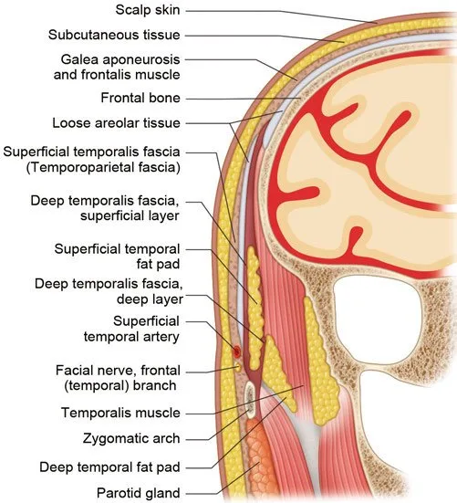 Fig 1. Forehead and temporal layered anatomy - the upper boundary of the periorbital region. (Watanabe, 2016, p. 154).
> Sources: [5] Watanabe 2016
Fig 1. Forehead and temporal layered anatomy - the upper boundary of the periorbital region. (Watanabe, 2016, p. 154).
> Sources: [5] Watanabe 2016 [C] [MEDLIB].
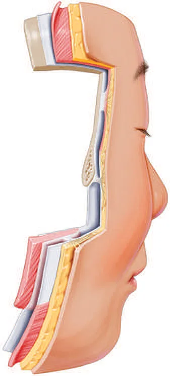 Fig 2. Layered forehead-brow-orbit transition in sagittal view. (Azizzadeh, 2018, p. 23).
> Sources: [13] Azizzadeh 2018
Fig 2. Layered forehead-brow-orbit transition in sagittal view. (Azizzadeh, 2018, p. 23).
> Sources: [13] Azizzadeh 2018 [C] [MEDLIB].
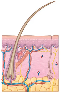 Fig 3. Generic skin layers; the eyelid is this diagram compressed to a fraction of a millimetre. (Smith, Gray's Surface Anatomy, 2018, p. 21).
> Sources: [10] Smith 2018
Fig 3. Generic skin layers; the eyelid is this diagram compressed to a fraction of a millimetre. (Smith, Gray's Surface Anatomy, 2018, p. 21).
> Sources: [10] Smith 2018 [C] [MEDLIB].
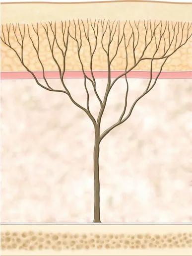 Fig 4. Retaining-ligament fibre architecture, skin to bone. (Kim, 2016, p. 32).
> Sources: [8] Kim 2016
Fig 4. Retaining-ligament fibre architecture, skin to bone. (Kim, 2016, p. 32).
> Sources: [8] Kim 2016 [C] [MEDLIB].
Consensus: the region is thin-skinned, ligament-anchored and septum-protected; deep volume is placed on the rim or in the ROOF, never superficially in the lid [3][4][8][17][47]. Discrepancy that changes the gesture: schools differ on whether the deep sulcus is filled directly on the rim or whether the target is the ROOF under the brow; both are described because they correct different deformities (D4.7) and are not averaged [18].
The orbicularis oculi in operational detail, because it is the muscle you weaken, the plane you cross and the pump you must not break. Its three portions behave differently: the orbital portion is the outer voluntary ring, the forced closer and the brow-tail depressor, and it is the crow's-feet and chemical-brow-lift target; the preseptal portion overlies the septum and drives the gentle blink; the pretarsal portion overlies the tarsus, drives the fine involuntary blink and houses the lacrimal pump (the Horner muscle around the canaliculi) [8]. Weakening the pretarsal fibres widens the aperture but can impair the pump and the closure, which is why the pretarsal point is gated by the snap test (D4.5, D4.8). Above, the orbital orbicularis interdigitates with the frontalis and the corrugator/depressor supercilii, so the brow sits at the mechanical equilibrium of one elevator against four depressors [14][25].
The lamellae of the upper lid, the surgeon's language you must share to refer well. The upper lid is read in an anterior lamella (skin + orbicularis), a middle lamella (orbital septum) and a posterior lamella (tarsus + levator aponeurosis + Müller's muscle + conjunctiva) [8][9]. The tarsus is the fibrous plate that gives the lid its rigidity; the levator palpebrae superioris is the striated elevator whose aponeurosis inserts into the anterior tarsus and skin (forming the lid crease); Müller's muscle is the sympathetically innervated smooth accessory elevator that contributes the last 1-2 mm of lift and is the target of the apraclonidine rescue in toxin ptosis (D4.10). A "droopy lid" is a posterior-lamella (levator) problem, a "heavy lid" is usually an anterior-lamella (skin) or brow problem, and the injector who cannot name the lamella cannot triage the referral [43].
Two deep fat pads that are constantly confused, and treated oppositely. The brow fat pad (galeal/sub-brow fat) is a superficial-plane pad just under the brow skin; the ROOF is a deep pad behind the orbicularis on the periosteum. Volume for brow-tail support is placed deep, in or on the ROOF, not in the superficial brow fat, because superficial brow volume migrates and lumps [3]. Behind both sits the septum and the true orbital fat, which is off-limits to filler.
Lymphatics and sensory nerves, because they explain the two commonest nuisances. The upper-lid lymphatics are sparse superficial precollectors that drain laterally to the preauricular and parotid nodes; they are fragile and easily obstructed by product, which is the anatomical basis of prolonged periorbital oedema (D4.10) [24]. Sensation is supraorbital and supratrochlear (V1) over the medial two-thirds and lacrimal (V1) laterally, which is why a supraorbital-nerve neuropraxia from a deep medial bolus numbs the forehead [6][7].
Trampa clásica: treating the upper-lid hollow as if it had a superficial fat plane. There is none over the tarsus; the only safe deep target is supraperiosteal on the rim or the ROOF, and a superficial deposit here is a visible, blue, long-lasting lump that needs hyaluronidase [1][17].
D4.3 · Vessels, nerves and the danger zone
The map, glanceable. Every artery of the upper periorbital region is a terminal branch of the internal-carotid ophthalmic system, which is why this region shares the glabella's blindness risk. Link to the full occlusion protocol in J2 — Vascular Occlusion and Hyaluronidase.
| Structure | Course in this region | Depth | Variant / frequency | What occludes if you inject into it |
|---|---|---|---|---|
| Supratrochlear artery (STA) | Exits the orbit at the superomedial rim ~1.7-2.2 cm from midline, ascends the medial brow/forehead | Deep (subgaleal/periosteal) at the rim, becomes subdermal as it climbs cephalad | Paired; dominant vertical forehead vessel | Ophthalmic → central retinal artery: retrograde embolism, blindness [23][31] |
| Supraorbital artery (SOA) | Exits the supraorbital notch/foramen at the mid-pupillary line (~2.5-2.7 cm from midline), runs superolaterally | Deep at the rim, superficial branch runs over frontalis | Notch in the majority; a closed bony foramen in a substantial minority (roughly a quarter to a third of orbits) [8][22] | Ophthalmic system: blindness, forehead/scalp necrosis [23] |
| Dorsal nasal artery | Medial canthal angle, anastomoses with the angular artery | Subcutaneous | Constant anastomosis to the facial system | Ophthalmic terminal: contributes to the medial-canthus watershed, blindness [23] |
| Supratrochlear / supraorbital nerves | Accompany their arteries out of the rim, sensory to forehead/scalp | With the vessels | Supraorbital nerve may split into superficial and deep divisions | Injury: forehead numbness, neuropraxia (not vascular) [6][7] |
| Frontal (temporal) branch of the facial nerve | Crosses the zygomatic arch, runs under the superficial temporal fascia toward the lateral brow/frontalis | Superficial in the temple (between SMAS and deep temporal fascia) | Motor; injury paralyses frontalis | Not a filler-occlusion risk; a motor-injury risk with deep temple/brow needling [6][7] |
| Superficial temporal artery, frontal branch | Ascends anterior to the ear, gives the frontal branch toward the lateral forehead | Subcutaneous over the temple | Tortuous, palpable | Local skin ischaemia; anastomoses toward the orbit [22] |
Why this region is a blindness zone, mechanistically. The STA, SOA and dorsal nasal are the distal terminals of the ophthalmic artery. An injection needle in one of them, if the plunger pressure exceeds the systolic pressure, drives filler retrograde up the column, and when pressure is released the bolus travels antegrade into the first branch it reaches, which can be the central retinal artery [23][31]. The retina is central nervous tissue: experimental and clinical data put its ischaemic tolerance at roughly 90 minutes, after which infarction is irreversible, which is why filler-induced visual loss is managed in minutes, not hours [31][32]. Fig 5 shows the supratrochlear/supraorbital neurovascular bundle being lifted at the superior orbital rim in a coronal-brow-lift dissection: the bundle a deep brow bolus can pierce.
The frequency facts that change the gesture: - The STA is subdermal in its upper course but deep on the rim; a deep supraperiosteal bolus on the rim and a superficial subdermal thread in the mid-forehead are both dangerous, for opposite reasons, and the safe plane is the one that is neither on top of nor tenting the vessel [22][23]. - The supraorbital foramen (closed bone) versus notch (open) matters: when the exit is a true foramen the vessel and nerve are fixed and cannot be displaced by a cannula, so the notch-versus-foramen variant is palpated before deep medial-brow work [8][22]. - The venous system of the region (supratrochlear and supraorbital veins draining to the superior ophthalmic vein) is the route by which periorbital filler can cause prolonged oedema and, rarely, is implicated in delayed inflammatory swelling; the venous map is as clinically relevant as the arterial for the swelling complication [24].
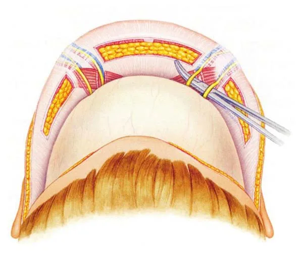 Fig 5. Supratrochlear/supraorbital bundle at the superior orbital rim (danger zone). (Seckel, Facial Danger Zones, p. 53).
> Sources: [6] Seckel
Fig 5. Supratrochlear/supraorbital bundle at the superior orbital rim (danger zone). (Seckel, Facial Danger Zones, p. 53).
> Sources: [6] Seckel [C] [MEDLIB]; [23] Cotofana 2022 [B].
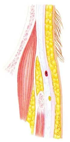 Fig 6. Temporal cross-section: motor nerve and artery planes lateral to the brow. (Seckel, Facial Danger Zones, p. 31).
> Sources: [6] Seckel
Fig 6. Temporal cross-section: motor nerve and artery planes lateral to the brow. (Seckel, Facial Danger Zones, p. 31).
> Sources: [6] Seckel [C] [MEDLIB].
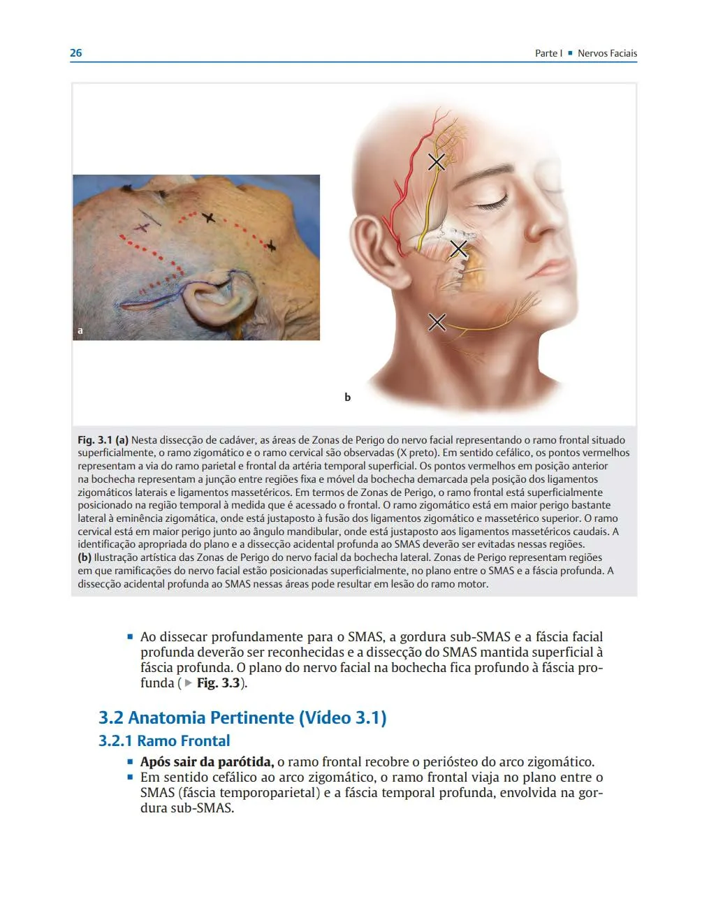 Fig 7. Frontal branch of the facial nerve and superficial temporal artery over the temple. (Rohrich, Facial Danger Zones, 2020, Fig 3.1).
> Sources: [7] Rohrich 2020
Fig 7. Frontal branch of the facial nerve and superficial temporal artery over the temple. (Rohrich, Facial Danger Zones, 2020, Fig 3.1).
> Sources: [7] Rohrich 2020 [C] [MEDLIB].
Consensus, the operating rules that follow from the anatomy [23][31][40]: 1. Rim guard. A fingertip pressed on the superomedial orbital rim during glabella/medial-brow injection compresses the STA against bone and reduces retrograde flow. 2. Deep and slow, or subdermal and threaded, never a fast bolus in between. Deep boluses go supraperiosteal against bone; superficial work is low-volume retrograde threading. 3. Small aliquots (≤0.1 mL), low pressure, needle moving or cannula. A moving tip cannot maintain an intra-arterial column; a blunt cannula displaces rather than pierces the vessel [40]. 4. Aspiration is a weak test, not a licence. A negative aspirate does not exclude intraluminal position; it is one layer of defence among several [40].
Discrepancy that changes the gesture: needle versus cannula for periorbital safety. One school holds the cannula is inherently safer because it displaces vessels; the cadaver-embolization school holds that a cannula under sustained pressure can still enter a fixed vessel and that no instrument makes a badly planned injection safe. Both positions are printed with their scenario and not averaged; the transferable conclusion is that the plane and the pressure matter more than the tool [23][40].
The venous side, because it drives the swelling and, rarely, the worst delayed events. The supratrochlear and supraorbital veins drain the region into the superior ophthalmic vein and thence the cavernous sinus; the angular vein at the medial canthus is the valveless bridge between the facial and orbital venous systems [24]. This valveless connection is why a medial-canthus or glabellar event can propagate against expected flow, and why the venous map matters for both the prolonged-oedema nuisance and the rare deep spreading infection. Practically, the dense superficial venous plexus of the lateral canthus is why crow's-feet toxin is the most ecchymosis-prone injection of the upper face and each point is compressed briefly (D4.8) [14].
The watershed and the anastomoses that make the region an internal-carotid outpost. The STA and SOA (internal-carotid/ophthalmic) anastomose at the forehead and medial canthus with the angular and superficial temporal branches (external-carotid); the dorsal nasal artery is the medial bridge [23]. This rich anastomosis is protective for skin perfusion but is exactly what allows a bolus to travel retrograde into the ophthalmic system and antegrade into the retina. The central retinal artery is the first intra-orbital branch of the ophthalmic; an embolus reaching it infarcts the whole retina, whereas a branch-level embolus gives a sectoral field loss, and both present with sudden painful visual change during injection [31][32].
Variant frequencies that change where the needle goes, stated as ranges from the anatomy literature [8][22]: the supraorbital exit is an open notch in the majority of orbits and a closed foramen in roughly a quarter to a third; when it is a foramen the neurovascular bundle is fixed and cannot be displaced by a cannula, so the medial brow is palpated first. The STA runs vertically about 1.7-2.2 cm from the midline and turns subdermal as it ascends, so the danger plane shifts from deep at the rim to superficial in the mid-forehead. The frontal branch of the facial nerve (motor) crosses the zygomatic arch and runs superficially over the temple toward the frontalis, so deep temporal or lateral-brow needling risks a motor palsy distinct from any vascular event, as Fig 6 (temporal cross-section) and Fig 7 (danger-zone map) document [6][7].
The prevention bundle, restated as a checklist because the anatomy only helps if it changes the gesture [40]: aspirate as one weak layer; inject with the needle moving or use a blunt cannula; keep each aliquot at or below 0.1 mL; keep the plunger pressure low; keep a fingertip rim guard on the superomedial rim during medial-brow and glabellar work; prefer deep-on-bone or superficial-threaded over a fast mid-plane bolus; and map with ultrasound in re-treatment or high-risk cases [15][23].
Trampa clásica: treating the medial brow and glabella as low-risk because the needle is "only going to muscle." The lateral corrugator point sits directly over the STA/SOA exit; a deep, fast, high-volume deposit there is the classic blindness case in the region [23][31].
D4.4 · Ageing of the region, and in what order
The staged model, glanceable. The upper periorbital region ages from the bone outward, and the order decides the treatment order.
| Stage | Tissue | What changes | Visible sign | Correct target |
|---|---|---|---|---|
| 1 | Bone | Superior orbital rim resorbs superomedially and inferolaterally; the aperture widens | The eye looks larger, the sulcus deepens, the brow loses its bony shelf | Supraperiosteal volume on the rim [8][1] |
| 2 | Ligament | Retaining adhesions stay fixed while everything around them descends | A step forms where a smooth transition used to be | Volume above, never on, the fixed ligament [8] |
| 3 | Deep fat | ROOF deflates under the brow; orbital-fat pads pseudoherniate through a lax septum | Brow-tail droop plus a full, hooded upper lid at once | ROOF augmentation; herniation is surgical [3][20] |
| 4 | Superficial fat / muscle | Brow fat pad and orbital soft tissue descend; orbicularis (the depressor) relatively over-acts | Brow descent, lateral hooding, deeper crow's feet at rest | Chemical brow lift + selective volume [25][11] |
| 5 | Skin | Dermal thinning, elastosis, dermatochalasis | Crêpe skin, static rhytides, redundant upper-lid skin | Skin quality; frank excess is blepharoplasty [46][20] |
The order matters because treating out of order fails. Filling the sulcus (stage 1-3 target) on a patient whose dominant problem is descended skin (stage 5) buys a fuller but still hooded lid; weakening the depressors (stage 4 target) on a patient whose brow has lost its bony shelf (stage 1) lifts a brow that has nothing to sit on. Fig 8 shows the bone-resorption arrows at the superomedial and inferolateral orbital rim; Fig 9 links ageing through the whole tissue stack; Fig 10 shows the clinical progression of the periorbital hollow across decades.
Tissue by tissue, the exhaustive account:
- Bone. The orbital aperture enlarges with age, and the resorption is not uniform: it concentrates in the superomedial and inferolateral quadrants [8]. The superomedial loss deepens the medial superior sulcus and undermines the head of the brow; the inferolateral loss (shared with the lower lid) widens the lateral orbit. The rim that a young brow rests on retreats, so the brow descends even before any soft-tissue change. This is why the deep, durable correction of the superior sulcus is supraperiosteal volume that rebuilds the bony shelf, not superficial filler under the skin [1][8].
- Deep fat, part 1: the ROOF. The retro-orbicularis oculi fat is a glide pad under the brow that gives the brow tail its fullness and support. Its deflation is an early and under-recognised contributor to the tired upper-lid look, and its restoration (deep, under the brow tail) can lift and defog the lid without touching the sulcus itself [3][20].
- Deep fat, part 2: orbital-fat pseudoherniation. As the orbital septum laxes, the preaponeurotic (nasal and central) fat pads bulge forward, producing the full, hooded upper lid. This is not a volume-loss problem and not a filler indication; it is the classic reason a hooded lid is referred, not injected [20].
- Superficial fat and muscle. The brow fat pad and the periorbital soft tissue descend; the orbicularis, the depressor of the brow tail, keeps its tone while the frontalis fatigues, so the depressor-elevator balance tips toward descent. This is the anatomical basis of the chemical brow lift: weaken the depressor, let the elevator win [25][14].
- Skin. Dermal collagen and elastin decline, the thinnest skin of the body becomes thinner and crêpe, static rhytides set, and redundant skin (dermatochalasis) accumulates. Skin is the layer filler and toxin cannot fix; it is the resurfacing and, when frank, the blepharoplasty indication [46][20].
⚠ The iatrogenic ageing pattern that mimics stage 1-3 and must be asked about: prostaglandin-associated periorbitopathy. Topical prostaglandin analogues for glaucoma (bimatoprost, latanoprost and family) produce orbital-fat atrophy, deepening of the superior sulcus, periocular hyperpigmentation and lash growth [46]. If a patient using a glaucoma eye-drop presents with a deep superior sulcus, that drop is the cause until proven otherwise; the change is often unilateral if the glaucoma is, and filling a pharmacologically driven, still-progressing atrophy is fighting a current that does not stop. This is salvaged directly from the periorbital consult and belongs in every upper-lid history.
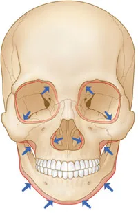 Fig 8. Age-related bone-resorption pattern; note the superomedial and inferolateral orbital-rim arrows. (Kim, 2016, p. 61).
> Sources: [8] Kim 2016
Fig 8. Age-related bone-resorption pattern; note the superomedial and inferolateral orbital-rim arrows. (Kim, 2016, p. 61).
> Sources: [8] Kim 2016 [C] [MEDLIB].
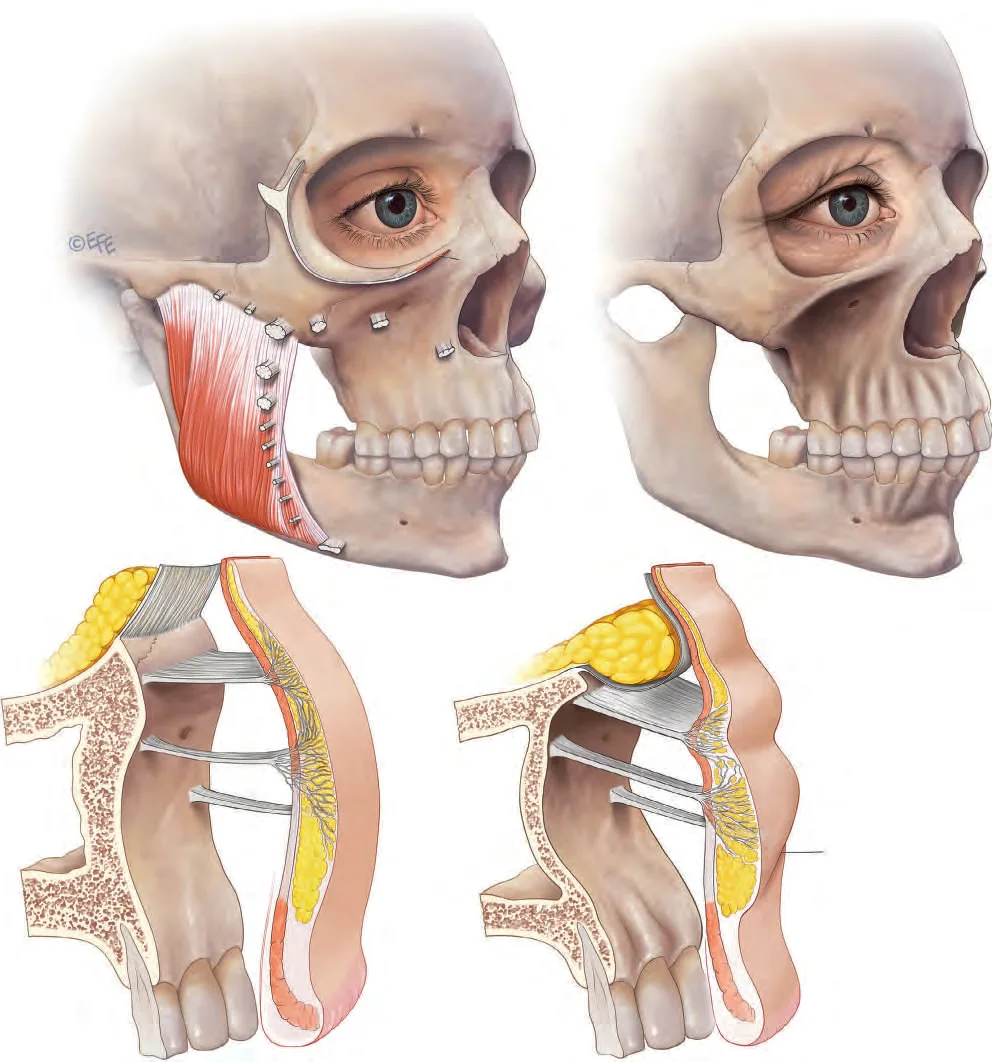 Fig 9. Ageing changes linked through the tissue layers. (Standring, Gray's Anatomy, 2016, p. 960).
> Sources: [9] Standring 2016
Fig 9. Ageing changes linked through the tissue layers. (Standring, Gray's Anatomy, 2016, p. 960).
> Sources: [9] Standring 2016 [C] [MEDLIB].
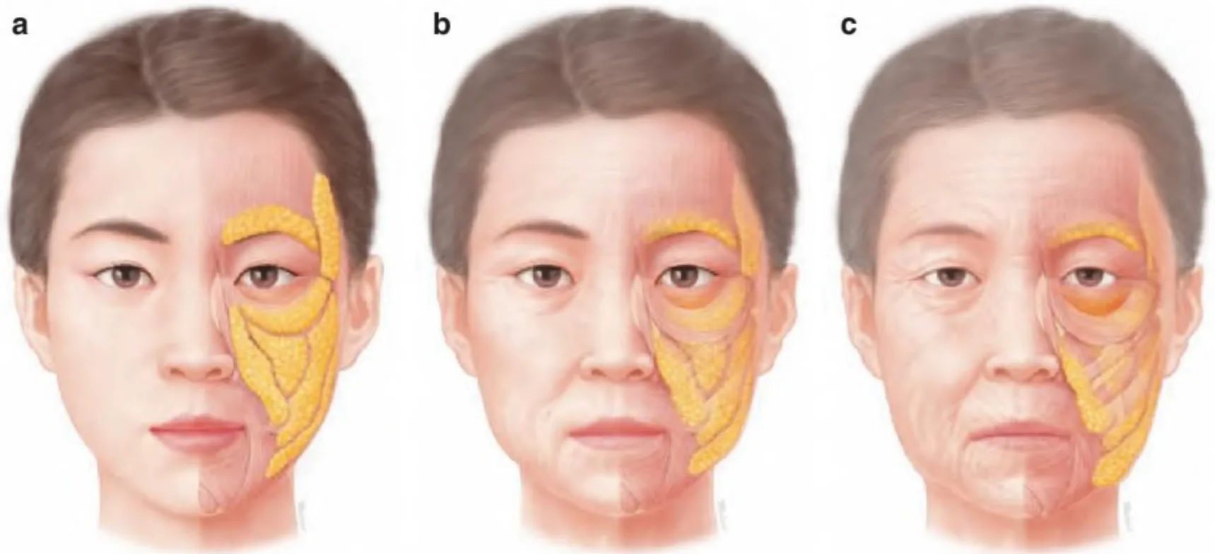 Fig 10. Periorbital ageing progression, young to old. (Anatomía Clínica de la Cara para Relleno y Toxina Botulínica).
> Sources: [12] Anatomía Clínica de la Cara
Fig 10. Periorbital ageing progression, young to old. (Anatomía Clínica de la Cara para Relleno y Toxina Botulínica).
> Sources: [12] Anatomía Clínica de la Cara [C] [MEDLIB].
Consensus: the region ages bone-first, skin-last, and the durable corrections are deep (rim, ROOF) while the surface problems (crêpe, redundancy) are skin and surgical work [1][8][20]. Discrepancy that changes the gesture: how much of the deep sulcus is bone versus fat loss, and therefore whether the target is supraperiosteal bone-rebuilding or a shallower fat-replacement; the two schools inject different planes and are treated separately in D4.7 [17][18].
The biomechanics behind the stages, because they explain why the order is fixed. Facial ageing is now framed biomechanically: the deep fat compartments deflate and the retaining ligaments weaken unevenly, so the soft-tissue envelope both loses support and descends, and the surface changes are downstream of the deep loss [11]. In the upper periorbital region this means the visible brow descent and lid hooding are late signs of an early, invisible bone-and-deep-fat loss, which is why the durable correction targets the deep layer and why treating the surface first fails.
Brow descent, quantified as far as the evidence allows. The brow descends with age, most at the tail, as the ROOF deflates and the orbital orbicularis (a depressor) keeps its tone against a fatiguing frontalis; the manual-brow-elevation test (D4.5) isolates how much of a "heavy lid" is really a dropped brow [25][29]. The chemical brow lift recovers a few millimetres at the tail; beyond that the rim has resorbed and only a surgical lift restores the shelf [29][30].
Why the aperture looks bigger, not smaller, with age here. The superomedial and inferolateral rim resorption widens the bony orbital aperture, so the eye appears larger and more skeletonised even as the lid skin becomes redundant; the two changes coexist and are treated differently (deep volume for the skeletonisation, surgery for the redundancy) [8]. Fig 8's arrows are the map of this widening.
Skin change specific to the thinnest skin of the body. Dermal collagen and elastin decline, solar elastosis accumulates, and the lid skin (already 0.3-0.6 mm) becomes crêpe and translucent, which both creates the static-rhytide and dermatochalasis problem and raises the Tyndall risk of any superficial filler [1][46]. This is the layer that resurfacing and, when frank, blepharoplasty own; neither filler nor toxin thickens redundant skin.
Upper versus lower lid, because the same patient ages both and they are treated in different chapters. The upper lid's dominant ageing signs are sulcus deepening, ROOF deflation, brow descent and dermatochalasis (this chapter); the lower lid's are the tear-trough groove, the malar mound/festoon and the double-convexity of pseudoherniation plus descent (D5). Sharing a patient does not mean sharing a plan: the upper hollow fills on the rim while the lower groove is anchored by the tear-trough ligament and carries the festoon exclusion, which is why the region is split by tier [20].
Trampa clásica: reading a full, hooded upper lid as volume loss and filling it. The fullness is pseudoherniated orbital fat (stage 3 septal laxity), and volume makes the hood heavier, not younger; the test is whether the lid is hollow (fill) or full (refer) [20].
D4.5 · Assessment: measure, photograph, test dynamically, scan
What gets measured, and why each number changes the plan.
| Measure | How | Normal / threshold | Decision it drives |
|---|---|---|---|
| MRD1 (margin-to-reflex distance 1) | Distance from the corneal light reflex to the upper-lid margin, eyes in primary gaze | ~4-5 mm; reduced = ptosis | Reduced MRD1 with normal skin = levator problem, refer; not a filler case [42][43] |
| Levator function / excursion | Excursion of the upper lid from downgaze to upgaze while fixing the brow with a thumb | ≥12-15 mm good; <8 mm poor | Distinguishes true (levator) ptosis from brow ptosis and grades surgical planning [43] |
| Tarsal platform show (TPS) | Visible lid skin between lash line and lid crease | Region-specific, note asymmetry | Guides where volume or lift is aimed; a low crease reads as heaviness |
| Dermatochalasis grade | Pinch of redundant upper-lid skin | Frank fold over lashes = excess | Frank excess is blepharoplasty, not injectable [20] |
| Brow position / manual lift | Note brow height vs rim; manually elevate the brow and re-look | If the lid improves on manual lift, the brow is the problem | Brow ptosis: toxin depressor lift if mild, surgical brow lift if marked [25][29] |
| Superior sulcus depth / A-frame | Inspect hollow, hollow shape (A-frame = medial deep) | Hollow with good skin = fill candidate | Deep sulcus = supraperiosteal/ROOF volume [18] |
| Lid fullness (hollow vs herniation) | Is the lid hollow or full/bulging? | Full = pseudoherniation | Hollow fills; full refers [20] |
The three-way upper-lid differential, salvaged and central. A "droopy lid" is one of three things, with three treatments, and confusing them is the classic error [43]:
| Dermatochalasis | True blepharoptosis | Brow ptosis | |
|---|---|---|---|
| Problem | Excess skin | Levator dysfunction | The brow has descended and drags the lid |
| Sign | Skin folds over the crease or lashes | MRD1 reduced: margin covers more pupil | Brow at or below the rim; forehead held in constant tension |
| Test | Skin pinch lifts a frank excess | Measure MRD1 and levator excursion (fixing the brow) | Manually elevate the brow: if the lid improves, the brow is part of the problem |
| Treatment | Upper blepharoplasty | Levator surgery, not skin resection | Toxin depressors if mild; brow lift if marked |
⚠ The scenario that is not aesthetic at all. A ptosis of recent onset, especially if asymmetric, that fluctuates through the day, or comes with diplopia, anisocoria or pain, is not treated: it is referred preferentially. The differential includes myasthenia gravis, Horner syndrome and third-nerve palsy, and some of these are emergencies [43]. See J8 — Contraindications and Special Populations.
Dynamic testing, because the region is dosed on function, not on the wrinkle at rest. - Frontalis compensation. A patient with dermatochalasis or lid ptosis constantly elevates the brows to see, using the frontalis as a crutch. Weakening it closes the visual field. Watch the forehead when the patient is not aware, and ask them to open the eyes without using the brows: constant frontalis recruitment is a strong relative contraindication to forehead toxin [14][26]. - Squeeze and animate. Ask for maximal squeeze (crow's feet mapping), brow raise (frontalis/brow elevator), frown (corrugator/procerus). The crow's-feet fan pattern (complete, inferior-predominant, superior-predominant) is read here and dictates the injection map (D4.8) [28]. - Snap and distraction test. Before any pretarsal toxin or lower-lid-directed work, test lid tone; slow recoil (laxity) contraindicates weakening the pretarsal orbicularis and flags a lid that will not tolerate margin change [14]. (The pretarsal point is a lower-lid gesture; the tone test still belongs in the upper-lid exam because it gates the crow's-feet extension.) - Pre-septal versus hollow palpation. Palpate whether the upper-lid fullness reduces on gentle globe retropulsion (fat herniation) or is a fixed hollow (fill candidate).
Photography, standardised. Frontal, oblique and lateral, fixed lighting, primary gaze and brow-raise, with a downlight view because the superior sulcus and the hollow change most with light direction and generate the most disputes without a baseline image [46]. Photograph the brow at rest and on maximal raise to document the elevator reserve.
Ultrasound, the modern addition the master course predates. High-frequency ultrasound and colour Doppler now map the STA/SOA/dorsal-nasal course and depth before a deep periorbital injection, and identify the tissue plane and any prior product [15][22]. Fig 11 shows the layer-by-layer ultrasound identification approach (temporal fossa illustration) that transfers directly to periorbital scanning: probe on the region, identify skin, muscle, fascia, fat and bone, and find the vessel before the needle does. Pre-injection Doppler vessel mapping is the assessment step with the highest safety yield in the highest-risk region [22][15].
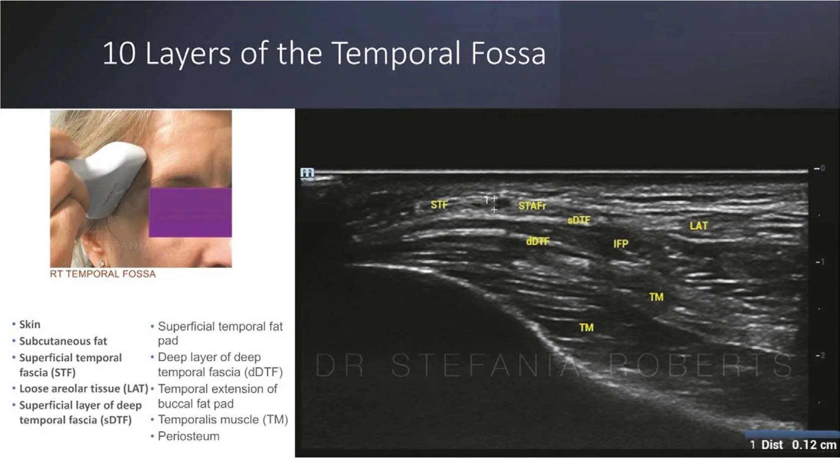 Fig 11. Layer-by-layer facial ultrasound, the pre-injection mapping method. (Malherbe, Ultrasound Protocol for Facial Aesthetics, 2024, p. 121).
> Sources: [15] Malherbe 2024
Fig 11. Layer-by-layer facial ultrasound, the pre-injection mapping method. (Malherbe, Ultrasound Protocol for Facial Aesthetics, 2024, p. 121).
> Sources: [15] Malherbe 2024 [C] [MEDLIB].
Consensus: measure MRD1 and levator function, do the three-way differential, test frontalis compensation, and photograph in multiple light directions before deciding anything [42][43][46]. Discrepancy that changes the gesture: whether pre-injection Doppler ultrasound should be routine or reserved for high-risk cases; the ultrasound school argues routine mapping in this ophthalmic-terminal region, the pragmatic school reserves it, and both positions are recorded [15][22].
Levator function grading, because it is what separates the referable from the treatable. Levator excursion is measured from full downgaze to full upgaze with the brow fixed by the examiner's thumb (to remove frontalis contribution): roughly >=12-15 mm is good, 5-11 mm is fair, and <5 mm is poor [43]. Good function with a reduced MRD1 suggests an aponeurotic (involutional) ptosis; poor function suggests a myogenic or congenital cause. Either way the correction is surgical, but the grading tells the oculoplastic surgeon which operation, and documenting it makes the referral useful rather than a shrug [43].
Exophthalmometry and the enophthalmos question. Before choosing between direct-sulcus and retrobulbar filler (D4.7), the globe position is assessed: a recessed globe (enophthalmos, measurable by exophthalmometry) plus a deep sulcus is the scenario the retrobulbar school targets, whereas a normally positioned globe with a pure sulcus hollow is a direct-sulcus case [18]. Reading the globe position is what makes that controversy a decision rather than a coin toss.
Photography protocol, region-specific. Standardised frontal, oblique and lateral views at fixed distance and lighting, in primary gaze and on maximal brow-raise, plus a view with a downlight to throw the sulcus and hollow into relief because the superior sulcus changes most with light direction and generates the most disputes without a baseline image [46]. Photograph the brow at rest and at maximal raise to document the frontalis reserve, and a maximal-squeeze view to map the crow's-feet fan (D4.8) [28]. Store the drop history in the record: a glaucoma prostaglandin is the commonest missed cause of a unilateral sulcus (D4.4) [46].
Ultrasound settings and what you look for. A high-frequency linear probe (broadly in the 15-22 MHz range for superficial facial work) resolves the lid layers, the septum, the fat pads and, on colour Doppler, the STA/SOA and any prior filler [15]. The pre-injection sweep answers three questions: where is the vessel, what plane is the target, and is there old product; Fig 11 shows the layer-identification method that transfers directly to the periorbital scan [15][22]. The abstention to record when ultrasound is unavailable is that the landmark-and-rim-guard technique is then the standard, not that mapping is optional in principle.
The pseudoptosis traps, because they mimic a levator problem and are not. A brow ptosis dragging the lid, a contralateral lid retraction making the normal side look ptotic, dermatochalasis overhanging the margin, and hypotropia (the lid follows a downward-deviated globe) all reduce the apparent MRD1 without a levator lesion, and each is separated by a specific manoeuvre: manual brow elevation for brow ptosis, covering the retracted side for pseudo-contralateral, skin retraction for dermatochalasis, and cover testing for hypotropia [43]. Naming the pseudoptosis prevents both the wrong referral and the wrong injection.
What is scanned, expanded. Beyond vessel mapping, ultrasound in this region documents the plane and integrity of prior filler (a nodule versus diffuse product), the septum and fat pads (hollow versus herniation), and the target depth before a deep bolus; colour Doppler distinguishes an artery from a vein before the needle enters either [15][22]. The scan is short, changes the plan when it finds old product or a superficial vessel, and its abstention (no ultrasound available) is recorded rather than assumed away.
The dynamic tests as a sequence, run in under two minutes. Primary-gaze MRD1; brow-raise for frontalis reserve; brow-fix downgaze-to-upgaze for levator excursion; maximal squeeze for the crow's-feet pattern; frown for the corrugator/procerus; manual brow elevation to unmask brow ptosis; and a snap/distraction test before any pretarsal-directed toxin. Each test changes a specific downstream decision, and skipping the sequence is how a brow ptosis gets injected as a lid hollow [43].
Trampa clásica: diagnosing "tired eyes" as volume loss without measuring MRD1 or lifting the brow. The commonest miss is a brow ptosis treated as a lid hollow, or a true levator ptosis injected instead of referred [43].
D4.6 · Goal and patient selection: who benefits, who does not, what is referred
The goal is restoration of the youthful upper-lid concavity and a supported brow, not the removal of skin or the elevation of a mechanically ptotic lid. The work is in selection; the injection is the easy part [18][20].
| ✅ Good candidate (upper periorbital volume / toxin) | ❌ Poor candidate → refer or defer |
|---|---|
| Genuine superior-sulcus hollow with good skin, minimal fat herniation, younger patient [18] | Frank dermatochalasis (redundant skin over the crease/lashes) → upper blepharoplasty [20] |
| Deep A-frame hollow, hollow reduces the "tired" look when volume is simulated | True blepharoptosis (MRD1 reduced, levator problem) → levator surgery [43] |
| Brow-tail droop that improves on manual elevation, brow still has bony support | Marked brow ptosis with a resorbed rim → surgical brow lift [29] |
| Crow's feet dynamic, good lid tone (snap test brisk) | Pseudoherniated orbital fat (full, bulging lid) → blepharoplasty; volume worsens the hood [20] |
| Post-blepharoplasty hollow from over-resection, realistic expectations | Post-blepharoplasty lid retraction / scleral show, lagophthalmos, dry eye → refer, do not fill |
| Wants partial, natural improvement over sessions | Recent, asymmetric, fluctuating ptosis ± diplopia/anisocoria → neurology, urgent [43] |
| No active glaucoma prostaglandin drop, or drop reviewed | On a prostaglandin analogue with a progressing sulcus → address the drug first [46] |
| Frontalis not used as a visual crutch | Uses the frontalis constantly to lift the lids to see → forehead toxin closes the field [14][26] |
Who benefits, stated positively. The patient this chapter treats has lost concavity, not gained skin: an early-to-mid superior-sulcus hollow, a deflated ROOF, a brow tail that has dropped but still sits on bone, and dynamic crow's feet. In selected cases, volumising the deep sulcus and supporting the brow gives a result that a blepharoplasty cannot, because surgery removes tissue and this problem is tissue lost, not tissue in excess; the literature frames this explicitly as a "fat or fillers" decision, and for the hollow (not the hood) the injectable can be the better operation [18][20]. Polynucleotides and skin boosters serve the thin-skin, crêpe patient whose problem is quality rather than volume [21].
Who does not, and where each goes: - Dermatochalasis (excess skin): upper blepharoplasty. Filler adds weight under skin that is already redundant; toxin does not lift skin. Neither is a dose problem [20]. - True blepharoptosis (levator): oculoplastic levator surgery. Chemical or filler correction is not on the table; apraclonidine is a diagnostic/temporary aid, not a treatment (D4.10). - Brow ptosis, marked, especially with a resorbed rim: surgical brow lift. Toxin depressor-lift and ROOF volume are for the mild case [29][30]. - Orbital-fat pseudoherniation (full hood): blepharoplasty with fat management. Volume is contraindicated; it makes the hood heavier [20]. - Recent-onset ptosis with red flags: neurology/ophthalmology, preferential referral. Myasthenia, Horner, third-nerve palsy [43]. - Prostaglandin periorbitopathy: coordinate with the prescribing ophthalmologist before treating a pharmacologically driven, progressing atrophy [46].
⚠ The post-blepharoplasty patient, four situations, salvaged from the periorbital consult:
| Situation | Injectable indicated? |
|---|---|
| Hollow from excessive fat resection | Yes, legitimate but technically hard: thin skin, altered planes, fibrosis. Low-hydrophilicity product, minimal volume, cannula, realistic consent |
| Lid retraction with scleral show | No. Mechanical/cicatricial. Volume does not correct it and adds weight to a lid that no longer rises. Refer |
| Visible or irregular scar | Depends: see M2 — Scar Aesthetics and Revision. Not a volume problem |
| Dry eye or lagophthalmos | No, and it is a contraindication to anything aggressive. Refer to ophthalmology first |
Consent, region-specific. State that the correction is partial and staged, that the eyelid swells and takes weeks to settle so it is not retouched early, that Tyndall and prolonged oedema are the expected complications of the region and hyaluronidase may be part of the plan, and that the deep injection carries a rare but real blindness risk (D4.10). Link to B2 — Patient Psychology and Selection and B3 — Ethics, Consent and Medicolegal.
Consensus: hollow fills, hood refers, skin resurfaces, and a recent neurological ptosis leaves the aesthetic clinic [20][43]. Discrepancy that changes the gesture: whether an early hooded lid with mild fat fullness should be volumised at the brow/ROOF to camouflage it or referred; the camouflage school augments the ROOF to lift the hood, the surgical school refers, and the deciding variable is how much true herniation is present [18][20].
The consent conversation, region-specific and scripted. The upper periorbital consult is where honest triage keeps the patient and false optimism loses them. A usable script: state which component each modality treats and which it cannot ("volume restores the hollow, it does not remove skin or lift a dropped lid"); state that correction is partial and staged over sessions; state that the eyelid swells and takes weeks to settle so it is not retouched early; state that Tyndall and prolonged oedema are the expected, reversible complications of this region; and state, in writing, the rare but real blindness risk of any deep injection near the orbit [31][40]. Link to B2 — Patient Psychology and Selection and B3 — Ethics, Consent and Medicolegal.
The expectation grid, because mismatched expectation is the region's commonest dissatisfaction.
| Patient wants | Reality | What to say |
|---|---|---|
| "Remove the hood" | Hood is skin/fat excess | Blepharoplasty; injectable makes it heavier [20] |
| "Open my eyes" (ptotic lid) | Levator problem | Oculoplastic surgery; not a dose [43] |
| "Lift my brow" (marked) | Rim resorbed | Surgical brow lift; toxin recovers only a few mm [29] |
| "Fill my hollow" (true hollow, good skin) | Achievable | Staged deep volume, under-correction first [18] |
| "Get rid of crow's feet at rest" | Static lines are dermal | Toxin softens dynamic lines; skin-quality/resurfacing for static [28][46] |
Anatomic and demographic variation that changes selection. The male brow sits lower and flatter and must not be feminised by an over-elevated tail; chemical brow lift in men is conservative and central-sparing [25]. Ethnic variation in lid crease height, tarsal platform show and orbital fat volume changes both the aesthetic goal and the baseline sulcus, and the plan is set against the patient's own architecture, not a single template [46]. The contact-lens or dry-eye patient tolerates orbicularis weakening poorly and is screened before any pretarsal-directed toxin [14].
Anticipating the complication at the selection stage, because the poor candidate declares the future complication. The patient with morning puffiness, allergic rhinitis, thyroid or renal disease, or a boggy poorly draining lid will convert a routine filler into months of periorbital oedema, so the fluid-retention history is a selection filter, not a footnote [34][46]. The patient with a bleeding tendency or on anticoagulation will bruise heavily in this venous region and is counselled and, where medically appropriate, timed around it [14]. The patient with prior unknown filler carries a hidden deposit that will lump or migrate when new product is added, so imaging precedes any addition [15]. Selecting well is largely declining the patient whose baseline predicts the region's known complications.
The unhappy-patient scenarios and how to prevent each at selection. The commonest periorbital dissatisfactions - "it made me puffy", "the blue is still there", "my hood is worse", "I still look tired" - trace to selection errors: filling a fluid-retaining or poorly draining lid, filling a vascular or pigmentary problem instead of a hollow, filling a hooded (not hollow) lid, or filling a brow/skin problem that was never a volume problem [20][34]. Each is prevented by the differential and the exclusion list above, which is why the region spends more effort on who not to treat than on how to treat.
The referral map, so "refer" is a destination, not a dismissal. Dermatochalasis and fat herniation to an oculoplastic or facial-plastic surgeon for blepharoplasty; true levator ptosis to oculoplastics for levator surgery; marked brow ptosis for a surgical brow lift; recent neurological ptosis to neurology/ophthalmology urgently; a progressing prostaglandin sulcus back to the prescribing ophthalmologist [20][43]. Naming the destination and writing the findings (MRD1, levator function) is what makes the referral land.
Trampa clásica: offering an injectable for dermatochalasis because saying "this is surgery" feels like losing the patient. The honest referral keeps the patient; the injectable that cannot work loses them after the money is spent [20].
D4.7 · Technique - the full grid
This block does not close with one technique; it closes with the full grid of options for the region, each row with its plane, tool, volume and evidence [17][18]. What is not used here is marked and justified, because the empty cell is a finding.
Product axis - every class, and its role in this region.
| Product class | Role in upper periorbital | Rheology / form used | Verdict here |
|---|---|---|---|
| HA, low G′ / low cohesivity / low hydrophilicity | Superior sulcus, thin-lid contour | Small particle, low-to-medium cross-linking, low water uptake | First-line. In the periorbital region water uptake matters more than G′: a hydrophilic gel looks perfect at 24 h and swells the lid at 3 weeks [17] |
| HA, medium-to-high G′ | ROOF / deep brow-tail support only | Firmer, projecting gel, deep on periosteum | Deep only, under the brow tail; never in the lid itself |
| CaHA (normal) | Brow/temporal support adjacent to region | Supraperiosteal, biostimulatory | Peri-regional (temple/brow), not in the lid |
| CaHA (hyperdiluted) | Peri-orbital skin quality, temple | 1:1 to 1:4 dilution, subdermal | Adjunct for skin/temporal support, not lid contour |
| PLLA | Global upper-face volume restoration | Deep, multi-session biostimulator | Peri-regional; not for the lid or acute sulcus shaping |
| PCL | Durable structural support (brow/temple) | Supraperiosteal | Peri-regional; caution given region vascularity |
| PMMA / permanent | None here | - | No aplica: permanent product in the thinnest, most complication-prone skin is indefensible; a Tyndall or nodule is then permanent |
| Autologous fat | Deep sulcus / global volume (surgical setting) | Micro/nanofat, deep | Surgeon's tool; relevant as the alternative to HA in the "fat or fillers" decision [20] |
| Polynucleotides (PN/PDRN) | Thin-lid skin quality, crêpe | Intradermal/superficial | First-line for quality, not volume [21] |
| Skin boosters (low-cross-link HA) | Skin hydration/quality | Intradermal micro-depots | Quality adjunct |
| Toxin (BoNT-A) | Crow's feet, chemical brow lift | see D4.8 | Core modality of the region |
Instrument axis.
| Instrument | Where | Note |
|---|---|---|
| Needle 30G, 13 mm | Deep supraperiosteal bolus on the rim, glabella/brow toxin | Direct bone contact; smallest aliquots, aspirate, low pressure |
| Needle 32G, 4-6 mm | Superficial skin-quality micro-depots, crow's feet toxin | Very superficial only |
| Cannula 25G, 40-50 mm | Sulcus/ROOF from a lateral entry | Blunt tip displaces vessels; the preferred deep tool for volume |
| Cannula 27G, 25-40 mm | Finer contour, thin patients | Flexible; slower, more control |
| With ultrasound guidance | Any deep periorbital bolus in a high-risk or re-treatment case | Maps STA/SOA and prior product before the needle [15][22] |
| Without ultrasound (landmark + rim guard) | Standard practice where US is unavailable | Requires strict rim-guard, aliquot and pressure discipline [40] |
Plane axis - which planes are used here and which are forbidden.
| Plane | Used here? | For what |
|---|---|---|
| Supraperiosteal | Yes, primary deep target | Superior sulcus on the rim, ROOF base |
| Deep fat (sub-orbicularis / ROOF) | Yes | Brow-tail support |
| SMAS (orbicularis) intramuscular | Toxin only | Never filler into the orbicularis (bruising, irregularity) |
| Superficial fat | Largely absent over the lid | No safe superficial fat plane over the tarsus |
| Subdermal | Peri-regional (temple/brow) only | Not in the lid |
| Intradermal | Skin-quality products only | Polynucleotides, boosters; never volumising filler (Tyndall) [1] |
Movement axis.
Bolus (deep, on bone, tiny) · microbolus (0.02-0.05 mL aliquots) · retrograde linear thread (cannula, sulcus) · fan (cannula, brow/temple support) · cross-hatch (peri-regional support, not lid) · serial puncture (skin-quality micro-depots) · tower/column (avoided in the lid: builds a visible ridge in thin skin) [17]. The region tolerates only the small, deep, slow movements; every large or superficial movement is a complication generator here.
The point-by-point technique, the core deliverable.
| Target | Plane | Entry | Direction | Tool | Volume / point | Total / side | Touch-up |
|---|---|---|---|---|---|---|---|
| Deep superior sulcus (A-frame) | Supraperiosteal on the superomedial rim | Lateral to the hollow, or direct perpendicular to bone | Perpendicular to bone, tip on periosteum | 30G needle (bone contact) or 25G cannula | 0.05-0.1 mL micro-aliquots, aspirate | 0.1-0.3 mL, deliberate under-correction | Second session at 2-4 weeks; the second session is planned, not a rescue |
| ROOF / brow-fat pad | Deep, under the brow tail on periosteum | Lateral brow entry | Retrograde, deep | 25G cannula | 0.1 mL retrograde | 0.2-0.4 mL | Reassess brow position at 2 weeks |
| Lateral brow / tail support | Supraperiosteal + deep | Lateral entry | Fan toward the tail | 25G cannula | micro-aliquots | 0.1-0.2 mL | With chemical brow lift (D4.8) |
| Peri-regional temple (indirect support) | Supraperiosteal (deep temporal) or subdermal | Single temple point | Bolus deep, or fan subdermal | 27-30G needle deep, or cannula | 0.2-0.5 mL deep bolus | per temple protocol | Supports the brow tail from behind |
Schools and named techniques, all of them: - MD Codes (de Maio) periorbital algorithm [17]: the region is supported indirectly - temple codes (the T1-T3 temporal sequence) and lateral-brow points restore the shelf the brow sits on, with cautious deep supraorbital volume; direct lid filling is deliberately minimal because the lid is the highest-complication surface. The algorithm is printed as an auditable code sequence, the same logic as the toxin maps [17]. - Direct supraperiosteal superior-sulcus technique: small supraperiosteal aliquots on the rim to rebuild the concavity; corrects the sulcus only, keeps product out of the orbit, respecting the mapped upper-lid safe zone [18][19]. - Retrobulbar / intra-orbital HA (named, controversial): injection of HA into the intraconal/retrobulbar space to push the globe forward and correct enophthalmos together with the superior sulcus, reported around 0.83 mL per mm of enophthalmos correction [18]. This deposits product inside the orbit and is the deliberate opposite of the "keep out of the orbit" school. - ROOF augmentation school: the target is the deflated ROOF under the brow, not the sulcus itself; lifts and defogs the hooded-but-hollow lid [3][20].
Non-injectable alternatives, and when the correct answer is NOT an injectable: - Upper blepharoplasty: frank dermatochalasis, orbital-fat herniation. Skin/fat excess is surgery [20]. - Levator surgery / ptosis repair: true blepharoptosis (reduced MRD1, levator dysfunction) [43]. - Surgical brow lift (endoscopic/temporal/direct): marked brow ptosis with a resorbed rim, beyond the reach of toxin depressor-lift [29]. - Autologous fat transfer: the surgeon's volume alternative in the "fat or fillers" decision for the deep sulcus [20]. - Laser / EBD skin resurfacing: the crêpe, static-rhytide component; non-ablative fractional, ablative fractional, or radiofrequency microneedling, always with internal corneal shields [46]. See G3 — Non-Ablative and Fractional Lasers. - Threads: brow-tail suspension is described but low-yield and short-lived in this mobile, thin region; rarely the correct first choice.
Discrepancy that changes the gesture, printed with both authors, never averaged: retrobulbar/intra-orbital HA [18] versus direct supraperiosteal superior-sulcus injection [18]. The retrobulbar school corrects enophthalmos plus sulcus by depositing HA inside the orbit (roughly 0.83 mL/mm of enophthalmos); the direct school refuses intra-orbital deposition and treats the sulcus concavity only on the rim. The deciding clinical variable is whether the patient has true enophthalmos (globe recession) in addition to the sulcus hollow; with pure sulcus and a normally positioned globe, the direct supraperiosteal approach is chosen and intra-orbital deposition is avoided.
Cannula entry and threading, step by step. From a lateral entry point (a 21-23G pilot needle makes the port), a 25G/40-50 mm cannula is advanced in the deep plane toward the superomedial rim, the tip is confirmed on bone, and product is laid in retrograde micro-aliquots while withdrawing; the operator never pushes a bolus with a stationary tip [17]. The blunt tip is chosen because it displaces rather than pierces the STA/SOA, but it does not make a fast high-volume deposit safe, and it still requires the rim guard and low pressure [23][40]. For the ROOF, a separate lateral-brow entry aims deep under the brow tail with the same retrograde discipline.
Ultrasound-guided injection, when available, as a sequence. Map the STA/SOA and any prior product with colour Doppler; identify the target plane; advance the cannula/needle under real-time view keeping the tip visible and off the vessel; deposit small aliquots and watch the tissue expand in-plane [15][22]. The same probe is the rescue tool if an occlusion is suspected (D4.10) [35].
Product choice by brand-behaviour, not brand-name. The lid rewards a low-hydrophilicity, low-to-medium G' HA that integrates smoothly and holds little water; a high-G' projecting gel belongs on the rim/ROOF for support, never in the lid skin; and a permanent product belongs nowhere in this region because its Tyndall or nodule is then permanent [17]. Hyaluronidase reversibility is itself the argument for HA over any non-reversible option here. Skin-quality problems take polynucleotides or low-cross-link boosters intradermally, not volumising gel [21].
Named techniques catalogued. The MD Codes periorbital algorithm treats the region indirectly through temple (the T1-T3 temporal sequence) and lateral-brow support with cautious deep supraorbital volume [17]; the direct supraperiosteal sulcus technique rebuilds the rim concavity [18]; the ROOF augmentation technique lifts the hollow-but-hooded lid from under the brow [3]; the retrobulbar/intra-orbital technique corrects enophthalmos plus sulcus by intra-orbital deposition (~0.83 mL/mm), the deliberate opposite of the keep-out-of-the-orbit rule [18]. Each is auditable and each is chosen by a specific finding (temple hollow, pure sulcus, hooded-hollow lid, true enophthalmos).
Touch-up protocol and the under-correction discipline. The region is deliberately under-corrected at the first pass and reviewed at 2-4 weeks; the second session is planned from the start, not a rescue, because periorbital oedema masks the true result for weeks and an early top-up is the usual cause of over-correction and lid puffiness [17]. The tower/column movement is avoided because it builds a visible ridge in thin skin.
Volume by point, region, session and the overfill ceiling. The region is defined by small numbers: per point 0.02-0.1 mL micro-aliquots; per side roughly 0.1-0.3 mL for the sulcus and 0.2-0.4 mL for the ROOF; per session the deliberate under-correction keeps the total low, and the ceiling is reached the moment the lid loses its concavity and starts to look full rather than restored [17][18]. Overfilling the upper periorbital region does not read as "more youthful"; it reads as a heavy, aged, hooded lid, and the correction is hyaluronidase, so the ceiling is respected by design and the plan is built around a planned second session rather than a complete first pass. Product placed against a fixed retaining ligament will bulge above the groove rather than efface it, so more volume against a ligament is wasted and visible, not corrective [8].
Product classes with representative behaviour, brands named as behaviour not as endorsement. For the lid contour the class is a soft, low-hydrophilicity, low-to-medium-G' cohesive HA (the "fine lines / redensity / skin" tier of the major HA ranges); for deep rim/ROOF support a firmer, higher-G' structural HA or a diluted biostimulator; for skin quality a low-cross-link booster HA or a polynucleotide, intradermally [17][21]. The exact commercial choice follows the rheology the region needs (water uptake first, projection second), not the brand's marketing tier, and equivalence across brands is by measured behaviour, not by name [17].
Trampa clásica: losing bone contact with the cannula and drifting into a superficial plane in the lid. Supraperiosteal or nothing here; a superficial deposit translucent through eyelid skin is the region's signature error and needs hyaluronidase to reverse [1][17].
D4.8 · Toxin of the region
The full toxin grid: muscle, points, units per point, safety distance, and the antagonist that is deliberately left alone. Doses in onabotulinumtoxinA units; brands are not interconvertible and are listed separately [14][16][25].
| Target muscle | Effect | Points | Units / point | Total | Plane | Safety distance | Antagonist NOT touched |
|---|---|---|---|---|---|---|---|
| Lateral orbital orbicularis oculi (crow's feet) | Soften lateral canthal lines | 3 per side (fan) | 4 U | 12 U/side, 24 U total | Superficial subcutaneous (visible papule = correct) | ≥1-1.5 cm lateral to the lateral canthus; off the anterior zygomatic arch | Zygomaticus major (below), lateral rectus (medial) [14] |
| Lateral orbicularis tail (chemical brow lift) | Elevate the brow tail | 1-2 per side | 1-2 U | 2-4 U/side | Subcutaneous, lateral brow end | Stay lateral, superficial | Frontalis - the sole brow elevator, never weakened at the lateral brow [25][29] |
| Corrugator supercilii (glabella) | Reduce vertical frown lines | 2 per side | 4 U | part of 20 U glabella | Intramuscular over the belly | Lateral point ≥1 cm above the bony rim | Frontalis (keep points high) [14][26] |
| Procerus (glabella) | Reduce horizontal root lines | 1 central | 4 U | part of 20 U glabella | Intramuscular | ≥1 cm above the rim | - |
| Depressor supercilii / medial orbicularis | Medial-brow depressor lift | 1 per side | 1-2 U | 2-4 U | Deep to the medial brow | ≥1 cm above the rim | Frontalis |
| Pretarsal orbicularis (lower lid, adjacent) | Widen aperture, soften roll | 1 per side | 1-2 U | 2-4 U | Superficial, 2-3 mm below the ciliary margin, pupillary line | Snap test first | Lower-lid support - do not treat a lax lid [14] |
Crow's feet, the region's approved indication, in full. The muscle is the lateral orbital portion of the orbicularis oculi, which is superficial, so the correct plane is superficial subcutaneous and a visible papule signals correct depth, not error [14]. The label pattern is 24 U total, 12 U per side, in three points of 4 U [16]. Fig 12 shows the classic lateral-canthal fan of injection points. The three clinical fan patterns, read on maximal squeeze (D4.5), each ask for a different map [28]:
| Pattern | Fan distribution | Adjustment |
|---|---|---|
| Complete | Superior, middle and inferior radii | Classic three-point fan |
| Inferior-predominant | Lines toward the cheek | ⚠ Lowering points nears the zygomaticus major: lip-drop risk. Keep low volume and very lateral |
| Superior-predominant | Lines toward the brow tail | Coordinate with the brow tail: treating high elevates; over-treating alters the arch |
A six-point pattern (adding points above and below the lateral canthal line) is used when rhytides sit both above and below the line; it stays lateral and superficial and respects the same distances [28].
Chemical brow lift, the depressor-elevator logic in full. The frontalis is the only elevator of the brow; the depressors are the corrugator, procerus, depressor supercilii and the orbital orbicularis (its lateral tail is the brow-tail depressor). Weaken the depressors and leave the elevator intact and the brow rises; weaken the elevator and the brow falls [14][25]. The chemical brow lift therefore places 1-2 U in the lateral orbicularis tail (a double-dose point at the very tail is described to maximise the lateral lift), plus the medial depressors, while the frontalis at the lateral brow is untouched [29][30]. This is the first-line treatment for a mild droopy brow tail, ahead of brow filler [25]. Reported brow-tail elevation with lateral-orbicularis toxin is on the order of a few millimetres and is the historical basis of the technique [29].
Glabella and medial brow, because they set the brow head and carry the ptosis risk. The glabellar complex (corrugator, procerus, depressor supercilii, medial orbicularis) is treated at 20 U total in five points of 4 U (two per corrugator, one procerus) [14]. The safety rule is geometric: the lateral corrugator point must be at least 1 cm above the supraorbital bony rim, because below that the toxin diffuses through the orbital septum to the levator palpebrae superioris and produces true blepharoptosis, the classic complication of the zone [14][26]. Keep the volume small (diffusion scales with injected volume, not only dose), aim the needle up and never toward the orbit, and localise the corrugator by palpation while the patient frowns rather than by the diagram [14].
Brand equivalence, printed separately and never converted with a fixed rule [14][16]:
| Brand (toxin) | Glabella dose | Note |
|---|---|---|
| Ona (Botox/Vistabel) | 20 U in 5 points of 4 U | Reference |
| Abo (Azzalure/Dysport) | 50 Speywood U in 5 points of 10 U | ⚠ Speywood units, not ona units |
| Inco (Bocouture/Xeomin) | 20 U in 5 points | Broadly 1:1 with ona but re-dose per brand |
| Prabo (Nuceiva/Jeuveau) | 20 U in 5 points | Re-dose per brand |
| Leti (Letybo) | 20 U in 5 points | Re-dose per brand |
The published ona:abo ratio oscillates between 1:2.5 and 1:3 depending on scenario, and both are printed with their context and neither is averaged [14]. All crow's-feet and brow figures above are in ona units.
Safety geometry of the region, consolidated [14][26][27]: 1. Crow's feet ≥1-1.5 cm lateral to the lateral canthus. Too medial and the toxin reaches the lateral rectus through the orbital rim, giving diplopia. 2. Crow's feet off the anterior zygomatic arch. Too low/anterior and it reaches the zygomaticus major, giving upper-lip droop and an asymmetric smile for weeks. 3. Lateral corrugator point ≥1 cm above the rim. Below that, septal diffusion to the levator gives true ptosis. The orbital septum otherwise shields the levator from crow's-feet spread, which is why superficial lateral injection is safe. 4. Keep off the mid-pupillary supraorbital line for any deep upper-lid-directed toxin: that is the levator's diffusion corridor. 5. Frontalis points stay high, above a line roughly 2 cm above the rim; the lower frontalis band is the brow depressor-by-elevator-loss and drops the brow if treated. Fewer units laterally if the brow tail is to be preserved [14]. 6. ⚠ Frontalis compensation is a relative contraindication: the dermatochalasis/ptosis patient using the frontalis to see loses visual field if it is weakened (D4.5) [26].
⚠ Spock (Mephisto) brow: excessive lateral brow-tail elevation from treating the central frontalis and sparing the lateral fibres; corrected with 1-2 U in the lateral frontalis above the point of maximal elevation, and prevented by distributing the pattern well from the start [14].
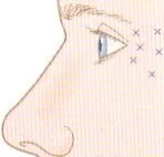 Fig 12. Typical lateral-canthal (crow's-feet) injection fan. (Carruthers, Toxina Botulínica, p. 52).
> Sources: [14] Carruthers
Fig 12. Typical lateral-canthal (crow's-feet) injection fan. (Carruthers, Toxina Botulínica, p. 52).
> Sources: [14] Carruthers [C] [MEDLIB]; [16] López del Val 2010 [C] [MEDLIB].
Consensus: crow's feet superficial and lateral, glabella intramuscular and high, depressors weakened and the frontalis respected [14][25][28]. Discrepancy that changes the gesture: the newest anatomical work maps the neuromuscular-junction distribution of the upper-face muscles and argues for targeting the NMJ-dense zones with fewer, better-placed units rather than the classic fixed pattern; the label-pattern school keeps the approved three-point map [27][28]. Both are printed; the deciding variable is operator experience and whether a modulated, NMJ-targeted result is sought.
Reconstitution, dilution and why volume equals diffusion. Toxin is reconstituted with preservative-free (or preserved, for comfort) saline without shaking, the date, time and dilution recorded [14]. In the periorbital region dilution matters as much as dose: diffusion scales with the injected volume, not only the units, so a very dilute glabellar injection increases the ptosis risk by spreading toward the levator, and a concentrated small-volume deposit is safer near the septum [14][26]. Crow's-feet points are superficial and small-volume by design; the visible papule confirms the plane [16].
Onset, duration and review, region-specific. Effect begins around days 2-4, peaks near two weeks, and lasts roughly 3-4 months for upper-face cosmetic use; the review and any retouch are at two weeks, never at day 7 when the effect is incomplete and retouching over-doses [14][25]. The chemical brow lift is judged at two weeks because the brow settles into its new depressor-elevator balance over that interval.
Injection technique, step by step, for the region's approved points. Mark with the patient sitting and animating; for crow's feet, tent the skin and inject superficially in the three-point fan >=1-1.5 cm lateral to the canthus, bevel up, tangential, then compress; for the glabella, palpate the corrugator while the patient frowns, inject the belly intramuscularly, the lateral point >=1 cm above the rim, needle aimed up and never toward the orbit; for the chemical brow lift, place 1-2 U in the lateral orbicularis tail keeping the frontalis untouched [14][25][29]. Compress every point in this vascular region.
Microbotox / intradermal toxin in the periorbital skin, status stated. Very dilute intradermal toxin is used off-label for fine periorbital skin quality and pore/oil reduction; the sweat/sebaceous-gland mechanism is well established but the skin-tightening claim is weak, the published dilutions vary too widely to average, and the technique sits at [D] for this indication, so consent must say so and the dilution used must be recorded [14]. It is a complement to skin-quality work, not a substitute for it.
Brow-shaping nuance, because millimetres change the face. Treating the central frontalis while sparing the lateral fibres over-elevates the tail (Spock brow); treating uniformly and low drops the whole brow; the aesthetic goal, especially in women, is a modest lateral-tail lift without arch distortion, and in men a flat, conservative result [14][25]. The lateral-tail double-dose point maximises the depressor-release lift, but only when the frontalis reserve is intact and the rim still supports the brow (D4.4) [29][30].
Brand table extended for the region's approved uses. All figures above are ona units; abo (Azzalure/Dysport) is dosed in Speywood units (glabella 50 U in five 10 U points) and is not converted with a fixed rule (ona:abo 1:2.5 to 1:3 by scenario); inco, prabo and leti are re-dosed per their own label rather than assumed 1:1 [14][16]. Crow's-feet and brow figures are printed only in ona units to avoid the by-side/total and cross-brand traps that have caused real dosing errors.
Trampa clásica: chasing a crow's-foot radius too low and anterior onto the zygomatic arch, hitting the zygomaticus major and dropping the lip; the fix is prevention, staying high, lateral and low-volume [14].
D4.9 · Combination and sequence
The order, glanceable. Full planning logic in L2 — Combination Sequencing and Treatment Planning.
| Step | Modality | Interval to next | Why in this order |
|---|---|---|---|
| 0 | Surgery if indicated (blepharoplasty, ptosis repair, brow lift) | wait ~3 months | Surgery changes the anatomy; planning volume on anatomy that will change wastes product [20] |
| 1 | Toxin (crow's feet, chemical brow lift, glabella) | 2 weeks | Settles brow position and lowers the filler volume the sulcus needs; a dermis that folds thousands of times a day re-engraves a treated wrinkle [25] |
| 2 | Deep filler (superior sulcus, ROOF) | same or next visit; deep before superficial | Structure first; the deep correction defines how much surface work is needed [17] |
| 3 | Skin quality (polynucleotides, boosters, microneedling) | parallel course, multiple sessions | A thicker dermis camouflages what is placed beneath and lowers Tyndall risk; better before volume than after [21][46] |
| 4 | Resurfacing (laser/EBD) if needed | ~2 weeks after toxin | Toxin first works with the healing skin, not against it; internal corneal shields mandatory [46] |
| - | Review | 2-4 weeks | The eyelid swells and settles slowly; retouch here, never at 7 days |
Why toxin goes first, salvaged and reinforced. Two sequence rules that few teach and that improve the result at no cost [25]: (1) toxin before filler and before resurfacing, because a dynamic dermis that creases constantly heals worse and re-etches the line, so relaxing it first is working with the tissue; and (2) skin quality before volume, because a thicker dermis hides what is placed beneath and reduces the Tyndall risk that defines this region. Toxin also reduces the crow's-feet dynamic pull on the lateral canthus, so the volume placed in the sulcus and ROOF settles into a quieter field.
Deep before superficial, always. The supraperiosteal and ROOF corrections define the scaffold; superficial or skin-quality work is layered on top only after the deep result is judged. Reversing the order means placing surface product against an unstable base and re-doing it once the deep volume settles [17].
Intervals that matter here specifically: - Toxin to filler: ~2 weeks, so the brow has reached its new position before the volume is judged. - Filler second session: 2-4 weeks; the region is deliberately under-corrected first and topped up, never fully corrected in one pass. - Review and retouch: 2-4 weeks, not 7 days. Periorbital oedema takes time to settle and an early retouch is the usual cause of over-correction. - Resurfacing after toxin: ~2 weeks.
⚠ Surgery in the plan, the three salvaged rules: 1. Surgery first. It changes the anatomy; volume planned on anatomy that will change is wasted [20]. 2. Wait about three months after eyelid surgery before assessing injectables on an operated lid. 3. Do not put toxin in the upper third of a patient under pre-surgical assessment. It changes brow and aperture position and falsifies the surgeon's evaluation, who may then resect too much. And if there is prior filler in the region, tell the surgeon; a forgotten deposit in the preperiosteal plane changes what the surgeon finds.
Hyaluronidase available at every session, not only for the vascular emergency but because correcting excess and prolonged oedema is an expected part of managing this region; the periorbital lid is where planned reversibility earns its place [40] (D4.10).
Consensus: surgery first, then toxin, then deep filler, then skin quality, with a slow review cadence [20][25][17]. Discrepancy that changes the gesture: whether toxin and deep filler can be done in the same visit or must be staged; the same-visit school treats an experienced, low-risk patient in one sitting, the staged school separates them so the brow settles before volume is judged, and the deciding variable is how much the brow position is expected to change with the toxin.
Energy devices and skin quality in the combined plan. Non-ablative fractional laser, ablative fractional laser and radiofrequency microneedling treat the crêpe, static-rhytide component that neither filler nor toxin addresses, and are sequenced about two weeks after toxin so the relaxed skin heals without re-creasing; internal metal corneal shields are mandatory for any device used inside the orbital rim, external goggles do not protect [46]. See G3 — Non-Ablative and Fractional Lasers. Skin boosters and polynucleotides run as a parallel multi-session course that thickens the thin lid dermis, improving camouflage of deep product and lowering Tyndall risk before volume rather than after [21].
Regenerative adjuncts and their place. Polynucleotides (PN/PDRN) are consolidating as the skin-quality lane for the thin, crêpe periorbital skin, distinct from volume, and recent work compares them directly with HA for the periorbital surface [21]. PRP is used similarly for skin quality in some practices; both are quality, not volume, and neither substitutes for the deep structural correction.
The annual plan, so sequence becomes a calendar. A representative year: surgery first if indicated, then a toxin session (crow's feet, chemical brow lift, glabella) with review at two weeks; deep filler (sulcus/ROOF) two weeks after the toxin has settled, under-corrected, with a planned top-up at 2-4 weeks; a parallel skin-quality course (three to four sessions); toxin repeated at 3-4 month intervals; filler reassessed at 9-12 months. The intervals are driven by the biology of each modality, not by the calendar of the clinic [25].
The cost-of-error asymmetry that sets the order. Under-treating costs one extra visit; over-treating the region costs a puffy lid for weeks (filler) or a dropped brow and closed field for months (toxin), so every interval in the plan favours staging and review over completeness in one pass [14][17]. This is the sequencing corollary of the region's under-correction rule.
⚠ The single most consequential sequencing error, restated. Do not place toxin in the upper third of a patient under pre-surgical assessment: it shifts brow and aperture position and falsifies the surgeon's measurement, risking over-resection, and prior filler in the region must be disclosed to the surgeon because a forgotten preperiosteal deposit changes the operative plane [20].
Maintenance and monitoring over years, because the region is a longitudinal relationship. Toxin is repeated at roughly 3-4 month intervals early and often lengthens as the treated muscles are chronically relaxed; deep filler is reassessed at 9-12 months, and the sulcus is re-treated only when the concavity genuinely returns, not on a fixed clock, because HA in the deep periorbital plane can persist and stacking product on residual product overfills the lid [17]. At each maintenance visit the same measures are repeated (MRD1, brow position, sulcus depth) so a slow drift toward ptosis, brow descent or over-fill is caught early, and the standardised photographs are compared side by side rather than from memory [43][46].
The interaction the plan must anticipate: toxin changes the filler requirement and vice versa. Relaxing the crow's-feet and depressor pull settles the lateral canthus and brow so the volume placed afterwards sits in a quieter field and less is needed; conversely, restoring the ROOF and sulcus can unmask a residual dynamic line that then wants toxin. The two modalities are planned together as one region programme, not as separate purchases, which is why the combined consult and the shared record matter [17][25].
Combination with the lower face and the rest of the upper third. The brow and glabella (D1) share the depressor-elevator system with this region, so upper-third toxin is usually planned as one map across forehead, glabella and crow's feet rather than piecemeal, respecting the frontalis-as-sole-elevator rule throughout [14][25]. Midface volume (D3) supports the lower orbit and can reduce the apparent upper hollow indirectly, so the whole periorbital-to-cheek unit is assessed before committing to the upper sulcus alone [17]. Full cross-region planning is in L2 — Combination Sequencing and Treatment Planning.
Trampa clásica: injecting toxin into the forehead or brow of a patient who is being worked up for blepharoplasty, changing the brow position and misleading the surgeon into over-resection. If surgery is on the table, the upper third is left untouched until after [20].
D4.10 · Region-specific complications
Not the generic bruise-and-swell list; the complications that happen here because of this anatomy. Generic vascular and toxin management live in J1 — Complications Overview through J8 — Contraindications and Special Populations; this block is the periorbital-specific set.
| Complication | Region-specific mechanism | Recognition | Management |
|---|---|---|---|
| Filler-induced visual loss / blindness | STA/SOA/dorsal-nasal are ophthalmic terminals; retrograde embolism to the central retinal artery | Immediate severe pain, sudden vision loss, ptosis, ophthalmoplegia, skin mottling | Emergency: stop, flood with hyaluronidase, ocular massage, transfer; retina tolerates ~90 min [31][32][35] |
| True blepharoptosis (toxin) | Corrugator/glabella toxin crosses the septum to the levator | Lid droop, reduced MRD1, 3-10 days after toxin | Apraclonidine 0.5% drops (Müller stimulation, raises lid ~1-2 mm) or phenylephrine; waits it out [14] |
| Diplopia (toxin) | Crow's-feet toxin too medial reaches the lateral rectus (or inferior oblique) | Double vision after lateral canthal toxin | Reassure, ophthalmology if persistent; self-limited, avoid by distance [14][27] |
| Brow heaviness / brow ptosis (toxin) | Frontalis over-treated, or compensating frontalis weakened | Heavy brow, closed field, loss of upper-field vision | Prevent by keeping points high; time; small lateral touch-up cannot re-elevate a dropped brow [14][26] |
| Lagophthalmos / dry eye (toxin) | Orbicularis over-weakening impairs lid closure | Incomplete closure, exposure symptoms | Lubrication, refer if corneal exposure; dose conservatively [14] |
| Smile asymmetry (toxin) | Crow's-feet toxin too low hits zygomaticus major | Drooped upper lip on smiling, weeks | Prevent (stay high/lateral); wait out [14] |
| Prolonged periorbital / malar oedema (filler) | Fragile lymphatic precollectors at the preseptal orbicularis level are obstructed by product | Persistent puffy lid/malar swelling, weeks to months | Hyaluronidase; the signature filler complication of the region [34][36] |
| Tyndall / blue discoloration (filler) | HA placed too superficially in translucent lid skin refracts light | Blue-grey hue over the deposit | Hyaluronidase; prevent by staying deep [1][36] |
| Subdermal nodule / visible lump (filler) | No superficial fat plane over the lid; product tents the thin skin | Palpable/visible lump | Hyaluronidase; massage rarely enough [17][36] |
| Penetrating globe injury (filler) | Needle path over the orbit enters the globe | Acute ocular pain, vision change at injection | Emergency ophthalmology; a described periocular-filler catastrophe [41] |
Filler-induced visual loss, the region's catastrophe, in operational detail. This is the highest-stakes lane in the highest-risk region, and it is managed in minutes [31][32].
- Recognise instantly. Disproportionate pain at injection, immediate or rapid vision loss, ptosis, ophthalmoplegia, and skin livedo/blanching along a vascular territory. Any of these during or after a brow/glabella/sulcus injection is visual-loss until disproven [31][32].
- Stop and flood. Stop injecting. Deliver high-dose hyaluronidase to the affected territory and periorbitally; reported flooding regimens use large pulsed doses (on the order of 500-1500 IU, repeated) into the periorbital and supratrochlear/supraorbital regions [36][38][39][40]. High-dose pulsed hyaluronidase is the accepted soft-tissue rescue; its ability to cross into the artery and dissolve an intravascular HA embolus is the debated part [33][36].
- Lower intraocular pressure and mobilise the embolus. Ocular massage, and measures to lower intraocular pressure (anterior-chamber paracentesis, timolol, acetazolamide, rebreathing) are described adjuncts while transferring [31][40].
- Systemic measures and transfer. Systemic steroids are used for the inflammatory/oedema component; arrange immediate transfer to ophthalmology. The ~90-minute retinal ischaemic window governs the urgency [31][32].
- Consensus guidance now exists. Recent consensus guidelines codify the recognition-and-management sequence for tissue-filler vascular events, and should anchor the clinic protocol [31].
⚠ CONTROVERSY, printed with both positions, never averaged: the retrobulbar hyaluronidase route. - Retrobulbar-injection school [33]: inject hyaluronidase into the retrobulbar space to attempt to reach the ophthalmic artery and dissolve the embolus at source; some series report benefit. - Peri/supraorbital + subcutaneous flooding school [35][36]: the retrobulbar route's efficacy is unproven, it is technically hazardous (globe, optic nerve), and hyaluronidase flooding of the peri/supraorbital soft tissue plus general resuscitation is preferred without the retrobulbar step. The deciding variables are operator skill with retrobulbar injection and access to imaging; both are recorded because the evidence does not settle it, and the clinic protocol should state which it will use before the emergency, not during it.
The currency the corpus predates: ultrasound-guided hyaluronidase. The master course and the older corpus books precede routine facial ultrasound, so this is flagged. Recent evidence supports ultrasound-guided hyaluronidase as first-line for impending filler-induced necrosis and vascular compromise: a 2025 systematic review with pilot meta-analysis reports superior outcomes with image-guided delivery, with pooled complete resolution around 94.6% and lower, better-targeted doses than blind flooding [35], and a 2024 dosing review reaches the same direction that ultrasound guidance increases efficacy [36]. Companion Doppler-ultrasound vessel mapping supports both prevention and targeted rescue [37]. This is the clearest example in the region where the external lane is mandatory and the corpus is a figure source, not a clinical reference. [MATERIAL GAP] in the own corpus, resolved by external evidence [35][36][37].
Toxin complications specific to the region, managed. - True ptosis from septal diffusion (the glabella/corrugator complication): apraclonidine 0.5% drops stimulate Müller's muscle and raise the lid roughly 1-2 mm as a symptomatic bridge; topical phenylephrine is an alternative. Neither reverses the toxin; they buy time while it wears off [14]. See J4 — Botulinum Toxin Complications. - Diplopia: from lateral-rectus (crow's feet too medial) or inferior-oblique diffusion; self-limited, ophthalmology if persistent [14][27]. - Brow ptosis/heaviness: frontalis over-treatment or weakening a compensating frontalis; prevented by keeping points high and screening frontalis compensation [26]. - Lagophthalmos/dry eye and smile asymmetry: dose conservatively, stay lateral and high; both are avoidable by geometry [14].
Filler complications specific to the region, managed. - Prolonged periorbital and malar oedema: the lid's lymphatic precollectors are fragile and sit at the preseptal orbicularis level; product obstructs them and the lid stays puffy for weeks to months. This is the region's commonest filler complication and it needs hyaluronidase, not time [34][36]. Choosing a low-hydrophilicity product and minimal volume is the prevention. - Delayed swelling / worsening festoons: adding a hygroscopic product to a compartment that already drains poorly worsens it persistently; a full argument for the festoon exclusion lives in D5, but the upper-lid corollary is that a boggy, poorly draining lid is not volumised. - Tyndall, nodules, lumps: all trace to superficial deposition in skin with no superficial fat plane; hyaluronidase reverses HA, which is the argument for HA over permanent products here [1][17][36].
Consensus: the region's two catastrophes are blindness (minutes, hyaluronidase, transfer) and the wrong operation; its commonest nuisances are oedema, Tyndall and toxin-diffusion ptosis, and all are reduced by deep placement, low volume, low-hydrophilicity HA and geometric toxin discipline [31][35][36]. Discrepancy that changes the gesture: blind high-dose flooding versus ultrasound-guided lower-dose hyaluronidase for impending necrosis; the ultrasound school reports better resolution at lower dose [35], the pragmatic school floods blind where ultrasound is unavailable [40], and the deciding variable is equipment and operator skill at the moment of the emergency.
Vascular occlusion staging, so the response matches the picture. Impending skin necrosis presents as immediate blanching then dusky livedo along a territory, with pain out of proportion; visual compromise adds sudden vision loss, ptosis and ophthalmoplegia [31][34]. Skin-level occlusion is managed with hyaluronidase flooding of the territory, warmth, massage and close review; visual-level occlusion is the 90-minute emergency of D4.1 [31][35]. Ultrasound-guided hyaluronidase now targets the occluded segment at lower total dose than blind flooding and reports higher complete-resolution rates, and is first-line where the equipment exists [35][36].
The management algorithm, condensed. Recognise (disproportionate pain, blanch/livedo, vision change) -> stop injecting -> flood the territory and periorbital region with high-dose hyaluronidase, repeated [36][38][39][40] -> for visual signs add ocular massage, intraocular-pressure-lowering measures and immediate ophthalmology transfer within the retinal window [31][32] -> image-guide the hyaluronidase if ultrasound is available [35] -> document and report. The clinic fixes in advance whether it will attempt the retrobulbar route, because the evidence is unsettled and the emergency is not the time to decide [33].
Delayed complications specific to the thin-skinned region. Delayed inflammatory nodules and delayed-onset oedema can appear weeks to months after HA in the lid, sometimes triggered by illness or dental work; biofilm and low-grade infection are in the differential and are managed distinctly from a simple over-fill, with hyaluronidase, antibiotics where indicated, and avoidance of repeated needling of an infected deposit (see J5 — Biofilm, Infection and Sterilisation) [36]. The reversibility of HA is again the argument for it over permanent products in a region prone to delayed events.
Bruising and ecchymosis, the region's near-universal minor event. The lateral canthal and periorbital skin has a dense superficial venous plexus, so crow's-feet toxin and any needle work here bruises readily; brief compression after each point, a fine needle, avoiding visible superficial veins, and pausing anticoagulants where medically appropriate reduce it, and the patient is warned it is expected, not a complication [14]. A periorbital haematoma from a deeper vessel is managed with pressure and reassurance and resolves over one to two weeks.
Infection, biofilm and the late red, tender nodule. A late, red, tender or fluctuant periorbital swelling weeks to months after filler is infection or biofilm until proven otherwise, not a simple over-fill, and it is not managed by repeated needling of the deposit; it takes appropriate antibiotics, sometimes incision, and hyaluronidase to remove the HA scaffold the biofilm sits on, with the full argument in J5 — Biofilm, Infection and Sterilisation [36]. Sterile technique and single-use product reduce the risk from the outset.
Toxin spread beyond the target, the mechanism restated as prevention. Every toxin complication of the region (ptosis, diplopia, lip-drop, brow heaviness, lagophthalmos) is unwanted spread to a neighbour muscle, and every one is prevented by the same three levers: correct distance, small volume (spread scales with volume, not only units), and correct depth [14][26][27]. There is no rescue that reverses toxin; apraclonidine buys 1-2 mm and time for ptosis, and the rest is waited out, which is why prevention by geometry is the whole game.
Prevention bundle, consolidated for the complication set. Low-hydrophilicity product and minimal volume prevent the oedema and Tyndall nuisances; deep-on-bone or subdermal-threaded placement with rim guard, small aliquots, low pressure, moving tip/cannula and pre-injection ultrasound prevent the vascular catastrophe; geometric toxin discipline (distances, high frontalis points, superficial lateral crow's feet) prevents ptosis, diplopia and lip-drop; and MRD1/differential screening prevents the wrong operation [14][17][31][43]. Every complication in the table above has a prevention line, and the prevention is cheaper than the reversal in every case.
Trampa clásica: attributing immediate post-injection pain and blurring to "just a bruise" and sending the patient home. In this region that is the presentation of retrograde embolism, and the clock is 90 minutes [31][32].
Step-by-step protocol - the upper periorbital session
Consultation and mapping, in order: 1. Translate the complaint. "Tired eyes", "hooded lid", "hollow", "droopy brow", "crow's feet" each point to a different tissue and a different treatment; do not treat the word [43]. 2. Directed history. Glaucoma drops (prostaglandin periorbitopathy), thyroid and renal disease and allergy (fluid retention and oedema risk), bleeding tendency and anticoagulants, dry eye and contact lenses, prior surgery, prior filler and which, and neuromuscular disease for toxin [14][46]. 3. Measure. MRD1, levator excursion with the brow fixed, tarsal platform show, dermatochalasis pinch, brow position with manual elevation, superior-sulcus depth, and hollow-versus-full palpation [42][43]. 4. Do the three-way differential. Dermatochalasis, true ptosis, brow ptosis; refer what is surgical and flag the neurological red-flags before any syringe [43]. 5. Animate. Squeeze (crow's-feet fan), brow-raise (frontalis reserve), frown (glabella); screen frontalis compensation [14][26]. 6. Ultrasound if available. Map STA/SOA and prior product; identify the plane [15][22]. 7. Photograph. Multi-view, multi-light, at rest and animated; store the drop history [46].
Treatment, in order: 8. Refer or defer anything surgical, neurological or drug-driven before injecting [20][43]. 9. Toxin first if indicated: crow's feet superficial and lateral, chemical brow lift on the depressors, glabella high and intramuscular, respecting every safety distance [14][25]. 10. Deep filler two weeks later: supraperiosteal sulcus or ROOF, low-hydrophilicity HA, cannula preferred, rim guard, small aliquots, deliberate under-correction [17][18]. 11. Skin quality in parallel: polynucleotides, boosters, resurfacing with internal corneal shields [21][46]. 12. Hyaluronidase and the visual-loss plan ready at every session [31][40]. 13. Review at 2-4 weeks, not day 7; retouch then; the second filler session is planned, not a rescue [17]. 14. Record per side: product, brand, lot, volume, plane, tool, entry; toxin brand, dilution, units per point and area; and the differential findings [46].
Coverage vs UPO
What the UPO master course teaches for this region (mainly under T8.2, botulinum toxin), the chapter's status, and what the atlas adds. Retrieved with the UPO collection prefix; a slide-only dose is never_sufficient_alone and the fastest-ageing lane [44][45].
| UPO topic | Status in chapter | What the atlas adds |
|---|---|---|
| Crow's-feet toxin dosing (24 U total, 12/side, 3x4 U) [44] | Covered (D4.8) | The three fan patterns, the six-point extension, the NMJ-distribution debate, brand non-convertibility (ona:abo 1:2.5-1:3) [27][28] |
| Glabella and frontalis dosing (20 U / 5x4 U) [44] | Covered (D4.8) | The septum-to-levator mechanism of ptosis, the >=1 cm rim rule with its reason, the Spock-brow correction [14][26] |
| Chemical brow lift concept [44] | Covered (D4.8) | The depressor-elevator physiology, the lateral-tail double-dose point, mm of elevation, first-line-over-filler ranking [25][29][30] |
| Upper-lid / brow anatomy | Partial | The fixed six-layer scaffold, the ROOF, the orbital septum firewall, the preaponeurotic fat pads, retaining ligaments [3][8][9] |
| Danger zones / blindness | Partial | STA/SOA course and depth, the 90-min retinal window, rim-guard mechanics, pre-injection Doppler, 2026 consensus [23][31][35] |
| Adverse events of fillers (Tejero) [45] | Partial | Region-specific set: lymphatic-precollector oedema, Tyndall in translucent skin, penetrating globe, retrobulbar controversy [34][41][33] |
| Superior-sulcus / ROOF filling | Not covered by UPO | The full product-plane grid, retrobulbar vs direct-sulcus controversy (0.83 mL/mm), MD-Codes periorbital algorithm [17][18] |
| Upper-lid ptosis differential | Not covered by UPO | MRD1, levator excursion, three-way differential, the neurological red-flags, referral criteria [42][43] |
| Ultrasound in assessment and rescue | Not covered (course predates it) | Doppler vessel mapping, US-guided hyaluronidase as first-line, pooled 94.6% resolution [15][35][37] |
| Prostaglandin periorbitopathy | Not covered by UPO | The glaucoma-drop cause of a deep, often unilateral, progressing superior sulcus [46] |
| Post-blepharoplasty injectable decisions | Not covered by UPO | The four post-surgical scenarios and which are treated vs referred [20] |
Rows UPO does not cover at all, made explicit: superior-sulcus and ROOF volumisation technique, the retrobulbar-hyaluronidase controversy, the MRD1 / levator / brow three-way differential, pre-injection ultrasound and ultrasound-guided rescue, prostaglandin periorbitopathy, and the post-blepharoplasty decision set. The UPO toxin material is solid for approved crow's-feet and glabella dosing and is cited as such; everything structural, vascular, surgical-boundary and ultrasound is atlas-added from the corpus books and the external lane, because the master course lane ages fastest and predates routine facial ultrasound [15][35].
Self-assessment
Ten active-recall questions, answers folded, built only from facts already stated above. No new claims.
- What is the label crow's-feet dose in ona units, and how is it split?
Answer
24 U total, 12 U per side, three points of 4 U, superficial subcutaneous [14][16] (D4.8).- Why must the lateral corrugator point sit at least 1 cm above the bony rim?
Answer
Below that, toxin diffuses through the orbital septum to the levator palpebrae and causes true blepharoptosis [14][26] (D4.8).- Which artery is the substrate for periorbital blindness, and what is the retinal ischaemic window?
Answer
The supratrochlear/supraorbital arteries (ophthalmic terminals) permit retrograde embolism to the central retinal artery; the retina tolerates roughly 90 minutes [23][31] (D4.3, D4.10).- In what order does the upper periorbital region age?
Answer
Bone (superomedial + inferolateral rim) -> ligament stays fixed -> deep fat (ROOF deflation, orbital-fat pseudoherniation) -> superficial fat/muscle descent -> skin thinning [1][8] (D4.4).- A patient on a glaucoma drop presents with a deep, unilateral superior sulcus. What is the likely cause?
Answer
Prostaglandin-associated periorbitopathy from bimatoprost/latanoprost: orbital-fat atrophy deepening the sulcus; address the drug before filling [46] (D4.4).- What three diagnoses does a "droopy upper lid" split into, and which single test separates brow ptosis?
Answer
Dermatochalasis (excess skin), true blepharoptosis (levator), brow ptosis (descended brow); manual brow elevation - if the lid improves, the brow is the problem [42][43] (D4.5).- Which muscle must never be weakened at the lateral brow, and why?
Answer
The frontalis - it is the sole brow elevator; weakening it drops the brow. The chemical brow lift weakens depressors only [14][25] (D4.8).- What is the region's commonest filler complication, and its mechanism?
Answer
Prolonged periorbital/malar oedema from obstruction of the fragile lymphatic precollectors at the preseptal orbicularis level; needs hyaluronidase [34][36] (D4.10).- Retrobulbar versus peri/supraorbital hyaluronidase for visual loss: what decides?
Answer
Evidence is unsettled; retrobulbar aims at the ophthalmic artery but is hazardous and unproven, peri/supraorbital flooding is safer; operator skill and imaging decide, and the clinic fixes its protocol in advance [33][35] (D4.10).- Why is toxin sequenced before filler and skin quality before volume?
Answer
Toxin settles brow position and lowers the volume the sulcus needs and lets treated wrinkles heal without re-creasing; a thicker dermis camouflages deep product and lowers Tyndall risk [21][25] (D4.9).What's new and trends
Dated table of what changed in this region in roughly the last two years. Every row cites a reference already used above; this section synthesises, it introduces no new claim.
| Year | Change | Direction | Maturity class | Ref |
|---|---|---|---|---|
| 2025 | Ultrasound-guided hyaluronidase for impending filler necrosis / vascular compromise | Toward image-guided, lower-dose, better-targeted rescue (pooled complete resolution ~94.6%) | clinically actionable now | [35] |
| 2024 | Hyaluronidase dosing review for filler complications | Consolidates dosing and supports ultrasound guidance increasing efficacy | clinically actionable now | [36] |
| 2023-2024 | Doppler-ultrasound vessel mapping and layer identification for facial aesthetics | Pre-injection mapping of STA/SOA and planes moving into routine high-risk practice | promising but not validated | [15][37] |
| 2026 | Consensus guidelines for tissue-filler vascular events | Codified recognition-and-management sequence for the clinic protocol | clinically actionable now | [31] |
| 2026 | Neuromuscular-junction distribution mapping of the upper face | Toward NMJ-targeted, fewer-unit toxin placement over the fixed pattern | preclinical/speculative | [27] |
| 2026 | HA fillers versus polynucleotides for the peri-orbital skin/quality problem | Polynucleotides consolidating as the skin-quality lane distinct from volume | promising but not validated | [21] |
| ongoing | Marketed non-hyaluronidase "filler-dissolving" or lymphatic-drainage devices for periorbital oedema | No controlled evidence; hyaluronidase remains the only proven reversal | unsupported commercial claim | [36] |
What did NOT change, and why the older references still stand. The approved toxin doses for the region are unchanged: crow's feet remain 24 U total (12/side, three 4 U points) and the glabellar complex 20 U in five 4 U points, and the Carruthers/label-era numbers remain the reference [14][16]. The core anatomy is stable: the orbital septum firewall, the levator-diffusion mechanism of glabellar-toxin ptosis, the STA/SOA ophthalmic-terminal blindness substrate and the ~90-minute retinal window are as described for years and are not superseded [23][26][31]. The MRD1 / levator / brow three-way differential and the referral red-flags are classic and unchanged [43]. What moved is not the doses or the anatomy but the tooling around them: ultrasound for mapping and rescue, consensus codification of the emergency, and a shift from fixed patterns toward NMJ-informed placement. Where a chapter fact rests on the UPO master lane, that lane ages fastest and is flagged in Coverage vs UPO.
Unexplored directions (AI speculation)
> Disclaimer. The items below are model-generated research directions, not clinical recommendations. Each is tagged [IA-ESPEC], carries no dose, product or actionable protocol, and states the cited anchor it springs from and the experiment that would settle it. [IA-ESPEC] is not evidence and is not [A-D].
[IA-ESPEC]Routine pre-injection Doppler as a visual-loss preventive. Anchor: ultrasound-guided delivery improves soft-tissue rescue outcomes and maps the STA/SOA before the needle [35][15]. Proposal: that routine pre-injection colour-Doppler mapping in every deep periorbital case, not just re-treatments, lowers the incidence of retrograde embolism. Expected effect: a lower rate of vascular occlusion and visual-loss events in mapped versus unmapped deep periorbital injections. Confounder: operator experience and case-mix differ between adopters and non-adopters of ultrasound, so a naive comparison confounds the tool with the injector. What would settle it: a prospective registry comparing occlusion/visual-loss rates in ultrasound-mapped versus landmark-only deep periorbital injections.[IA-ESPEC]ROOF augmentation versus chemical brow lift for durable brow-tail position. Anchor: ROOF deflation unsupports the brow tail and chemical brow lift elevates it a few millimetres [3][29]. Proposal: that deep ROOF volume gives more durable brow-tail elevation than repeated depressor toxin. Expected effect: greater brow-tail height retained at 12 months for ROOF volume than for toxin alone. Confounder: baseline rim resorption and brow ptosis severity differ between patients and drive the ceiling of any lift, so groups must be matched on rim support. What would settle it: a randomised comparison measuring brow-tail position at 6 and 12 months for the two modalities alone and combined.[IA-ESPEC]Reversal of the sulcus in prostaglandin periorbitopathy. Anchor: prostaglandin analogues deepen the superior sulcus by orbital-fat atrophy, often unilaterally [46]. Proposal: that switching or stopping the drop partially reverses the sulcus, changing who needs filler at all. Expected effect: a measurable reduction in superior-sulcus depth after supervised drug withdrawal or switch. Confounder: ongoing intrinsic ageing and glaucoma progression also change orbital fat, so the untreated fellow eye must serve as the internal control. What would settle it: a cohort measuring sulcus depth before and after a supervised drug switch, with the untreated eye as internal control.[IA-ESPEC]NMJ-targeted crow's-feet placement. Anchor: the upper-face neuromuscular-junction distribution has now been mapped [27]. Proposal: that placing toxin over NMJ-dense zones matches label-dose efficacy with fewer units and less unwanted diffusion (less diplopia/lip-drop). Expected effect: equal wrinkle-severity reduction at a lower total unit dose, with fewer off-target events. Confounder: individual NMJ maps vary and the assessor's wrinkle scoring is subjective, so blinded rating and per-face controls are needed. What would settle it: a split-face trial of NMJ-guided versus the standard three-point pattern on efficacy and off-target rates.[IA-ESPEC]An in-clinic "code blindness" drill. Anchor: the retina tolerates roughly 90 minutes and outcome is time-dependent [31][32]. Proposal: that a standardised rehearsed emergency drill shortens time-to-first-hyaluronidase and improves visual outcomes. Expected effect: shorter measured time from recognition to first hyaluronidase dose after drill implementation. Confounder: event rarity and recall bias in real cases, and the Hawthorne effect during observation, inflate apparent improvement, so simulated timed scenarios are the cleaner measure. What would settle it: a simulation study measuring team response time and correctness before and after drill implementation.
When there is genuinely nothing to propose beyond these, the section still exists; here there is, because the region sits exactly on the frontier between the stable anatomy and the moving tooling.
§ Safety
The region's safety contract, consolidated. Numbered citations retained as the gate requires [31][35][40].
Absolute stop / refer, do not inject: - Recent-onset, asymmetric or fluctuating ptosis with diplopia, anisocoria or pain: neurological emergency differential (myasthenia, Horner, third-nerve palsy) [43]. - Active neuromuscular disease (myasthenia, Eaton-Lambert, ALS) for toxin: absolute contraindication [14]. - Frank dermatochalasis, true levator ptosis, orbital-fat pseudoherniation: surgical, not injectable [20][43]. - Lid retraction / scleral show / lagophthalmos / dry eye post-blepharoplasty: refer, do not fill [20]. - Permanent fillers in the lid: indefensible in the thinnest, most complication-prone skin.
Relative / condition-first: - On a prostaglandin glaucoma drop with a progressing sulcus: address the drug first [46]. - Frontalis used as a visual crutch: forehead toxin closes the field [26]. - Bleeding diathesis or anticoagulation: the region bruises heavily; compress each point [14]. - Prior unknown filler in the region: image (ultrasound) before adding [15].
The three red lines, and the mechanics that prevent each: 1. Blindness. Deep injections on bone or subdermal threads only, never a fast bolus in between; rim guard on the superomedial rim; aliquots <=0.1 mL; low pressure; moving needle or cannula; ultrasound where available; aspiration as one weak layer, not a licence [23][40]. Have a written emergency plan and hyaluronidase ready every session [31]. 2. Toxin diffusion (ptosis / diplopia). Distance, not dose: lateral corrugator >=1 cm above the rim, crow's feet >=1-1.5 cm lateral to the canthus, off the anterior zygomatic arch, small volume [14][26]. 3. The wrong operation. Measure MRD1, do the three-way differential, lift the brow manually, and refer what is surgical before any syringe [43].
Emergency kit for the region (visual-loss protocol): high-dose hyaluronidase in quantity, the written recognition-and-management sequence per current consensus, means to lower intraocular pressure, and a named ophthalmology transfer route; the 90-minute window means the kit is prepared before, not during, the emergency [31][35][40].
Documentation every session: product/brand, lot, volume per side, plane, tool, entry point; toxin brand, dilution, units per point and per area; the ptosis/differential findings; and standardised photographs in multiple light directions [46]. The lid is the region that changes most with light and generates the most disputes without a baseline image.
References
- Cotofana S, et al. Anatomy of the Aging Face: A Review. Facial Plast Surg 2016. DOI 10.1055/s-0036-1582234.
[B][MEDLIB] - Cotofana S, et al. Anatomy of the Facial Fat Compartments and their Relevance in Aesthetic Surgery. J Dtsch Dermatol Ges 2019. DOI 10.1111/ddg.13737.
[B][MEDLIB] - Schenck TL, et al. Functional Anatomy of the Superficial Fat Compartments of the Face. Plast Reconstr Surg 2018. DOI 10.1097/PRS.0000000000004364.
[B][MEDLIB] - Radlansky RJ. Atlas Ilustrado de Anatomia Clínica da Face.
[C][MEDLIB] - Watanabe K, et al. Anatomy for Plastic Surgery of the Face, Head, and Neck. Thieme 2016.
[C][MEDLIB] - Seckel BR. Facial Danger Zones: Avoiding Nerve Injury in Facial Plastic Surgery. Thieme.
[C][MEDLIB] - Rohrich RJ, et al. Facial Danger Zones (Zonas Faciais de Perigo). 2020.
[C][MEDLIB] - Kim HJ, et al. Clinical Anatomy of the Face for Filler and Botulinum Toxin Injection. Springer 2016.
[C][MEDLIB] - Standring S. Gray's Anatomy. 41st ed. Elsevier 2016.
[C][MEDLIB] - Smith A, et al. Gray's Surface Anatomy and Ultrasound. Elsevier 2018.
[C][MEDLIB] - Freytag DL, et al. Understanding Facial Aging. 2022.
[B][MEDLIB] - Anatomía Clínica de la Cara para Relleno y Toxina Botulínica.
[C][MEDLIB] - Azizzadeh B, et al. Master Techniques in Facial Rejuvenation. 2nd ed. Elsevier 2018.
[C][MEDLIB] - Carruthers J, Carruthers A. Botulinum Toxin (Procedures in Cosmetic Dermatology). Elsevier.
[C][MEDLIB] - Malherbe DC, et al. Ultrasound Protocol for Facial Aesthetics. Springer 2024.
[C][MEDLIB] - López del Val LJ, et al. Toxina Botulínica: Aplicaciones. Elsevier 2010.
[C][MEDLIB] - de Maio M. Periorbital Filling With The MD Codes Algorithm: A Narrative Review and a Practical Guide. J Cosmet Dermatol 2021. PMID 34623020 · DOI 10.1111/jocd.14491.
[B] - Rejuvenation of the deep superior sulcus in the eyelid. J Cosmet Dermatol 2016. PMID 27095130 · DOI 10.1111/jocd.12221.
[B] - Detection of the Safe Zone for Upper Eyelid Hyaluronic Acid Filler Injection. 2022. PMID 36092216.
[B] - Fat or fillers: the dilemma in eyelid surgery. 2024. PMID 38898854.
[B] - Hyaluronic Acid Fillers Versus Polynucleotides for Under-Eye Rejuvenation. 2026. PMID 42452433.
[B] - The Superficial Temporal Artery: Anatomy and Clinical Significance. 2022. PMID 36799316.
[B] - Visualizing the Individual Arterial Anatomy of the Face. 2022. PMID 35517577.
[B] - Exploring the venous supply of the face: an illustrated overview. 2024. PMID 39691476.
[B] - An Update on the Cosmetic Use of Botulinum Toxin: The Pattern of Practice. 2022. PMID 35622575.
[B] - Avoiding Complications on the Upper Face Treatment With Botulinum Toxin. 2022. PMID 34341857.
[B] - The Neuromuscular Junction Distribution in the Upper Face. 2026. PMID 42130073.
[B] - Treatment of crow's feet lines and forehead lines with botulinum toxin. 2023. PMID 37499083.
[B] - Temporal Brow Lift Using Botulinum Toxin A: An Update. 2003. PMID 14504491.
[B] - Eyebrow elevation using chemomodulation and myomodulation. 2025. PMID 41288638.
[B] - Consensus Guidelines for the Management of Tissue Filler-Induced Vascular Adverse Events. 2026. PMID 41490283.
[A] - Visual loss after aesthetic facial filler injection: a literature review. 2022. PMID 34852044.
[B] - Effectiveness of retrobulbar hyaluronidase in the treatment of filler-induced visual loss. 2022. PMID 35787383.
[B] - Hyaluronic Acid Dermal Filler-Associated Vascular Occlusion. 2026. PMID 42087426.
[B] - Boey JJE, et al. Superior Outcomes with Ultrasound-Guided Hyaluronidase for Impending Filler-Induced Facial Skin Necrosis: A Systematic Review and Pilot Meta-Analysis. Aesthetic Plast Surg 2025. PMID 39881073 · DOI 10.1007/s00266-025-04707-0.
[A] - Kroumpouzos G, Treacy P. Hyaluronidase for Dermal Filler Complications: Review of Applications and Dosing Recommendations. JMIR Dermatol 2024. PMID 38231537 · DOI 10.2196/50403.
[B] - Lee W, et al. Doppler Ultrasound for Vessel Mapping in Hyaluronic Acid Filler Injection. 2023. PMID 37564711 · DOI 10.1055/s-0043-1770078.
[B] - Multidose Hyaluronidase Administration as an Optimal Procedure for filler vascular occlusion. 2023. PMID 36770671.
[B] - Early hyaluronidase use in preventing skin necrosis after dermal filler treatment. 2018. PMID 31001410.
[B] - The 10-Point Plan 2021: Updated Concepts for Improved Procedural Safety During Facial Filler Treatments. 2021. PMID 34276222.
[A] - Penetrating globe injury following periocular hyaluronic acid filler. 2026. PMID 41809727.
[B] - Use of electronic health records to examine margin-to-reflex distance. 2026. PMID 42262631.
[B] - Eyelid disorders and morphologies among older individuals. 2025. PMID 40994591.
[B] - UPO Sorted - Toxina Botulínica (T8.2, Dr. Fernández). Máster de Medicina Estética UPO.
[D][MEDLIB] - Efectos Adversos de Rellenos (Dra. Tejero, 2024). Máster de Medicina Estética UPO.
[D][MEDLIB] - Medicina Estética Facial: Rejuvenecimiento no quirúrgico.
[C][MEDLIB] - Vieira Braz A, et al. Atlas de Anatomia e Preenchimento Global da Face. 2017.
[C][MEDLIB]
Verification: Chapter D4 (Upper Periorbital Region and Eyelid), authored EN-canonical from the generated brief. Corpus retrieval: 10 subchapter runs evaluation/runs/D4.1-D4.10.jsonl (medrag, k=8, figure-k=6, generic + aesthetic-regenerative overlay, 2026-08-24); Voyage rerank was 429-throttled so several facets fell back to unranked RRF and were mined on retrieved context, not on score. External lane mandatory and used for the currency and dose/contraindication facets: filler-induced visual loss and 2026 consensus [31][32][33][34], high-dose and ultrasound-guided hyaluronidase [35][36][38][39][40], upper-face toxin and NMJ mapping [25][26][27][28], brow-lift toxin [29][30], superior-sulcus and safe-zone filler [17][18][19], MRD1/ptosis [42][43]. Figures: 12, all opened with Read before captioning and referenced in prose (Figs 1-4 anatomy, 5-7 danger zone, 8-10 ageing, 11 ultrasound, 12 crow's-feet toxin). Salvage: D2.3 + D2.4 (upper-eyelid three-way differential, MRD1, crow's-feet patterns, prostaglandin periorbitopathy, post-blepharoplasty scenarios) and C3.2 (periorbital toxin dosing: crow's feet 24 U/12 per side/3x4 U, 1-1.5 cm lateral, pretarsal 1-2 U, apraclonidine +1-2 mm) integrated; tear-trough / lower-lid facts intentionally routed to D5, not lost. Added nothing beyond the 10-block template (coverage complete without a new subchapter). [MATERIAL GAP] declared and externally resolved: ultrasound-guided hyaluronidase is absent from the own corpus (course predates routine facial ultrasound) and rests on [35][36][37]. Conflicts preserved, never averaged: retrobulbar vs peri/supraorbital hyaluronidase; retrobulbar vs direct-sulcus filler; ona:abo 1:2.5-1:3.
Navigate
Prev: D3 — Midface and Cheeks · Next: D5 — Tear Trough and Infraorbital Region · Domain: D — Region-by-Region - Face & Head