A2 · Skin Biology, Photoaging & Aging Science
> Currency and provenance — 31 references · median 2019, range 2007-2025, 29 % from 2022 on · provenance: verified external 26 % (8) · MEDLIB corpus 74 % (23, of which 1 from the UPO master's).
Domain: A — Foundational Science · The skin as the target organ of every energy-based and injectable procedure. Why tissue degrades, in what order, and which biological response each tool is meant to trigger. A1 — Anatomía facial y regional.es gives the macro-architecture; this gives the micro-architecture and its failure modes.
Governing idea: intrinsic aging and photoaging are different processes with different histology, and treating them as one axis is how the wrong tool gets chosen. Dermal atrophy with collagen loss calls for biostimulation; solar elastosis with accumulated matrix damage calls for resurfacing. They are not interchangeable. The whole chapter is built to keep those two axes separate at the chair.
Subchapters
- [x] A2.1 — Skin architecture: epidermal layers, keratinocyte biology, stratum corneum barrier
- [x] A2.2 — Dermis: collagen types I/III, elastin, glycosaminoglycans, and ECM remodeling
- [x] A2.3 — Intrinsic aging: genetics, telomere biology, cellular senescence, and hormonal influences
- [x] A2.4 — Extrinsic aging and photoaging: UVA/UVB, visible/infrared radiation, ROS, AP-1, and MMPs
- [x] A2.5 — Pigmentation: melanogenesis, melasma, post-inflammatory hyperpigmentation, and differential diagnosis
- [x] A2.6 — Skin microbiome and barrier function in aesthetic patients: established evidence versus speculation
- [x] A2.7 — Wound healing cascade: hemostasis, inflammation, proliferation, remodeling, and failure modes
In 30 seconds
| Number that changes a decision | Value | Consequence |
|---|---|---|
| Epidermal turnover, young adult | ≈ 28 days [1] | Judge no retinoid before 12 weeks (≥ 3 cycles) |
| Stratum corneum acid mantle | pH 4.5–5.5 [5] | Alkaline cleansers/chronic antiseptics prolong barrier recovery |
| Type I : type III collagen | I ≈ 80 %, III ≈ 15 % [1][7] | Excess III = scar; goal of biostimulation is organized type I, not "more collagen" |
| Dermal collagen loss | ≈ 1 %/year from decade 3 ⚠ [8] | Order-of-magnitude, not a hard rate — printed with its uncertainty |
| Post-menopausal collagen loss | ≈ 30 % in first 5 years ⚠ [14] | Accelerated early window; magnitude, not a citeable point estimate |
| UVB / UVA / visible light | 280–320 / 320–400 / 400–500 nm [18][20] | Visible light drives melasma; a UV-only SPF does not protect it |
| Photoaging cascade | UV → ROS → AP-1/NF-κB → ↑MMP-1, -3, -9 + ↓TGF-β [8][9][18] | Cumulative, non-compensable damage |
| Barrier repair before any procedure | 2–4 weeks emollients + gentle cleansing [2] | Disrupted barrier = no aggressive treatment |
| Biostimulation / resurfacing result | 6–12 months (remodeling to 12–24 mo) [27] | Judging a biostimulator at 4 weeks judges inflammation |
| Isotretinoin window | current or < 6 months [24] | Defers ablative resurfacing and dermabrasion |
| Fitzpatrick IV–VI | any injury can cause PIH [21][23] | Patch test, visible-light filter, conservative parameters |
Red lines: never resurface over an active barrier defect · never apply energy to an unrecognized melasma (Wood's lamp/dermoscopy first) · never promise microbiome modulation (promise barrier restoration, which is measured) · never judge a collagen-inducing procedure before remodeling.
> Tags: [A] guideline/consensus with year · [B] primary or review literature with PMID/DOI · [C] monograph/textbook · [D] slide or opinion, never sufficient alone · [MEDLIB] own corpus · [MODELO] model reasoning, never a figure · ⚠ disputed/extrapolated figure · (P) model's own reasoning.
A2.1 · Skin architecture: epidermal layers, keratinocyte biology, stratum corneum barrier
| Layer (surface → deep) | Composition | Aesthetic relevance |
|---|---|---|
| Stratum corneum | 15–20 layers of anucleate corneocytes + intercellular lipids (ceramides ≈ 50 %, cholesterol ≈ 25 %, free fatty acids ≈ 15 %) [1][6] | The barrier. Decides penetration of every topical and tolerance to peels |
| Stratum lucidum | Only in palms/soles (thick skin); eleidin | Explains why acral peels behave differently |
| Stratum granulosum | Keratohyalin granules (profilaggrin), lamellar bodies | Factory of the lipid cement and of natural moisturizing factor (NMF) |
| Stratum spinosum | Desmosomes (desmoglein/desmocollin), Langerhans cells | Cohesion; target of keratolytic peels; cutaneous immune surveillance |
| Stratum basale | Stem keratinocytes + melanocytes (≈ 1:10 to basal keratinocytes) + Merkel cells | Turnover origin; all regeneration starts here |
Epidermal turnover ≈ 28 days in a young adult and slows progressively with age [1][3]. That number is the clock for every topical protocol: a retinoid needs ≥ 3 complete cycles, so it is not assessed before 12 weeks; a peel's visible re-epithelialization tracks the same cycle. The Fig 1 panel maps the five compartments and their resident non-keratinocyte cells.
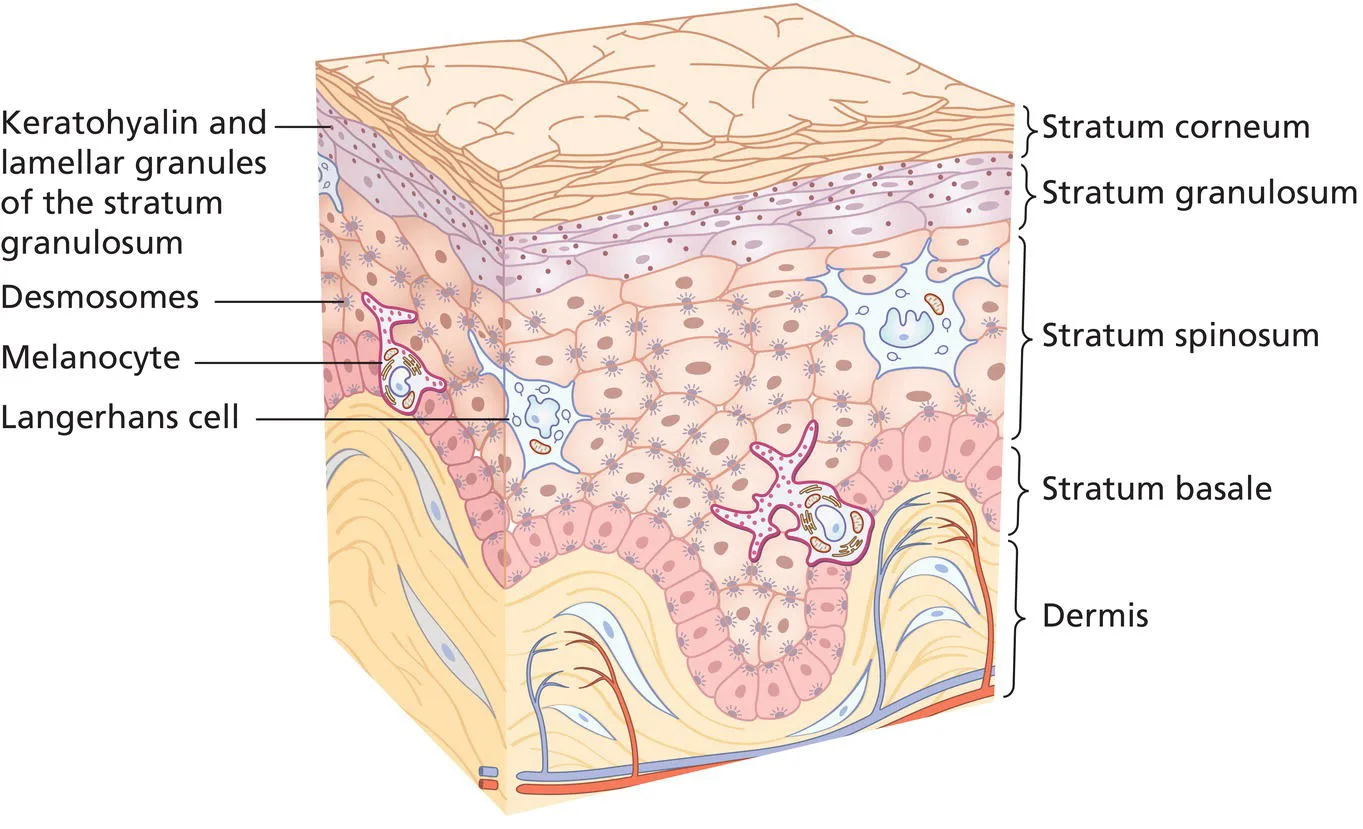 Fig 1. Diagram of the epidermis — stratum corneum, granulosum, spinosum, basale over the dermis, with keratohyalin and lamellar granules, desmosomes, a basal melanocyte and a Langerhans cell — (Draelos, 2009, p. 77).
> Sources: Draelos — Cosmetic Dermatology [2009]
Fig 1. Diagram of the epidermis — stratum corneum, granulosum, spinosum, basale over the dermis, with keratohyalin and lamellar granules, desmosomes, a basal melanocyte and a Langerhans cell — (Draelos, 2009, p. 77).
> Sources: Draelos — Cosmetic Dermatology [2009] [C][MEDLIB] [1].
The barrier is brick-and-mortar, not a film. Corneocytes (bricks) are embedded in a lamellar lipid mortar whose three pillars are ceramides, cholesterol and free fatty acids; deplete any one and transepidermal water loss (TEWL) rises [1][6]. Three properties matter clinically:
- Acid mantle, pH 4.5–5.5 [5]. The acidic surface is not decoration: it activates the lipid-processing enzymes (β-glucocerebrosidase, acid sphingomyelinase) that mature the mortar, and it restrains pathogen overgrowth. Alkaline soaps and chronic antiseptics push pH up and prolong barrier recovery.
- Antimicrobial peptides (AMPs) — cathelicidin (LL-37), β-defensins — are secreted into the same lipid matrix; they are part of the barrier, not an accessory [5]. Skin immune competence develops over early life and remains sensitive to how the barrier is treated in adulthood [4].
- Natural moisturizing factor (NMF) — filaggrin-derived amino acids, urea, lactate, PCA — holds water in the corneocyte. Filaggrin loss-of-function (the atopic phenotype) is the archetypal reason a patient stings with every active.
Dermo-epidermal junction (DEJ). The basement membrane (type IV/VII collagen, laminin) anchors epidermis to dermis through interdigitating rete ridges and dermal papillae. With age the papillae flatten, which cuts the exchange surface, makes the skin more fragile and shearable, and yields poorer graft take and slower wound healing [1][8]. This flattening is the histological reason older skin tolerates a deep peel less well — the same wounding depth reaches a thinner, less vascular interface.
Keratinocyte biology in one line: a basal stem cell divides, its daughter commits and migrates upward, expresses successive keratins (K5/K14 basal → K1/K10 suprabasal), extrudes its lipids at the granular layer, then dies into a corneocyte. Every resurfacing device and every peel is a controlled acceleration of exactly this program.
Consensus: barrier integrity is assessed and, if defective, repaired before any procedure — across cosmeceutical, laser and peeling schools [1][2][6]. Discrepancy: how long to repair — 2 weeks (device-forward practices) vs 4 weeks (cosmeceutical-forward). Decide by the clinical signs (scaling, stinging, baseline erythema), not the calendar [2].
Corneocyte and cornified envelope — the brick in detail. Terminal differentiation replaces the plasma membrane with a cornified envelope of cross-linked structural proteins (involucrin, loricrin, small proline-rich proteins) sealed by transglutaminase-1; loss-of-function in transglutaminase-1 is lamellar ichthyosis, the extreme of a defective envelope. Corneocytes are held by corneodesmosomes whose ordered proteolysis (kallikreins KLK5/KLK7, restrained by the LEKTI inhibitor) drives invisible daily desquamation; the same proteases are pH-sensitive, which is the second reason surface pH is load-bearing. Chemical peels are, mechanistically, an acceleration of corneodesmosome cleavage and controlled coagulation to a chosen depth.
Non-keratinocyte residents — density and role.
| Cell | Location | Density / ratio | Function | Aesthetic relevance |
|---|---|---|---|---|
| Melanocyte | Basal | ≈ 1:10 basal keratinocytes; 1 per ≈ 36 keratinocytes served | Melanin synthesis and transfer | Pigment disorders (A2.5); density fairly constant across races, activity differs |
| Langerhans cell | Suprabasal/spinous | ≈ 2–4 % of epidermal cells | Antigen presentation | Depleted transiently by UV → local immunosuppression, part of photocarcinogenesis |
| Merkel cell | Basal, touch-rich sites | Sparse | Mechanoreception (slow-adapting) | Fine touch; not a target, but the origin of Merkel-cell carcinoma |
| Resident T cells | Epidermis + dermis | ~2× the T cells of blood | Immune surveillance/memory | Basis of the barrier's immune arm [4] |
Keratin map — the differentiation clock made molecular. Basal cells express K5/K14; on suprabasal commitment they switch to K1/K10; hyperproliferative or wounded epidermis expresses K6/K16/K17. A device or peel that "resurfaces" is forcing basal K5/K14 cells to repopulate and re-run this program; the visible re-epithelialization tracks that molecular switch.
Regional thickness governs procedure depth. Epidermis runs from ≈ 0.05 mm on the eyelid to ≈ 0.8–1.5 mm on palms/soles; total skin from ≈ 0.5 mm (eyelid) to > 4 mm (back). The same laser fluence or peel concentration reaches a different fraction of the dermis by region — the biological reason a peri-ocular peel is not a back peel at the same strength.
Penetration routes — why an "active" may do nothing. A topical crosses the SC by the intercellular lipid route (dominant for most actives), the transcellular route, or the transappendageal (follicular/sweat) shunt. Permeation follows Fick's law: flux rises with concentration gradient, lipophilicity (log P ~1–3 optimal) and a small molecular weight (the "500 Dalton rule" of thumb). This is why an intact, healthy barrier legitimately limits penetration — and why a disrupted one over-absorbs and stings.
Barrier metrics that make the assessment objective: TEWL (normal facial ≈ 6–15 g/m²/h; rises with barrier defect), corneometry (capacitance hydration), surface pH (4.5–5.5), and sebumetry. These convert "sensitive skin" from a complaint into a measurement and gate the go/no-go for a procedure.
DEJ in detail. The junction is a layered structure: keratinocyte hemidesmosomes (integrin α6β4, BP180/BP230) → lamina lucida (laminin-332) → lamina densa (type IV collagen) → anchoring fibrils (type VII collagen) into the papillary dermis. Autoimmune or genetic failure of these proteins causes the blistering diseases (bullous pemphigoid, epidermolysis bullosa) — the clinical proof of how much mechanical and exchange function this thin interface carries, and why its age-related flattening degrades graft take and healing [1][8].
Appendages are the healing reserve, not decoration. Eccrine glands (thermoregulatory, ubiquitous), apocrine glands (axilla/anogenital), sebaceous glands (holocrine, sebum, densest on face/scalp) and hair follicles all open through the epidermis. The hair follicle is a stem-cell niche (bulge) and the principal source of keratinocytes that re-surface a partial-thickness wound — the biological reason facial skin (follicle-dense) re-epithelializes faster than follicle-poor sites and why deep resurfacing that destroys adnexa heals as a scar, not as skin. Sebum contributes squalene, wax esters and free fatty acids to the surface film and feeds the lipophilic microbiota (A2.6).
What a moisturizer actually does — three mechanism classes.
| Class | Examples | Mechanism | Use |
|---|---|---|---|
| Occlusive | Petrolatum, dimethicone, lanolin | Physically block TEWL (petrolatum ↓ TEWL > 98 %) | Acute barrier repair, post-procedure |
| Humectant | Glycerin, hyaluronic acid, urea, PCA | Draw water into the SC (mimic NMF) | Hydration; can worsen if used alone in dry air |
| Emollient/lipid | Ceramides, cholesterol, fatty acids | Replace the mortar; a physiologic 3:1:1 ratio speeds repair | Barrier restoration [6] |
Barrier repair (the 2–4 week pre-procedure step) is exactly this: reduce TEWL with occlusion, refill the corneocyte with humectant, and replace the deficient lipid pillars.
Aging of the epidermis and barrier — quantified.
| Feature | Young | Aged | Consequence |
|---|---|---|---|
| Turnover | ≈ 28 days | Slows (↑ ~30–50 %) | Dull, retained corneocytes; slower peel recovery |
| DEJ | Interdigitated rete/papillae | Flattened | Fragility, shear, poorer graft take [8] |
| SC lipids / NMF | Replete | ↓ | Xerosis, higher baseline TEWL |
| Surface pH | 4.5–5.5 | Rises toward neutral | Impaired enzyme function, dysbiosis risk |
| Langerhans density | Normal | ↓ | Reduced immune surveillance |
Consensus (added): every energy/peel plan is calibrated to regional epidermal/dermal thickness and to an assessed barrier, not to a single machine setting [1][18].
Peel depth is defined by which of these layers is reached — the layer map is the peel map:
| Peel depth | Layer reached | Typical agent (example) |
|---|---|---|
| Very superficial | Stratum corneum | Low-strength AHA, salicylic acid |
| Superficial | Epidermis to basal layer | Glycolic 20–70 %, Jessner, TCA 10–20 % |
| Medium | Papillary → upper reticular dermis | TCA 35 %, TCA + Jessner |
| Deep | Mid-reticular dermis | Phenol-croton oil |
Depth chosen determines healing time and complication profile (A2.7): the deeper the wound relative to the layers above, the longer remodeling and the higher the PIH/scar risk — the direct clinical payoff of knowing the architecture.
The epidermis is an endocrine and delivery organ, not only a wall. Keratinocytes perform the first step of vitamin D synthesis (7-dehydrocholesterol → previtamin D3 under UVB) — the physiologic counterweight to photoprotection, answered in A2.4 by supplementation rather than sun. The barrier is also the surface every procedure manipulates for delivery: microneedling and ablative fractional laser transiently create microchannels that raise permeation of applied actives by orders of magnitude (laser/needle-assisted drug delivery), which is a benefit (efficacy) and a risk (uncontrolled absorption, granuloma from the wrong product) at once. The same barrier whose integrity limits a serum is deliberately breached to make a procedure work — and must then be repaired.
Barrier phenotypes to classify before choosing aggressiveness.
| Phenotype | Signs | Procedure readiness |
|---|---|---|
| Healthy barrier | No scaling/stinging, normal TEWL | Full range per indication |
| Atopic/sensitive (filaggrin-low) | Stinging, xerosis, flexural history | Repair 2–4 wk; conservative, patch test |
| Rosacea-barrier | Flushing, baseline erythema, burning | Barrier + rosacea therapy first; avoid heat |
| Oily/seborrheic | High sebumetry, comedones | Watch post-procedure acne/folliculitis (A2.6) |
| Photodamaged-thin/atrophic | Thin, fragile, telangiectasia | Slow healing; match tool to histology (A2.3/A2.4) |
| Acutely disrupted (post-procedure/over-actives) | Scaling, stinging, ↑TEWL | No new insult until repaired |
Classifying the barrier converts "the patient wants a peel today" into a defensible go/no-go.
Trampa clásica: reading a stinging, scaling face as "sensitive skin needing a stronger active." It is a disrupted barrier; adding acids or retinoid raises TEWL further and converts irritation into a post-inflammatory problem. Signature: burning on application, worsening erythema, no improvement over weeks. Repair first (2–4 weeks emollients, gentle low-pH cleansing), then reintroduce actives.
A2.2 · Dermis: collagen types I/III, elastin, glycosaminoglycans, and ECM remodeling
| Component | Proportion / role | Change with age | Aesthetic lever |
|---|---|---|---|
| Type I collagen | ≈ 80 % of dermal collagen; tensile strength [1][7] | ↓ ≈ 1 %/year from decade 3 ⚠ [8] | The target of biostimulation and resurfacing |
| Type III collagen | ≈ 15 %; fine "young"/reticular collagen [1] | Relative proportion rises in repaired tissue → scar | Excess III = immature repair, not a goal |
| Elastin / oxytalan | Elastic recoil; fibrillin scaffold | Not usefully re-synthesized in adults; loss ≈ permanent | Prevention (photoprotection) beats correction |
| GAGs / hyaluronic acid | Water binding, dermal turgor and volume | Falls markedly | Rationale for skin boosters A3 — Ácido hialurónico: bioquímica, reticulación y reología.es |
| Fibroblast | Synthesizes all of the above + MMP/TIMP | Replicative senescence → less synthesis, more MMP [8][12] | The cell every device tries to re-activate |
| Ground substance | Proteoglycans (decorin, versican) | Disorganized in photoaging | Governs fiber spacing and hydration |
The type I : type III ratio is the marker of repair quality, not the amount of collagen [1][7]. Tissue rich in type III is scar-like; the goal of biostimulation is mature, cross-linked, well-oriented type I. This distinction runs through the whole atlas: A4 — Bioestimuladores: ciencia de materiales.es is the materials science of driving that ratio the right way. Fig 2 shows the normal papillary/reticular architecture the treatments try to restore; Fig 3 schematizes the fibroblast-driven assembly of collagen the biostimulators aim to re-induce.
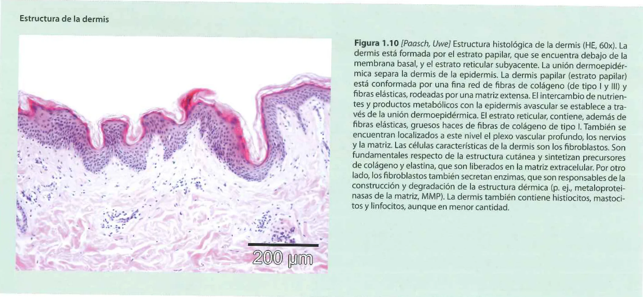 Fig 2. Histologic structure of the dermis (H&E, 60×): papillary dermis under the basement membrane (fine type I/III collagen and elastic fibers) over reticular dermis (thick type I bundles); the fibroblast is the resident synthetic cell, also the source of MMPs — (Rubin, Atlas Ilustrado de peeling, Fig 1.10).
> Sources: Atlas Ilustrado de peeling — Mark G. Rubin
Fig 2. Histologic structure of the dermis (H&E, 60×): papillary dermis under the basement membrane (fine type I/III collagen and elastic fibers) over reticular dermis (thick type I bundles); the fibroblast is the resident synthetic cell, also the source of MMPs — (Rubin, Atlas Ilustrado de peeling, Fig 1.10).
> Sources: Atlas Ilustrado de peeling — Mark G. Rubin [C][MEDLIB] [11].
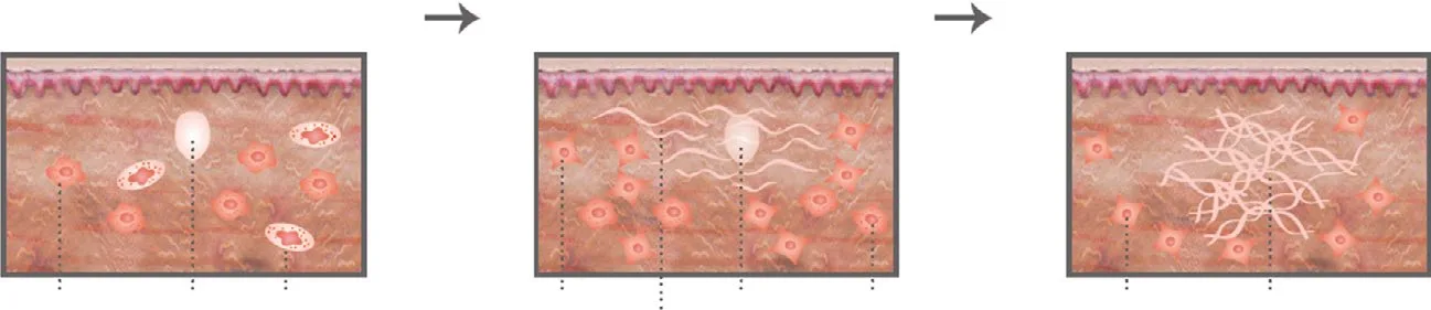 Fig 3. The process of collagen formation and the role of resident cells and proteins in skin over time — progressive fiber accumulation in the dermal matrix — (Piccolo, 2025, p. 122).
> Sources: Piccolo — A.R.T. Autologous Regenerative Therapy in Aesthetic Medicine [2025]
Fig 3. The process of collagen formation and the role of resident cells and proteins in skin over time — progressive fiber accumulation in the dermal matrix — (Piccolo, 2025, p. 122).
> Sources: Piccolo — A.R.T. Autologous Regenerative Therapy in Aesthetic Medicine [2025] [C][MEDLIB] [10].
Collagen homeostasis is a balance of synthesis and degradation, and both drift with age [8][9]. On the synthesis side, fibroblasts in aged and photoaged skin over-express CCN1 (CYR61), which suppresses type I procollagen and induces collagenase (MMP-1); retinoic acid reverses this — it reduces CCN1, raises procollagen and lowers MMP-1 — which is the molecular reason topical retinoids improve both chronologically aged and photoaged skin [9]. On the degradation side, fibroblast senescence tilts the MMP/TIMP balance toward proteolysis [8][12]. The dermis a treatment meets is therefore not a static scaffold but a slowly self-digesting one; the therapeutic goal is to shift the equilibrium toward organized synthesis.
Elastin is the asymmetry that governs strategy. Type I collagen turns over and can be re-induced; the mature elastic network cannot be usefully rebuilt in an adult, and UV-driven elastotic material is disorganized, not functional [8][18]. That asymmetry is why photoprotection is treatment and not advice: it prevents an essentially irreversible loss.
GAGs and the water compartment. Hyaluronic acid binds up to ~1000× its weight in water; its decline flattens dermal turgor and contributes to the "crepey" quality of aged skin. Skin boosters and some biostimulators act partly by restoring this water-binding compartment and partly by mechanically stimulating fibroblasts [7][10].
Consensus: the outcome that matters for any collagen-inducing procedure is organized type I collagen, judged over months, not fiber quantity at weeks [1][7][27]. Discrepancy: whether elastin can be meaningfully restored — marketing claims of "elastin regeneration" vs the histologic evidence that adult elastogenesis is minimal. Decide against the claim: promise collagen and hydration, frame elastic laxity as largely preventable [8].
The collagen family is more than I and III. Each type has a location and a failure signature the clinician meets:
| Collagen | Location | Role | Clinical correlate |
|---|---|---|---|
| I | Reticular dermis (≈ 80 %) | Tensile strength | Target of biostimulation/resurfacing; falls with age |
| III | Papillary dermis, early repair (≈ 15 %) | Compliance, scaffolding | Excess = immature scar; high in fetal/early wounds |
| IV | Basement membrane (lamina densa) | DEJ integrity | Breached in melasma → dermal pigment drop [21] |
| VII | Anchoring fibrils | Epidermis-dermis anchorage | Failure = epidermolysis bullosa; UV-sensitive |
| V / VI | Interstitial, fibril regulation | Fibril diameter, spacing | Microfibrillar organization, aging disarray |
| XVII (BP180) | Hemidesmosome | Adhesion | Bullous pemphigoid antigen |
Collagen synthesis is a vitamin-C-dependent assembly line. Fibroblasts transcribe pro-α chains → prolyl/lysyl hydroxylation (requires ascorbate as co-factor — the biochemical basis of topical vitamin C in anti-aging) → triple-helix procollagen → secretion → cleavage to tropocollagen → self-assembly into fibrils → lysyl-oxidase (copper-dependent) cross-linking into mature fibers. Two clinical corollaries: ascorbate deficiency (scurvy) collapses synthesis, and pathological over-cross-linking (glycation, A2.3) stiffens the network [15].
The elastic fiber system is three tiers, not one. Oxytalan (fibrillin microfibrils, superficial/vertical in papillary dermis) → elaunin (elastin-poor, mid) → mature elastin fibers (reticular, horizontal). Photoaging degrades this into amorphous solar elastosis (A2.4); intrinsic aging thins and fragments it. Adult elastogenesis is minimal, which is the asymmetry that makes elastic laxity largely a prevention problem, not a correction one [8].
Ground substance and GAGs — the water and spacing compartment.
| GAG / proteoglycan | Function | Age change |
|---|---|---|
| Hyaluronic acid | Binds ~1000× its weight in water; volume/turgor | Falls markedly → crepey skin; rationale for boosters A3 — Ácido hialurónico: bioquímica, reticulación y reología.es |
| Dermatan / chondroitin sulfate | Fiber hydration and spacing | Redistributed/disorganized |
| Decorin | Regulates collagen fibril diameter | Falls; fragmented decorin in aged dermis |
| Versican / biglycan | Elastic-fiber assembly, hydration | Rises in elastotic (photoaged) dermis |
The MMP/TIMP balance is the degradation dial.
| Enzyme | Substrate | Trigger |
|---|---|---|
| MMP-1 (collagenase-1) | Type I/III collagen | UV/AP-1, CCN1, senescence [8][9] |
| MMP-2, MMP-9 (gelatinases) | Denatured collagen, type IV BM | UV, inflammation |
| MMP-3 (stromelysin-1) | Proteoglycans, activates pro-MMP-1 | UV/AP-1 |
| TIMP-1..4 | Inhibit MMPs | Fall relatively with age → net proteolysis |
Fibroblast heterogeneity matters for outcome. Papillary fibroblasts (superficial) and reticular fibroblasts (deep) are distinct populations; the papillary population, which maintains the fine superficial matrix and the DEJ, is preferentially lost with age. This is a mechanistic reason superficial skin quality is hard to restore and why biostimulators aim to repopulate and re-activate fibroblasts, not merely add filler volume [8][10]. Replicative senescence (A2.3) reduces synthetic capacity and tilts these cells toward the SASP, raising MMP output [12].
The aging dermis — quantified changes that justify each tool.
| Change with age | Direction | Aesthetic correlate |
|---|---|---|
| Dermal thickness | ↓ (thinning) | Fragility, transparency, visible vessels |
| Type I collagen | ↓ ≈ 1 %/year from decade 3 ⚠ [8] | Loss of firmness |
| Fibroblast number/synthesis | ↓ | Reduced repair capacity |
| MMP activity | ↑ (net proteolysis) | Ongoing matrix loss [8] |
| Elastic network | Fragmented (intrinsic) / elastotic (extrinsic) | Laxity, texture change |
| Hyaluronic acid / water | ↓ | Crepey, dehydrated quality |
| Dermal microvasculature | Rarefied | Pallor, slower healing, cooler skin |
| Subcutaneous fat compartments | Redistributed/atrophic | Volume loss, deepened folds (A1 — Anatomía facial y regional.es) |
How each modality is meant to move the type I : type III ratio the right way — every device in the atlas is, biologically, a fibroblast stimulus or a controlled wound aimed at organized type I:
| Modality | Primary stimulus | Dominant remodeling driver |
|---|---|---|
| Topical retinoid | ↓CCN1, ↑procollagen, ↓MMP-1 [9] | Molecular re-tuning without wounding |
| Microneedling | Micro-injury, PDGF/TGF-β release | Proliferation → remodeling [27] |
| Radiofrequency / HIFU | Dermal thermal coagulation | Neocollagenesis over months |
| Non-ablative/ablative laser | Controlled wound to a chosen depth | Full healing cascade (A2.7) |
| Biostimulators (PLLA, CaHA) | Foreign-body/fibroblast stimulation | Type I neocollagenesis [10] (A4 — Bioestimuladores: ciencia de materiales.es) |
All share one clock: the result is remodeling-dependent and read at 6–12 months, not at the inflammatory peak [27].
Consensus (added): topical vitamin C (ascorbate) is grounded in its role as a hydroxylation co-factor, not marketing; it supports the same synthesis step every biostimulator depends on [6][9]. Discrepancy (added): reticular vs papillary targeting — deep dermal remodeling devices vs superficial skin-quality approaches. Decide by whether the deficit is structural laxity (deep) or fine texture/DEJ quality (superficial) [8].
Hyaluronic acid turnover — fast, and why that matters for boosters. Dermal hyaluronan turns over rapidly (half-life of hours to ~1–2 days for native HA), degraded by hyaluronidases and free radicals. Native (unmodified) HA in a skin booster is therefore transient hydration, while its fibroblast-mechanostimulation and the water compartment it restores are the durable levers — the biochemical reason cross-linking (the subject of A3 — Ácido hialurónico: bioquímica, reticulación y reología.es) exists and why "more HA" is not the same as "lasting volume." Decorin and the small proteoglycans set collagen fibril diameter and spacing; their loss with age is part of why aged dermis both holds less water and organizes fibers less well [7].
Normal dermis vs scar — the matrix comparison that defines the goal.
| Feature | Normal dermis | Scar |
|---|---|---|
| Collagen orientation | Basket-weave, multidirectional | Parallel, aligned to tension |
| Type I : III | High I | Relatively high III (immature) |
| Elastic fibers | Present, organized | Reduced/absent |
| Vascularity | Normal plexus | Initially hypervascular (red) → pale |
| Adnexa (follicles, glands) | Present | Absent (why deep wounds scar) |
| Cellularity | Low, quiescent fibroblasts | High early; myofibroblasts contract |
Every collagen-inducing tool is aiming for the left column — organized, multidirectional type I with restored elastic and vascular components — not merely for density. Mechanical tension across a healing dermis biases it toward the right column (aligned, hypertrophic collagen), which is why tension-off techniques and support matter in scar outcome (A2.7).
Trampa clásica: promising "more collagen" and showing an early result. Early density is edema and type III (inflammatory repair), not the mature type I that carries the cosmetic benefit. Signature: impressive week-4 photo that regresses by month 3. Consent to a 6–12 month horizon and photograph on that schedule.
A2.3 · Intrinsic aging: genetics, telomere biology, cellular senescence, and hormonal influences
Intrinsic (chronological) aging is programmed, oxidative-metabolism driven, and uniform across the whole body surface — which is why it is studied in sun-protected skin (inner arm, buttock) [8][13]. Its clinical signature is thin, dry, lax skin with fine wrinkles, without marked dyschromia and without elastosis. It responds to biostimulation and hydration and tolerates aggressive resurfacing poorly.
| Driver | Mechanism | Clinical/therapeutic read |
|---|---|---|
| Genetics / skin type | Constitutive pigment, fibroblast reserve, filaggrin status | Sets the ceiling of what any protocol can achieve |
| Telomere attrition | Shortening per division; replicative limit in fibroblasts/keratinocytes [16] | Conceptual basis of "cellular clock"; a biomarker, not yet a lever |
| Cellular senescence + SASP | Growth-arrested cells secrete IL-6, MMPs, ROS — pro-inflammatory, not dead [12][13] | Basis of senolysis; links to K1 — Anti-Aging Medicine & Longevity.es |
| Inflammaging | Chronic low-grade inflammation as a shared aging driver [13] | Systemic angle: diet, metabolic control, sleep |
| Hormonal (estrogen) | Post-menopausal 17β-estradiol fall → ↓collagen, ↓GAG, ↑MMP, ↓fibroblast function [14] | Accelerated early window; topical/systemic estrogen debated |
| Glycation (AGEs) | Non-enzymatic collagen–sugar cross-links → rigid, yellow, remodeling-resistant fibers [15] | Links diet/diabetes to skin quality |
Cellular senescence is the unifying mechanism. A senescent cell does not die; it arrests and turns pro-inflammatory, expressing the senescence-associated secretory phenotype (SASP) — IL-6, IL-8, MMPs and ROS — and its chronic accumulation degrades the surrounding matrix and recruits more senescence [12][13]. The most reliable single marker distinguishing a senescent cell from a merely cycle-arrested one is senescence-associated β-galactosidase (SA-β-gal); a persistent DNA-damage response and SASP/ROS accumulation are the accompanying hallmarks [12]. Senotherapeutics that reduce SASP (quercetin, fisetin, naringenin, apigenin in preclinical work) are promising but not yet a validated clinical protocol for skin — the honest current status is target identified, translation pending [12].
Fig 4 contrasts the two aging arms at the biochemical level — the single most useful schematic in this chapter, because it shows where they converge: extrinsic (solar/environmental) drives elastase inhibition and abnormal elastin deposition toward elastosis, intrinsic (oxidative metabolism) drives ROS → MAPK (AP-1/NF-κB) → MMP activation, and both suppress TGF-β signaling and collagen synthesis, so both end in reduction of dermal matrix components.
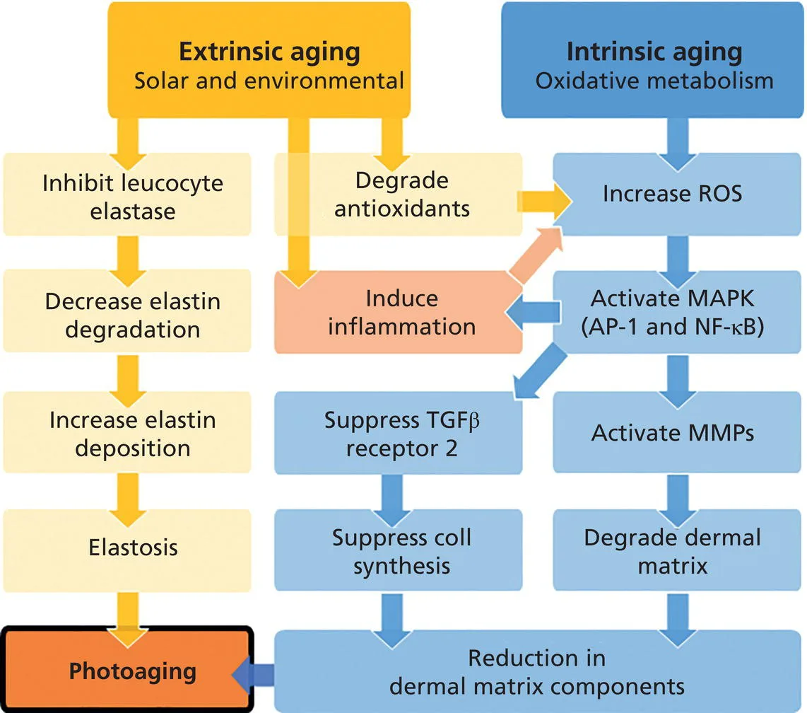 Fig 4. Extrinsic aging (solar/environmental) → inhibit leucocyte elastase, alter elastin turnover → elastosis; intrinsic aging (oxidative metabolism) → ↑ROS → activate MAPK (AP-1, NF-κB) → activate MMPs → degrade dermal matrix; both induce inflammation, suppress TGF-β receptor 2 and collagen synthesis → reduction in dermal matrix components — (Draelos, 2009, p. 1119).
> Sources: Draelos — Cosmetic Dermatology [2009]
Fig 4. Extrinsic aging (solar/environmental) → inhibit leucocyte elastase, alter elastin turnover → elastosis; intrinsic aging (oxidative metabolism) → ↑ROS → activate MAPK (AP-1, NF-κB) → activate MMPs → degrade dermal matrix; both induce inflammation, suppress TGF-β receptor 2 and collagen synthesis → reduction in dermal matrix components — (Draelos, 2009, p. 1119).
> Sources: Draelos — Cosmetic Dermatology [2009] [C][MEDLIB] [1].
Telomeres and the "aging clock." Telomere length shortens with replicative age and correlates with biological aging; a systematic review of physical activity and telomere length confirms the association but not a simple causal dose that a clinician can prescribe [16]. Treat telomere length as a research biomarker, not a chairside target.
Hormonal aging is the one intrinsic driver with a treatable window. Estrogen loss after menopause accelerates collagen loss, reduces GAGs and dermal vascularity, raises MMP activity and impairs wound healing and barrier function [14]. The figure that circulates — ≈ 30 % of dermal collagen lost in the first 5 post-menopausal years ⚠ — comes from small older series and is repeated without its confidence interval; use it as a magnitude for counseling, not as a point estimate [14]. Topical estrogen reverses several markers, but the skin effect of current low-dose systemic hormone therapy is not well established, and prescribing hormones for a skin indication is outside aesthetic scope [14].
Glycation is the dietary/metabolic link. Reducing sugars cross-link dermal collagen into advanced glycation end products (AGEs) that are stiff, yellow-tinged and resistant to remodeling; AGE accumulation rises with age and is amplified by UV, and it stiffens exactly the type I network that biostimulation is trying to rebuild [15]. This is the mechanistic bridge between glycemic control/diabetes and measurable skin quality, and the rationale for anti-glycation actives — though clinical anti-glycation efficacy in skin is still early [15].
Consensus: intrinsic aging is managed by biostimulation, hydration, photoprotection and metabolic optimization; it is not a resurfacing indication in the absence of elastosis [1][8][17]. Discrepancy: systemic vs topical anti-aging — the longevity/inflammaging school (senolysis, metabolic control [13]) vs the classic dermatologic school (topical retinoid + photoprotection [8]). These are complementary, not exclusive; decide by the patient's systemic risk profile and expectations.
The hallmarks of aging, read in the skin. The 2023 update expanded the canonical list to include chronic inflammation, dysbiosis and disabled macroautophagy alongside genomic instability, telomere attrition, epigenetic alteration, loss of proteostasis, deregulated nutrient sensing, mitochondrial dysfunction, cellular senescence, stem-cell exhaustion and altered intercellular communication [13]. Skin is a visible readout of several at once: telomere attrition and senescence in fibroblasts/keratinocytes, stem-cell exhaustion (fewer papillary fibroblasts, slower turnover), mitochondrial ROS, and inflammaging as the connective theme [8][12][13].
Telomere and telomerase biology. Telomeres are TTAGGG repeats capping chromosomes; each division shortens them (end-replication problem) until a critical length triggers replicative senescence via p53/p21 and p16^INK4a. Somatic skin cells have low telomerase, so this clock runs; UV accelerates attrition through oxidative single-strand breaks. Length correlates with biological age and lifestyle (physical activity, smoking) but the association is not a prescribable dose, which is why it stays a research biomarker, not a chairside target [16].
Senescence has three doors, one exit. Triggers: replicative (telomere), oncogene-induced, and stress-induced premature senescence (UV, ROS, therapy). The exit is a stable growth arrest with a persistent DNA-damage response and the SASP. SASP mediators degrade matrix and spread senescence to neighbours (bystander effect):
| SASP class | Examples | Skin consequence |
|---|---|---|
| Pro-inflammatory cytokines | IL-6, IL-8, IL-1β | Inflammaging, matrix breakdown |
| Proteases | MMP-1, -3 | Collagen/elastin degradation |
| Growth factors/chemokines | GRO, VEGF, CCN1 | Aberrant remodeling, ↓procollagen [9] |
| Reactive species | ROS | Oxidative feed-forward |
Best single discriminator of a senescent (vs cycle-arrested) cell: SA-β-galactosidase; supporting hallmarks are the persistent DDR and SASP/ROS accumulation [12].
The endocrine axis is broader than estrogen.
| Hormone | Skin effect of decline/excess | Aesthetic read |
|---|---|---|
| Estrogen ↓ (menopause) | ↓collagen, ↓GAG, ↓vascularity, ↑MMP, impaired healing/barrier [14] | The one treatable intrinsic window; topical estrogen reverses markers, systemic effect unproven |
| Androgens | Sebum, follicle behaviour; relative androgen dominance post-menopause | Sebaceous/acneiform shift; hair patterning |
| Thyroid ↓ | Dry, coarse, cool skin; myxedema | Screen unexplained xerosis |
| GH/IGF-1 ↓ (somatopause) | Reduced dermal thickness | Longevity angle; not a skin prescription |
| Cortisol ↑ (chronic stress/steroids) | Collagen catabolism, atrophy, delayed healing | Iatrogenic dermal thinning from topical steroids |
The circulating figure — ≈ 30 % of dermal collagen lost in the first 5 post-menopausal years ⚠ — is a magnitude from small older series without its confidence interval; counsel with it, do not cite it as a point estimate [14].
Glycation is measurable. The Maillard reaction cross-links collagen into AGEs (carboxymethyl-lysine, pentosidine, glucosepane) that fluoresce; skin autofluorescence is a validated non-invasive AGE surrogate. AGEs stiffen and yellow the type I network, resist MMP remodeling, and accumulate faster with hyperglycemia and UV — the bridge from glycemic control to skin quality and the rationale for anti-glycation actives (whose clinical efficacy is still early) [15].
Progeroid syndromes are the natural experiments. Hutchinson-Gilford progeria (LMNA/progerin) and Werner syndrome (WRN helicase) accelerate several hallmarks and produce premature cutaneous aging, sclerodermoid change and lipodystrophy — extreme models that confirm the genomic-instability/senescence axis drives skin aging [8][13].
Intrinsic vs extrinsic — the side-by-side that decides the tool (bridges to A2.4).
| Axis | Intrinsic (chronological) | Extrinsic (photoaging) |
|---|---|---|
| Distribution | Whole body, uniform | Sun-exposed only |
| Wrinkles | Fine, at rest | Coarse, deep, geometric |
| Color | Pale, even | Mottled dyschromia, lentigines, telangiectasia |
| Dermis | Atrophic, thin | Solar elastosis (accumulated) |
| Epidermis | Thin, flat DEJ | Atypia, dysmaturation, keratoses |
| Dominant biology | Senescence, telomere, hormones, glycation | ROS → AP-1 → MMP [8] |
| Responds best to | Biostimulation, hydration, hormonal review | Resurfacing, energy, pigment-directed care |
| Reversible? | Partially (retinoid, biostimulation) | Prevention >> correction (elastin) |
Modifiable vs non-modifiable intrinsic drivers — where counseling has leverage.
| Non-modifiable | Modifiable (lifestyle levers) |
|---|---|
| Chromosomal/genetic set-point | Glycemic control (↓ glycation/AGE) [15] |
| Telomere baseline | Smoking cessation (↓ MMP, ↑ perfusion) |
| Skin phototype | Sleep, chronic stress (↓ cortisol catabolism) |
| Menopause timing | Physical activity (telomere association) [16] |
| Progeroid genetics | Nutrition (protein, vitamin C, antioxidants) |
The clinical value of separating these is honest expectation-setting: the non-modifiable ceiling is real, and the modifiable levers are systemic, slow and additive to (not a substitute for) topical/procedural care [13][16].
Consensus (added): intrinsic aging is a systemic-plus-topical management problem — photoprotection, retinoid, hydration, metabolic/glycemic optimization and (where indicated) hormonal review; not a wounding indication absent elastosis [8][14][17]. Discrepancy (added): senolytics for skin — the longevity school (SASP reduction as a future lever [12][13]) vs the position that no skin-specific senolytic protocol is validated. Decide against clinical use for now; the target is real, the protocol is not [12].
Oxidative-stress and mitochondrial theory — the shared engine. Intrinsic aging is driven substantially by endogenous reactive oxygen species from oxidative metabolism; mitochondrial DNA (lacking histone protection and efficient repair) accumulates damage, raising ROS in a feed-forward loop that activates the same MAPK/AP-1 → MMP axis that UV drives extrinsically (Fig 4). This convergence is why antioxidant strategies are argued for both aging arms and why the intrinsic/extrinsic split is about where damage starts, not about two unrelated pathways [8][13].
Trampa clásica: attacking intrinsically aged, atrophic, dyschromia-free skin with an aggressive ablative resurfacing "because it looks old." Atrophic skin with a flattened DEJ heals slowly and scars more readily; the indication was biostimulation and volume, not wounding. Signature: prolonged erythema and delayed healing after a treatment that was never matched to the histology.
A2.4 · Extrinsic aging and photoaging: UVA/UVB, visible/infrared radiation, ROS, AP-1, and MMPs
| Radiation | Wavelength | Penetration | Dominant damage |
|---|---|---|---|
| UVB | 280–320 nm | Epidermis | Direct DNA lesions (cyclobutane pyrimidine dimers) → mutation, erythema, burn, carcinogenesis [18] |
| UVA | 320–400 nm | Reticular dermis | ROS → indirect DNA/matrix damage; elastosis; immediate pigment darkening; passes through window glass [18] |
| Visible light / HEV | 400–500 nm | Dermis | Persistent pigmentation in Fitzpatrick IV–VI; drives melasma — a UV-only filter does not protect it [20] |
| Infrared-A | 760–1440 nm | Deep dermis/subcutis | Thermal/oxidative stress, MMP-1 induction [19] |
The cascade, in one line: UV → ROS → activation of AP-1 and NF-κB → ↑MMP-1, MMP-3, MMP-9 → collagen degradation → imperfect repair → accumulated solar scarring [8][18]. In parallel, AP-1 suppresses TGF-β signaling and type I procollagen synthesis, so the skin degrades more and builds less at the same time [9]. That dual action is why photodamage is cumulative and cannot be compensated by synthesis. Fig 5 shows the membrane-to-nucleus half of that loop; Fig 6 shows the acute-to-chronic tissue consequence.
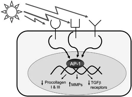 Fig 5. UV irradiation induces membrane signaling that activates AP-1 in the nucleus → ↓ procollagen I & III, ↑ MMPs, ↓ TGF-β receptors — the transcriptional switch of photoaging — (Lim, Photodermatology, 2007, p. 114).
> Sources: Lim — Photodermatology [2007]
Fig 5. UV irradiation induces membrane signaling that activates AP-1 in the nucleus → ↓ procollagen I & III, ↑ MMPs, ↓ TGF-β receptors — the transcriptional switch of photoaging — (Lim, Photodermatology, 2007, p. 114).
> Sources: Lim — Photodermatology [2007] [C][MEDLIB] [18].
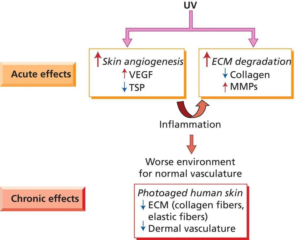 Fig 6. Acute effects of UV — ↑ skin angiogenesis (↑VEGF, ↓TSP) and ↑ ECM degradation (↓collagen, ↑MMPs) → inflammation → chronic photoaged skin with ↓ECM (collagen and elastic fibers) and ↓ dermal vasculature — (Draelos, 2009, p. 126).
> Sources: Draelos — Cosmetic Dermatology [2009]
Fig 6. Acute effects of UV — ↑ skin angiogenesis (↑VEGF, ↓TSP) and ↑ ECM degradation (↓collagen, ↑MMPs) → inflammation → chronic photoaged skin with ↓ECM (collagen and elastic fibers) and ↓ dermal vasculature — (Draelos, 2009, p. 126).
> Sources: Draelos — Cosmetic Dermatology [2009] [C][MEDLIB] [1].
Histology of photoaging is distinct from intrinsic aging: solar elastosis (amorphous basophilic elastotic material replacing normal dermal collagen), epidermal atypia and dysmaturation, atrophy alternating with foci of hyperplasia, telangiectasia and mottled dyschromia [8][18]. Fig 7 is the elastotic dermis under the microscope; Fig 8 is what that histology looks like on a face — coarse, deep, geometric wrinkling that no filler corrects because the substrate itself is degraded.
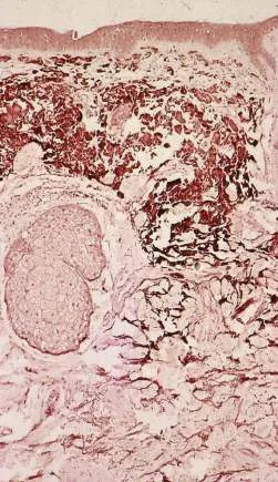 Fig 7. Photoaged skin: accumulation of abnormal basophilic (elastotic) material in the dermis — the histologic hallmark of solar elastosis — (Cosmetic Medicine & Surgery, 2016, p. 31).
> Sources: Cosmetic Medicine & Surgery [2016]
Fig 7. Photoaged skin: accumulation of abnormal basophilic (elastotic) material in the dermis — the histologic hallmark of solar elastosis — (Cosmetic Medicine & Surgery, 2016, p. 31).
> Sources: Cosmetic Medicine & Surgery [2016] [C][MEDLIB] [24].
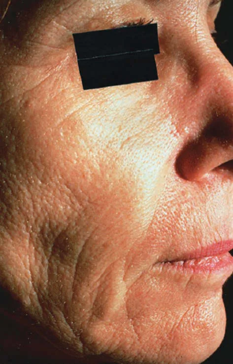 Fig 8. Photoaged skin in a 55-year-old woman: coarse wrinkling, laxity and mottled dyschromia — the clinical face of the histology in Fig 7 — (Baran, Textbook of Cosmetic Dermatology, 2017, p. 77).
> Sources: Baran — Textbook of Cosmetic Dermatology [2017]
Fig 8. Photoaged skin in a 55-year-old woman: coarse wrinkling, laxity and mottled dyschromia — the clinical face of the histology in Fig 7 — (Baran, Textbook of Cosmetic Dermatology, 2017, p. 77).
> Sources: Baran — Textbook of Cosmetic Dermatology [2017] [C][MEDLIB] [7].
> Practical consequence — separate A2.3 from A2.4 at every consultation. An aged face is a mixture, never purely one or the other. Assess and record two axes independently: (a) laxity + volume loss + fine wrinkling → intrinsic; (b) texture + dyschromia + elastosis + telangiectasia → extrinsic. Treat each with its own tool — biostimulation/volume for the first, resurfacing/energy/pigment-directed care for the second. Layering both is the subject of L2 — Combination Protocols & Layered Rejuvenation.es.
Photoprotection is the treatment of extrinsic aging. Broad-spectrum coverage of UVB, UVA, UVA1, visible light and short IR-A is what prevents extrinsic aging; a UV-only product leaves the visible-light and IR arms unopposed [19]. Because inorganic nanoparticle filters (micronized zinc oxide, titanium dioxide) do not block visible light, protection against HEV requires a tinted filter — iron oxides ± pigmentary titanium dioxide, which must be visible on the skin to work [20]. For melasma and Fitzpatrick IV–VI, the tinted (iron-oxide) filter is not optional; a transparent broad-spectrum sunscreen under-protects exactly the patients most prone to pigment [20]. The popularized claim that "≈ 80 % of visible aging is extrinsic and therefore preventable" ⚠ is repeated without a robust locatable primary source; use it as an argument for daily photoprotection, not as data [8].
Retinoids are the correction arm. By lowering CCN1 and MMP-1 and restoring procollagen, topical retinoic acid/retinol partially reverses established photodamage over months — the same mechanism described in A2.2, applied here to sun-damaged rather than chronologically aged dermis [9].
Consensus: daily broad-spectrum photoprotection (UV + visible light) is the base of every anti-aging and pigment protocol, prescribed, not suggested [19][20]. Discrepancy: whether infrared-A protection is clinically necessary — antioxidant-containing/IR-labeled products (exposome school [19]) vs "UV + visible light is enough." Decide by phenotype and exposure: high occupational/actinic load and melasma justify the broader product [19][20].
Grade photoaging before treating it — Glogau.
| Glogau | Age band (typical) | Features | Typical strategy |
|---|---|---|---|
| I mild | 20s–30s | No keratoses, little wrinkling, minimal/no makeup | Photoprotection, antioxidants, retinoid |
| II moderate | 30s–40s | Early lentigines, wrinkles in motion, palpable keratoses | + superficial peels, light devices |
| III advanced | 50s–60s | Persistent dyschromia, telangiectasia, wrinkles at rest | + medium resurfacing, fractional |
| IV severe | 60s–70s+ | Yellow-gray color, wrinkles throughout, actinic keratoses/skin cancer | Ablative resurfacing, oncologic surveillance |
Fitzpatrick phototype sets the risk ceiling.
| Type | Constitutive color | Sun response | Melanoma/PIH risk balance |
|---|---|---|---|
| I | Very fair | Always burns, never tans | ↑skin-cancer, low PIH |
| II | Fair | Burns, tans minimally | ↑skin-cancer |
| III | Medium | Sometimes burns, tans | Intermediate |
| IV | Olive | Rarely burns, tans easily | ↑PIH |
| V | Brown | Very rarely burns | ↑↑PIH, melasma |
| VI | Dark brown/black | Never burns | ↑↑PIH, lower photoaging, later presentation |
Photobiology metrics that define a sunscreen. MED (minimal erythema dose) anchors SPF (UVB/erythema protection). UVA is graded by PPD (persistent pigment darkening) / UVA-PF and the Japanese PA+ to PA++++; the EU "UVA in a circle" mark requires UVA-PF ≥ 1/3 of SPF. A high SPF with poor UVA balance still lets the dermis-penetrating, elastosis-driving UVA through — the metric that matters for aging, not just burning [18][19].
Filter classes and what each does.
| Class | Examples | Covers | Gap |
|---|---|---|---|
| Inorganic (mineral) | Zinc oxide, titanium dioxide | Broad UV; ZnO best UVA | Micronized/nano forms do not block visible light |
| Organic (chemical) UVB | Octinoxate, octisalate, homosalate | UVB | Little/no UVA |
| Organic UVA | Avobenzone, Mexoryl SX/XL, Tinosorb S/M, bemotrizinol | UVA/broad | Some photo-unstable alone (avobenzone) |
| Tinted (pigmentary) | Iron oxides ± pigmentary TiO₂ | Visible light + UV | Must be visible/colored to work [20] |
The exposome — aging beyond UV. Solar UV is the dominant extrinsic driver, but air pollution (PAHs via the aryl-hydrocarbon receptor), tobacco smoke, heat and possibly high-energy visible ("blue") light from sun (screen exposure is negligible by dose, a common overstatement) add oxidative and inflammatory load; this is why exposome-oriented daily products pair broad-spectrum filters with antioxidants [19]. Tobacco independently produces a characteristic accelerated facial aging (elastosis, sallow color, deep perioral rhytides).
The DNA-damage half of photocarcinogenesis. UVB creates cyclobutane pyrimidine dimers (CPDs) and 6-4 photoproducts directly; unrepaired, these are the signature C→T "UV mutations" in p53 that seed actinic keratoses and squamous/basal cell carcinoma. UVA generates "dark CPDs" for hours after exposure via melanin-linked chemiexcitation — a reason post-sun antioxidant strategies and continued protection are argued [18]. Field cancerization (multiple keratoses over sun-damaged skin) is the clinical expression, and any resurfacing plan in Glogau III–IV must include lesion surveillance.
Vitamin D — the balance argument answered. Broad-spectrum sunscreen use has not been shown to cause clinically meaningful vitamin-D deficiency in real-world use; supplementation, not sun exposure, is the safe correction, so the "sunscreen causes deficiency" argument does not justify under-protection [19].
Application reality — the reason "SPF 50" under-delivers. SPF is measured at 2 mg/cm²; real-world application is typically 0.5–1 mg/cm², and protection falls roughly exponentially with under-application, so a labeled SPF 50 applied thinly behaves like ~SPF 10–20. The practical prescription is the "two-finger" / ~¼ teaspoon for the face, reapplied every 2 hours of exposure and after sweating/swimming. A perfect filter chemistry under-applied is a common cause of "my sunscreen doesn't work" — and of persistent melasma despite treatment [19][20].
Pigment-darkening kinetics — three overlapping responses to UVA/visible light. Immediate pigment darkening (IPD) — seconds to minutes, oxidation of existing melanin; persistent pigment darkening (PPD) — hours, the basis of UVA grading; delayed tanning (DT) — days, true neomelanogenesis after UVB. Visible light adds a sustained pigmentation in Fitzpatrick IV–VI that outlasts UV-only tanning and is not blocked by transparent filters [20] — the kinetic reason iron-oxide tint is required for pigment-prone skin.
Heat is an independent extrinsic driver, not only wavelength. Infrared-A and conducted heat induce MMP-1 and can flare melasma regardless of the UV filter, which is why hot occupations, saunas, cooking heat and poorly cooled devices matter in a photoaging/pigment history — a point that recurs as a safety rule in A2.5 [19][21].
Consensus (added): grade with Glogau, phototype with Fitzpatrick, and prescribe a broad-spectrum (UVB + UVA + visible-light) filter matched to phototype and pigment risk as the base of every extrinsic-aging plan [18][19][20]. Discrepancy (added): how much visible-light/IR and antioxidant coverage is necessary — exposome-maximalist (tinted + antioxidant + IR claims [19]) vs UV-plus-visible-light-sufficient. Decide by actinic load and pigment phenotype [19][20].
Documenting photodamage and field cancerization. In Glogau III–IV, the same UV that produced the cosmetic complaint produced field cancerization: multiple actinic keratoses, dyskeratosis and a raised squamous/basal-cell risk over the treatment area. Any resurfacing plan there records baseline lesions, treats or refers keratoses first, and schedules dermatologic surveillance — an aesthetic procedure over an unexamined actinic field can mask or delay a skin cancer. Standardized, cross-polarized and UV photography documents dyschromia and subclinical pigment that plain light misses, and anchors the before/after the patient will judge [18][24].
Trampa clásica: treating deep actinic rhytides and elastosis with volume alone. Filler lifts, but the degraded, elastotic substrate still reads as "old skin"; the missing tool was resurfacing plus photoprotection. Signature: a well-projected face that still looks weathered because texture and dyschromia were never addressed.
A2.5 · Pigmentation: melanogenesis, melasma, post-inflammatory hyperpigmentation, and differential diagnosis
Melanogenesis. Tyrosinase (the rate-limiting enzyme) converts tyrosine → DOPA → dopaquinone, which branches to eumelanin (brown-black, photoprotective) or pheomelanin (red-yellow, pro-oxidant) [21][23]. Melanin is packaged in melanosomes and transferred through dendrites to the surrounding keratinocytes: one melanocyte serves ≈ 36 keratinocytes — the epidermal melanin unit. Fig 9 shows that unit: the melanocyte body with its organelles feeding pigment up the dendrites into the neighbouring keratinocytes. Every depigmenting strategy targets one step of this loop — tyrosinase (hydroquinone, arbutin, kojic acid), transfer (niacinamide), or turnover (retinoids, exfoliation).
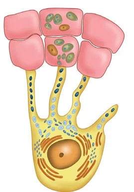 Fig 9. The melanocyte synthesizes melanosomes and distributes them via dendrites to the keratinocytes it serves — the epidermal melanin unit, the target of every lightening agent — (Alam, Cosmetic Dermatology for Skin of Color, 2009, p. 134).
> Sources: Alam — Cosmetic Dermatology for Skin of Color [2009]
Fig 9. The melanocyte synthesizes melanosomes and distributes them via dendrites to the keratinocytes it serves — the epidermal melanin unit, the target of every lightening agent — (Alam, Cosmetic Dermatology for Skin of Color, 2009, p. 134).
> Sources: Alam — Cosmetic Dermatology for Skin of Color [2009] [C][MEDLIB] [23].
| Entity | Dominant mechanism | What decides treatment |
|---|---|---|
| Melasma | Hyperactive melanocyte plus a vascular component and a disrupted/pendulous basement membrane; hormonal + UV + heat triggers [21] | A chronic, relapsing disease. Heat worsens it — poorly set ablative lasers, RF and hair removal make it worse |
| PIH (post-inflammatory) | Inflammation → melanin release ± dermal drop-out; epidermal or dermal [21][22] | Dermal PIH lasts months–years; treat the inflammation first |
| Solar lentigo | Cumulative UV, local melanocytic hyperplasia [24] | Responds well to targeted energy; mind the differential |
| Ephelides (freckles) | Genetic, UV-inducible melanogenesis without ↑ melanocyte number | Cosmetic only; recur with sun |
| Café-au-lait / naevi | Developmental melanocytic | Not an energy target without diagnosis |
| Exogenous ochronosis | Paradoxical darkening from chronic high-dose hydroquinone | Stop hydroquinone; do not escalate it |
Melasma is not "a dark patch to laser." Its mechanism is triple — an overactive melanocyte, an abnormal (often breached) basement membrane letting pigment fall into the dermis, and a dermal vascular component — which is why it recurs and why heat-based devices can flare it [21]. The basement-membrane and vascular involvement explain the therapeutic ceiling: purely epidermal lightening reaches only part of the disease. Fig 10 is the clinical malar pattern; management is a multimodal, indefinite program — rigorous photoprotection (UV and visible light, tinted [20]), topical tyrosinase inhibitors (triple combination, oral/topical tranexamic acid), and only cautious, low-energy resurfacing once the disease is quiet [21].
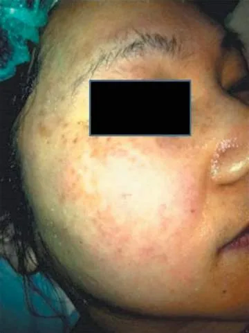 Fig 10. Melasma — symmetric, poorly demarcated brown hyperpigmentation of the malar region, a chronic relapsing disorder aggravated by UV, heat and hormones — (Cosmetic Medicine & Surgery, 2016, p. 138).
> Sources: Cosmetic Medicine & Surgery [2016]
Fig 10. Melasma — symmetric, poorly demarcated brown hyperpigmentation of the malar region, a chronic relapsing disorder aggravated by UV, heat and hormones — (Cosmetic Medicine & Surgery, 2016, p. 138).
> Sources: Cosmetic Medicine & Surgery [2016] [C][MEDLIB] [24].
Post-inflammatory hyperpigmentation is the iatrogenic risk of this whole atlas. Any inflammatory insult — acne, a peel, microneedling, an ill-set laser — can release melanin; epidermal PIH fades over weeks–months, dermal PIH over months–years [21][22]. The rule is to treat the inflammation first and add injury last; escalating aggression on PIH perpetuates it. Fig 11 shows a discrete PIH lesion, the outcome to avoid iatrogenically.
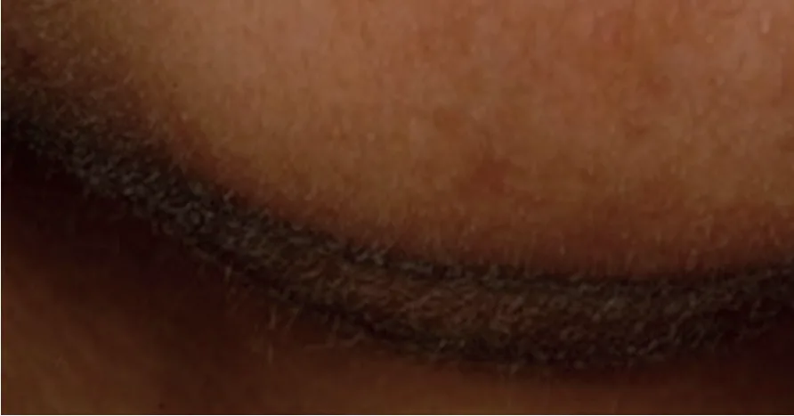 Fig 11. Post-inflammatory hyperpigmentation — brown macular discoloration following a prior inflammatory insult; epidermal pigment fades faster than dermal — (Rigopoulos, Hyperpigmentation, 2018, p. 288).
> Sources: Rigopoulos — Hyperpigmentation [2018]
Fig 11. Post-inflammatory hyperpigmentation — brown macular discoloration following a prior inflammatory insult; epidermal pigment fades faster than dermal — (Rigopoulos, Hyperpigmentation, 2018, p. 288).
> Sources: Rigopoulos — Hyperpigmentation [2018] [C][MEDLIB] [22].
Differential diagnosis before energy (the step that prevents most pigment disasters): distinguish melasma vs solar lentigo vs PIH vs ephelides, and screen the pigmented lesion for atypia (asymmetry, irregular border, colour variegation, evolution) — a melanoma treated as a "spot" is the catastrophic error. Wood's-lamp accentuation suggests epidermal pigment (more amenable), while non-accentuation suggests a dermal component (slower, more device-cautious) [22]. Dermoscopy separates lentigo from an early melanocytic lesion.
Clinical golden rule — Fitzpatrick IV–VI: any injury can trigger PIH, so patch-test, use a visible-light (iron-oxide) filter, favour conservative parameters and longer intervals, and pre/post-treat with tyrosinase inhibitors [20][21][23]. This is developed in B6 — Ethnic, Racial & Cultural Considerations in Face & Body Reshaping.es and G4 — Vascular, Pigment & IPL.es; oral/topical melasma pharmacology extends into peels and topicals [25].
Consensus: melasma is a chronic disease managed medically first, with photoprotection (UV + visible light) as non-negotiable base; energy is adjunctive and cautious [20][21]. Discrepancy: device role in melasma — low-fluence QS/1064 nm and non-ablative fractional (device school) vs strictly medical management (dermatologic-conservative school). Decide by phototype and disease activity; never apply heat-heavy devices to active melasma or in Fitzpatrick IV–VI without a plan for PIH [21].
Melanogenesis is a regulated circuit, not just tyrosinase. UV → keratinocyte p53 → POMC → α-MSH → melanocyte MC1R (Gs/cAMP) → MITF → transcription of tyrosinase, TRP-1 and TRP-2 (DCT). Parallel signals: stem-cell factor (SCF)/c-kit and endothelin-1 (ET-1) from keratinocytes and dermal vessels amplify melanogenesis — the molecular reason melasma has a vascular component and responds partly to anti-angiogenic/vascular measures. MC1R polymorphisms (red-hair/fair-skin variants) bias synthesis toward pro-oxidant pheomelanin and raise skin-cancer risk. Every lightening strategy maps to one node:
| Target node | Agents | Mechanism |
|---|---|---|
| Tyrosinase | Hydroquinone, arbutin, kojic acid, azelaic acid, cysteamine | Enzyme inhibition |
| MITF/cAMP upstream | Niacinamide (also blocks transfer), some botanicals | Reduced signaling/transfer |
| Melanosome transfer | Niacinamide, soy trypsin inhibitors | PAR-2 blockade |
| Turnover/dispersion | Retinoids, AHAs, exfoliation | Faster epidermal clearance |
| Vascular/plasmin | Tranexamic acid (oral/topical) | ↓plasmin → ↓VEGF/ET-1, ↓melanocyte activation |
| Antioxidant | Vitamin C, thiamidol, glutathione | ROS/oxidation reduction |
Melasma has depth and pattern classes that change prognosis. By depth (Wood's-lamp/dermoscopy): epidermal (accentuates, better prognosis), dermal (does not accentuate, refractory), mixed (most common). By pattern: centrofacial, malar, mandibular. Severity is tracked with the MASI/mMASI score (area × darkness × homogeneity). The basement-membrane breach that lets pigment fall into the dermis is why "epidermal-only" logic under-treats and why aggressive energy can worsen it [21].
Melasma treatment ladder (medical first; the whole point of A2.5).
- Rigorous photoprotection — UV and visible light, tinted/iron-oxide, reapplied; non-negotiable base [20].
- Topical — triple combination (hydroquinone + retinoid + corticosteroid, e.g. Kligman-type) as first-line; non-hydroquinone routes (azelaic acid, cysteamine, thiamidol, kojic/arbutin) for maintenance or hydroquinone-intolerant.
- Oral tranexamic acid — adjunct for refractory/extensive disease after screening for thromboembolic risk (a prescribing decision, cross-referenced, not dosed here) [21].
- Procedures last and cautious — superficial peels, low-fluence 1064 nm, non-ablative fractional only once disease is quiet; heat-heavy and ablative devices can flare it [21].
Extended hyperpigmentation differential (before any energy).
| Entity | Clue | Trap |
|---|---|---|
| Solar lentigo | Sharp, sun-exposed, sable dermoscopy | Confused with early lentigo maligna |
| Ephelides | Small, UV-fluctuating, familial | Cosmetic only |
| Drug-induced (minocycline, amiodarone, antimalarials, chemo) | History; blue-gray hue | Will not respond to lightening; stop drug |
| Exogenous ochronosis | Blue-black, caviar-like on chronic hydroquinone | Escalating hydroquinone worsens it |
| Riehl melanosis / pigmented contact dermatitis | Reticulate face, cosmetic allergen | Patch test, allergen removal |
| Ashy dermatosis / erythema dyschromicum perstans | Gray macules, trunk | Not an energy target |
| Hori nevus / nevus of Ota | Bilateral (Hori) dermal blue-gray | Dermal — needs specific laser, not lightening |
| Poikiloderma of Civatte | Reticulate red-brown lateral neck, spares submental shadow | Vascular + pigment; IPL cautious |
| Melanoma / lentigo maligna | ABCDE, dermoscopic atypia | Biopsy, never laser |
Screen every pigmented lesion for atypia before treating; a melanoma lasered as a "spot" is the catastrophic error [22][24]. Note for completeness the hypopigmentation differential a clinician must not confuse with treated dyschromia — vitiligo, idiopathic guttate hypomelanosis, pityriasis alba, post-inflammatory hypopigmentation — which are worked up, not "lightened."
Diagnostic tools before treatment — what each shows.
| Tool | Epidermal pigment | Dermal pigment | Note |
|---|---|---|---|
| Wood's lamp | Accentuates (sharper, darker) | Does not accentuate | Depth triage in melasma/PIH [22] |
| Dermoscopy | Regular network, brown | Blue-gray dots/globules (dermal) | Separates lentigo vs melanocytic atypia |
| Reflectance confocal / biopsy | — | Confirms depth/diagnosis | When malignancy is possible |
Energy-device caution grid — the pigment-safety matrix.
| Phototype | Melasma present | Reasonable | Avoid |
|---|---|---|---|
| I–III | No | IPL, Q-switched, fractional per lesion | — |
| I–III | Yes | Low-fluence 1064 nm, cautious NAFL after control | High-fluence IPL/ablative on active disease |
| IV–VI | No | Conservative fractional, longer wavelengths, test spot | Aggressive IPL (PIH risk) |
| IV–VI | Yes | Medical management first; energy last, low energy | Heat-heavy/ablative, hair-removal over patch |
Counsel melasma as a chronic disease, in writing. The consultation that prevents disappointment states three facts: it relapses (a control-and-maintain condition, not a cure), heat and light (not only sun) trigger it, and procedures come last after months of medical control. Framing energy as first-line is the single most common route to a worse, more refractory patient [21].
Consensus (added): melasma is chronic, medical-first, photoprotection-anchored; energy is adjunctive and phototype-cautious [20][21][23]. Discrepancy (added): oral tranexamic acid role — routine adjunct for moderate/severe (pro-TXA school) vs reserve-for-refractory (conservative school). Decide by extent, chronicity and thrombotic risk screening [21].
PIH prevention protocol — the practical distillation for Fitzpatrick IV–VI. Before any inflammatory procedure: pre-treat with a tyrosinase inhibitor for 2–4 weeks, confirm a quiet barrier, and test-spot. During: choose the least inflammatory effective setting, longer wavelengths, longer intervals. After: strict photoprotection (UV + visible light, iron-oxide), bland barrier recovery, and early anti-inflammatory/lightening if erythema lingers. The governing principle is that in higher phototypes inflammation itself is the pigment risk, so the whole plan minimizes and shortens inflammation rather than maximizing "power" [20][21][23].
Trampa clásica: lasering a melasma as if it were a lentigo. Heat and mistimed energy flare melasma and can seed dermal PIH; the two entities that look superficially similar demand opposite caution. Signature: transient clearing followed by a darker, more refractory rebound. Wood's lamp/dermoscopy before any energy, every time.
A2.6 · Skin microbiome and barrier function in aesthetic patients: established evidence versus speculation
This is the thinnest block in the theme, in the own corpus and in the applied clinical literature, and it is written as a state-of-the-field rather than a protocol — deliberately, instead of overselling it. The retrieval confirmed the gap: the microbiome facet returned the lowest scores of the chapter (A2.6 mechanism_foundations 0.80 but dose_parameters 0.44, technique_mapping 0.51), and no corpus figure exists for it (proven below in Verification).
| Claim | Status | Aesthetic use today |
|---|---|---|
| Skin hosts site-specific communities | Established [B] [5] |
Explains why face ≠ back ≠ flexures for post-procedure care |
| Acid pH 4.5–5.5 + AMPs are part of the barrier | Established [B] [5] |
Preserve them: low-pH, non-alkaline cleansing |
| Dysbiosis associated with acne, rosacea, atopic dermatitis | Association robust; causality/reversal not [4][5] | Do not promise a "microbiome cure" |
| Barrier disruption = transient microbiome disruption | Plausible, mechanistically supported [4] | Part of the rationale for post-procedure barrier care |
| "Topical probiotic" modulates the skin microbiome | Not established (cosmetic, not pharmacologic category) | [MODELO] promise barrier restoration, which is measured (TEWL, corneometry) |
| Gut–skin axis modulates aging | Emerging, mostly systemic/preclinical [26] | Longevity/nutrition angle, not a topical claim |
What is established [5]: the skin surface is a nutrient-poor, acidic, desiccated habitat that nonetheless carries a diverse, site-specific microbiota — Cutibacterium acnes dominant in sebaceous zones (face, back, chest), Staphylococcus and Corynebacterium in moist zones (flexures), and greater diversity in dry zones. The acid mantle (pH 4.5–5.5) and antimicrobial peptides are structural components of the barrier, not accessories, and cutaneous immunity is shaped over early life by the barrier–microbiome interface and remains modifiable by how the barrier is treated in adulthood [4][5]. Dysbiosis is reproducibly associated with acne, rosacea and atopic dermatitis — but the association being robust does not make causality or therapeutic reversal established [4][5].
What matters in aesthetic practice — what can actually be applied today:
- Every barrier-breaching procedure is a transient microbiome disruption, not just a wound. Peels, microneedling and ablative laser transiently perturb the resident community; this is part of why post-procedure acne flares and folliculitis occur, and why the sebaceous face behaves differently from dry limbs after the same procedure [4][5]. Match aftercare to the site.
- Alkaline cleansers and chronic antiseptics prolong barrier — and community — recovery by pushing surface pH up and blunting the lipid-processing enzymes that rebuild the mortar [2][6]. Post-procedure, favour bland, low-pH, non-antiseptic cleansing unless there is a specific infection.
- "Topical probiotic" is a cosmetic category, not a pharmacologic one.
[MODELO](P) With the available evidence, do not promise microbiome modulation; promise barrier restoration, which is objectively measurable by TEWL and corneometry [2][6]. The measurable claim is defensible; the microbiome claim is not. - The gut–skin axis is an emerging, mostly systemic story. The microbiome-aging relationship (including the distinctive microbiota of centenarians and its anti-inflammatory/antioxidant metabolic pathways) is a longevity and nutrition topic with preclinical and epidemiologic support, not a topical aesthetic lever [26]. It belongs to K1 — Anti-Aging Medicine & Longevity.es, flagged here so the two are not conflated.
Gap statement (P): this subchapter will be upgraded with an external lane the moment trials with a clinical outcome — not only 16S/metagenomic sequencing — demonstrate that a topical intervention reverses dysbiosis and changes a skin phenotype. Until then, the honest position is: barrier science is actionable; microbiome modulation is a promise the evidence does not yet support.
Dysbiosis-disease associations — robust, but association is not causation.
| Condition | Microbial association | Actionable today |
|---|---|---|
| Acne | Loss of C. acnes strain diversity, phylotype IA1 shift (not overgrowth per se) [5] | Treat acne on its evidence base; avoid diversity-destroying chronic antibiotics |
| Rosacea | Demodex/Bacillus oleronius signals, barrier dysfunction | Barrier care + disease-specific therapy, not "probiotics" |
| Atopic dermatitis | S. aureus dominance, ↓diversity in flares [4] | Restore barrier, manage flares; emollient-first |
| Aging skin | ↓diversity, ↑pH shifts (early data) | Preserve acid mantle; no topical "anti-aging microbiome" claim |
The pattern is consistent — dysbiosis tracks disease — but no aesthetic topical is shown to reverse dysbiosis and change the phenotype, which is the boundary between mechanism and marketing [4][5].
Post-procedure infection differential — a barrier breach is an open door. After any barrier-breaching procedure, "acne-like" or inflammatory eruptions have distinct causes that demand opposite treatments:
| Presentation | Likely cause | Wrong move |
|---|---|---|
| Pustules, honey crust | Bacterial (S. aureus) impetiginization | Ignoring → cellulitis |
| Grouped vesicles, pain, dermatomal-ish | HSV reactivation (esp. after ablative/peels) | Treating as "acne"; needs antiviral prophylaxis |
| Monomorphic pustules, itch, trunk/face | Pityrosporum (Malassezia) folliculitis | Antibacterials (worsen it); needs antifungal |
| Comedonal/inflammatory acne flare | Occlusion + microbiome perturbation | Over-aggressive re-treatment |
Recognizing these is barrier-and-microbiome science applied — and a reason occlusive aftercare, HSV prophylaxis for deep resurfacing, and the right anti-microbial choice matter [4][5].
Defensible vs indefensible claims (P) — the honest sales line.
| Defensible (measurable) | Indefensible (unproven) |
|---|---|
| "Restores barrier — lower TEWL, higher hydration" | "Balances/heals your microbiome" |
| "Low-pH, respects the acid mantle" | "Probiotic serum treats rosacea" |
| "Reduces post-procedure barrier recovery time" | "Reverses dysbiosis" |
| "Supports the skin's own defenses (barrier)" | "Detoxifies bad bacteria" |
Consensus: protect the barrier (low-pH cleansing, lipid replacement, avoid over-antisepsis) — this is where microbiome science and classic barrier care agree and where the evidence is solid [2][4][6]. Discrepancy: whether topical "microbiome" products do anything beyond moisturization — cosmetic-marketing claims vs the absence of clinical-outcome trials. Decide against the claim; sell barrier metrics, not sequencing [5].
The community by site — established composition.
| Site type | Dominant taxa | Aesthetic note |
|---|---|---|
| Sebaceous (face, back, chest) | Cutibacterium acnes, Malassezia (fungal) | Lipid-rich; where post-procedure acne/folliculitis flare [5] |
| Moist (flexures, nares) | Staphylococcus, Corynebacterium | Higher bacterial load |
| Dry (forearm, leg) | Mixed, most diverse | Lower biomass, greater β-diversity |
The barrier is four barriers, not one — and only the first three are solidly actionable:
- Physical — brick-and-mortar SC, corneodesmosomes, tight junctions of the granular layer.
- Chemical — acid mantle (pH 4.5–5.5), lipids, NMF, AMPs [5].
- Immune — Langerhans and resident T cells, innate sensing; matures across early life and stays barrier-dependent [4].
- Microbial — the resident community competitively excluding pathogens and tuning immunity [5]. The evidence supports its existence and associations; it does not yet support reversing dysbiosis topically to change a phenotype.
Objective barrier measures — sell these, not sequencing.
| Metric | What it reads | Use |
|---|---|---|
| TEWL | Water flux through SC | Barrier integrity/recovery |
| Corneometry | SC hydration (capacitance) | Moisturization efficacy |
| Surface pH | Acid mantle | Cleanser suitability |
| Sebumetry | Sebum output | Site/product matching |
| 16S/shotgun sequencing | Community composition | Research, not a clinical claim |
Two organisms every aesthetic clinician meets. Cutibacterium acnes is a commensal whose specific phylotypes (loss of strain diversity, phylotype IA1 predominance) — not mere presence — associate with acne; this is why "kill all C. acnes" with chronic antibiotics is both ineffective long-term and a resistance driver. Malassezia (a lipophilic yeast) underlies pityrosporum folliculitis, which is commonly misdiagnosed as post-procedure "acne" and worsens with antibacterials — an important post-treatment differential [5].
Antibiotics and antiseptics reshape, and delay. Chronic topical antibiotics select resistance and reduce community diversity; alkaline soaps and prolonged antiseptic use raise pH and slow barrier (and community) recovery [2][6]. Post-procedure, prefer bland low-pH cleansing and reserve antiseptics/antibiotics for a specific indication.
Prebiotic / probiotic / postbiotic — definitions and status (P).
| Term | Meaning | Skin evidence status |
|---|---|---|
| Prebiotic | Substrate favouring desired commensals | Cosmetic; barrier benefit plausible, phenotype change unproven |
| Probiotic (topical) | Live organisms applied to skin | No validated clinical-outcome trials; regulatory grey zone |
| Postbiotic | Non-viable microbes/metabolites (lysates, ferments) | Some barrier/soothing signals; not microbiome "modulation" |
The honest label: these can act as moisturizers/soothers with measurable barrier effects; they are not shown to modulate the skin microbiome to a clinical outcome [5]. The regulatory line matters too: a live-organism product making a therapeutic microbiome claim would be a drug, not a cosmetic — the absence of that approval pathway for topical skin probiotics is itself evidence of where the science stands.
Procedure-specific perturbation (P), matched to aftercare.
| Procedure | Perturbation | Aftercare emphasis |
|---|---|---|
| Superficial peel | SC/acid-mantle disruption | Low-pH rebuild, photoprotection |
| Microneedling | Punctures the physical + microbial barrier | Sterile technique, bland recovery |
| Ablative/fractional laser | Wide barrier breach + wound | Occlusive/emollient, infection watch (bacterial, HSV, Candida) |
The gut–skin axis — bounded. The microbiome-aging relationship is largely systemic: centenarian gut microbiota carry anti-inflammatory and antioxidant metabolic pathways and may modulate immune aging, positioning diet/gut-directed strategies as a longevity approach rather than a topical aesthetic one [26]. It connects to inflammaging (A2.3) and to K1 — Anti-Aging Medicine & Longevity.es; it is flagged here precisely so a systemic hypothesis is not sold as a topical claim.
Cleanser and aftercare selection — where microbiome science is already actionable.
| Choice | Barrier/microbiome effect | Preferred when |
|---|---|---|
| Syndet, low-pH (≈ 5.5) | Preserves acid mantle and lipids | Default; post-procedure |
| True soap (alkaline) | Raises pH, strips lipids, slows recovery | Avoid on face/barrier repair |
| Chronic antiseptic wash | ↓diversity, resistance selection | Only for a specific infection |
| Bland emollient (ceramide-dominant) | Refills mortar, lowers TEWL | Barrier repair, post-procedure |
| Occlusive (petrolatum) | ↓TEWL > 98 %, wound-friendly | Ablative recovery |
This is the actionable core of A2.6: manage the barrier with objective measures, and let the microbiome claims stay in the research column [2][5][6].
Trampa clásica: marketing "microbiome balancing" as a treatment and setting an expectation the product cannot meet. Signature: a patient disappointed that a "probiotic serum" did not fix rosacea, or a pityrosporum folliculitis worsened by an antibacterial "acne" product. Reframe to barrier restoration with measurable metrics (TEWL, corneometry); treat the disease (rosacea, acne, atopic dermatitis) on its own evidence base.
A2.7 · Wound healing cascade: hemostasis, inflammation, proliferation, remodeling, and failure modes
Every ablative, fractional, needling, peeling and biostimulating procedure is a controlled wound, so its result obeys the wound-healing schedule — and its complications are that schedule failing [27].
| Phase | Chronology | Main actors | Aesthetic implication |
|---|---|---|---|
| Hemostasis | 0 – minutes | Platelets, fibrin, PDGF/TGF-β release | Rationale for PRP (F2 — PRP, Polynucleotides (PN-PDRN) & Exosomes.es) |
| Inflammation | 0 – 3/5 days | Neutrophils → macrophages (M1 → M2), TNF-α/IL-1β/IL-6 | Prolonged = bad scar; the M1→M2 switch is the turning point [27] |
| Proliferation | ≈ 3 days – 3 weeks | Fibroblasts, type III collagen, angiogenesis (VEGF), re-epithelialization | Biostimulation acts here; granulation tissue forms |
| Remodeling | 3 weeks – 12–24 months | III → I, fibre alignment, MMP/TIMP balance, myofibroblast contraction | The final result of a laser or a scar is not judged before ≈ 1 year |
The phases overlap; they are not sequential boxes. Fig 12 lays out the cell and mediator cast across days 0 → 365; Fig 13 shows the same as overlapping response curves on a log-time axis — the visual that fixes why remodeling dominates the long tail. Fig 14 contrasts the two possible outcomes from the same insult: regeneration (restored architecture) versus repair (a collagen scar).
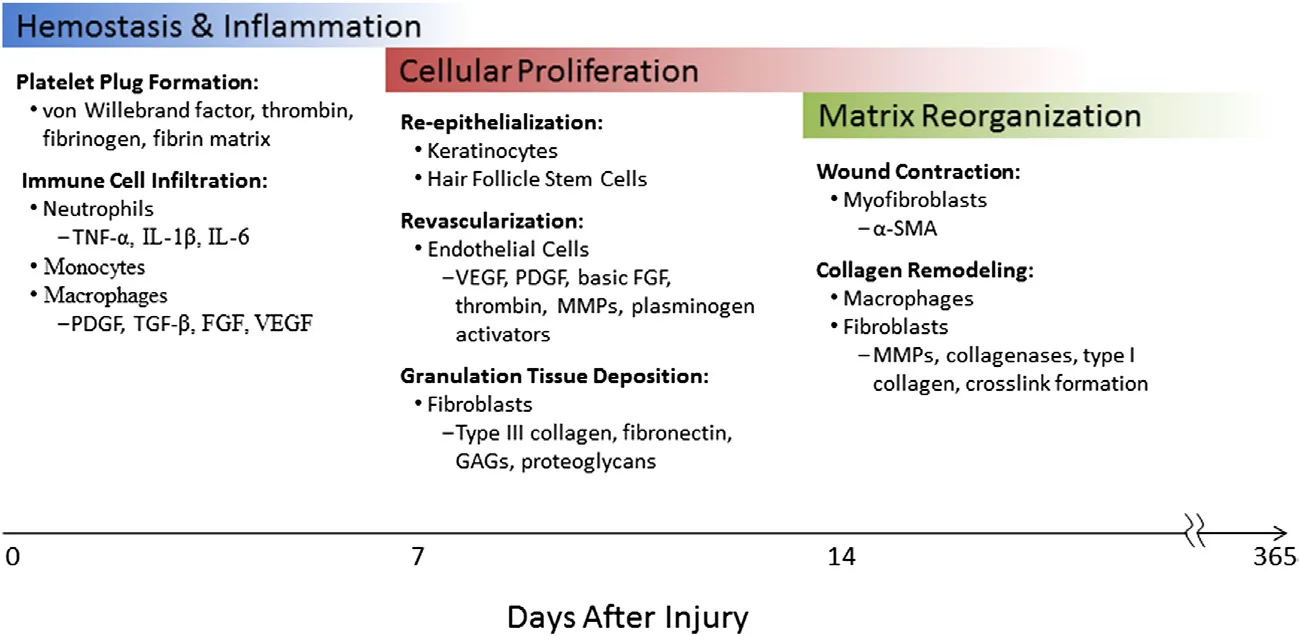 Fig 12. Cellular and molecular course of normal wound healing — Hemostasis & Inflammation (platelet plug; neutrophils, monocytes, macrophages; PDGF, TGF-β, FGF, VEGF), Cellular Proliferation (re-epithelialization by keratinocytes/hair-follicle stem cells; revascularization; granulation with type III collagen, fibronectin, GAGs), Matrix Reorganization (myofibroblast contraction, α-SMA; collagen remodeling by MMPs to type I with crosslinks) across days after injury — (Albanna, Skin Tissue Engineering, 2016, p. 27).
> Sources: Albanna — Skin Tissue Engineering and Regenerative Medicine [2016]
Fig 12. Cellular and molecular course of normal wound healing — Hemostasis & Inflammation (platelet plug; neutrophils, monocytes, macrophages; PDGF, TGF-β, FGF, VEGF), Cellular Proliferation (re-epithelialization by keratinocytes/hair-follicle stem cells; revascularization; granulation with type III collagen, fibronectin, GAGs), Matrix Reorganization (myofibroblast contraction, α-SMA; collagen remodeling by MMPs to type I with crosslinks) across days after injury — (Albanna, Skin Tissue Engineering, 2016, p. 27).
> Sources: Albanna — Skin Tissue Engineering and Regenerative Medicine [2016] [C][MEDLIB] [28].
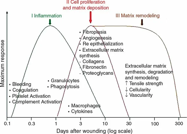 Fig 13. The phases of wound healing as overlapping curves against days after wounding (log scale): I Inflammation (bleeding, coagulation, granulocytes, macrophages) → II Cell proliferation and matrix deposition (fibroplasia, angiogenesis, re-epithelialization) → III Matrix remodeling (synthesis/degradation, ↑ tensile strength, ↓ cellularity, ↓ vascularity) extending past day 300 — (Krakowski, Scar Book, 2017, p. 212).
> Sources: Krakowski — Scar Book: Formation, Mitigation, Rehabilitation and Prevention [2017]
Fig 13. The phases of wound healing as overlapping curves against days after wounding (log scale): I Inflammation (bleeding, coagulation, granulocytes, macrophages) → II Cell proliferation and matrix deposition (fibroplasia, angiogenesis, re-epithelialization) → III Matrix remodeling (synthesis/degradation, ↑ tensile strength, ↓ cellularity, ↓ vascularity) extending past day 300 — (Krakowski, Scar Book, 2017, p. 212).
> Sources: Krakowski — Scar Book: Formation, Mitigation, Rehabilitation and Prevention [2017] [C][MEDLIB] [30].
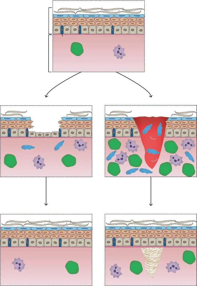 Fig 14. From an intact skin unit, injury resolves either by regeneration (restored epidermis and dermal architecture) or by repair (re-epithelialized surface over a dermal collagen scar) — the fork that decides the cosmetic outcome — (Ogawa, Total Scar Management, 2020, p. 11).
> Sources: Ogawa — Total Scar Management [2020]
Fig 14. From an intact skin unit, injury resolves either by regeneration (restored epidermis and dermal architecture) or by repair (re-epithelialized surface over a dermal collagen scar) — the fork that decides the cosmetic outcome — (Ogawa, Total Scar Management, 2020, p. 11).
> Sources: Ogawa — Total Scar Management [2020] [C][MEDLIB] [29].
Three consequences that get forgotten:
- The result of a collagen-inducing procedure appears after remodeling, not during it. Promising a biostimulator result at 4 weeks is promising inflammation and edema, not the mature type I collagen that carries the benefit. Consent to a 6–12 month horizon (remodeling continues 12–24 months) and photograph on that schedule [27].
- A hypertrophic scar or keloid is remodeling that never closed — excess type III, an M1→M2 transition that stalled, insufficient MMP-mediated turnover, prolonged inflammation [27][29]. Keloids extend beyond the original wound margins and are more frequent in darker phototypes; hypertrophic scars stay within margins. The same biology that under-remodels here is what a scar-revision plan tries to restart (M2 — Scar Aesthetics & Revision.es).
- Re-epithelialization ≠ healing. A wound "closed" at 7 days is still remodeling at 12 months; tensile strength keeps rising and pigment/erythema keep resolving long after the surface looks intact [27]. This is the biological basis for strict photoprotection for months, not weeks, after any resurfacing — pigment laid down during active remodeling becomes PIH.
Failure modes to recognize: prolonged inflammation (chronic wound, poor M1→M2 switch) → atrophic or hypertrophic scarring; excessive fibroplasia → hypertrophic scar/keloid; impaired proliferation (diabetes, smoking, corticosteroids, radiation, aged flattened DEJ) → dehiscence and delayed closure; infection → converts a controlled wound into an uncontrolled one and seeds scarring [27][28]. Isotretinoin — current or within < 6 months — historically defers ablative resurfacing and dermabrasion for concern over impaired re-epithelialization; recent evidence has softened this for superficial procedures, but the conservative window is still standard for deep resurfacing [24].
Consensus: judge any wounding procedure's final result at ≈ 12 months, protect from UV throughout remodeling, and optimize healing factors (glycemic control, stop smoking, no unnecessary corticosteroids) beforehand [27][28]. Discrepancy: the isotretinoin waiting period — the classic 6-month deferral (conservative/medicolegal school) vs newer data permitting superficial peels/microneedling on or soon after isotretinoin (contemporary evidence school). Decide by procedure depth: hold the window for ablative/deep, individualize for superficial [24].
Growth factors — who signals what, and the injectable rationale.
| Factor | Source | Main action | Aesthetic relevance |
|---|---|---|---|
| PDGF | Platelets, macrophages | Chemotaxis, fibroblast/SMC proliferation | Core of PRP rationale F2 — PRP, Polynucleotides (PN-PDRN) & Exosomes.es |
| TGF-β1 | Platelets, macrophages | Fibroblast → myofibroblast, matrix synthesis | Pro-fibrotic; excess → hypertrophic scar |
| TGF-β3 | — | Anti-scarring isoform | Fetal-type, scarless-healing interest |
| VEGF | Macrophages, keratinocytes | Angiogenesis | Granulation vascularity |
| FGF-2 / KGF (FGF-7) | Fibroblasts, keratinocytes | Angiogenesis, re-epithelialization | Repair support |
| EGF | Platelets, keratinocytes | Keratinocyte migration/proliferation | Re-epithelialization |
| IGF-1 | Fibroblasts, serum | Cell survival, matrix | Regenerative adjuncts |
Hemostasis in one line: vascular spasm → platelet plug (von Willebrand factor, GP receptors) → coagulation cascade → fibrin matrix that is both a haemostatic seal and the provisional scaffold and growth-factor reservoir the next phases build on — the biology PRP and fibrin-based products exploit [27][28].
Tensile strength recovers slowly — the number behind "don't judge early." A wound has only ≈ 5 % of final tensile strength at 1 week, ≈ 20 % at 3 weeks, ≈ 50–60 % by 6 weeks–3 months, plateauing at ≈ 70–80 % of unwounded skin — never 100 % — over months of remodeling [27]. This is why sutured wound support and activity restriction extend past visible closure, and why a scar keeps changing color and pliability for a year.
Scar phenotypes — recognize the failure mode.
| Scar | Feature | Biology | Note |
|---|---|---|---|
| Atrophic | Depressed (ice-pick, boxcar, rolling) | Net matrix loss / tethering | Acne sequela; needs volumizing/resurfacing |
| Hypertrophic | Raised, within original margins | Excess type III, stalled remodeling | May regress over time |
| Keloid | Raised, beyond margins, recurs | Prolonged TGF-β, ↓apoptosis, genetic | ↑ in darker phototypes; excision alone recurs |
| Contracture | Tightening across a joint/free margin | Myofibroblast over-contraction | Functional impact |
Factors that impair healing — screen before wounding.
| Systemic | Local |
|---|---|
| Diabetes/hyperglycemia (AGE, microangiopathy) | Infection (converts controlled → uncontrolled wound) |
| Smoking (vasoconstriction, hypoxia) | Ischemia/poor perfusion |
| Age (flattened DEJ, slower turnover) [8] | Foreign body, necrotic tissue |
| Malnutrition (protein, vitamin C, zinc) | Radiation-damaged tissue |
| Corticosteroids/immunosuppression | Repeated trauma/tension |
| Isotretinoin (< 6 months) for deep procedures [24] | Desiccation (moist healing is faster) |
Closure intentions. Primary (approximated edges — a clean excision), secondary (left to granulate and contract — an ablative field or open wound), tertiary/delayed primary (closed after a clean-up interval). Most aesthetic wounding heals by secondary intention over a raw surface, which is why moist recovery and photoprotection through re-epithelialization matter so much.
Fetal scarless healing — the aspirational model. Early-gestation fetal skin heals without scar: high hyaluronic acid, a TGF-β3 > TGF-β1 profile, and minimal inflammation. It is the biological argument that scar is a consequence of the adult inflammatory program, not an inevitability — and the rationale behind anti-scarring and regenerative strategies [27][29].
Chronic wounds are healing stuck in inflammation — persistent M1 macrophages, high MMP/low TIMP, senescent fibroblasts, bacterial biofilm — the mirror image of hypertrophic scar (over-repair) and a reminder that both extremes are the same schedule failing [27].
Phase-by-phase — what goes wrong and the aesthetic sign.
| Phase | Failure | Aesthetic sign |
|---|---|---|
| Hemostasis | Anticoagulation, fragile aged vessels | Bruising, hematoma after injectables/procedures |
| Inflammation | Prolonged / stalled M1→M2, infection | Persistent redness, delayed healing, worse scar [27] |
| Proliferation | Impaired (diabetes, smoking, ischemia) | Delayed re-epithelialization, dehiscence |
| Proliferation | Excess fibroplasia | Hypertrophic scar / keloid [29] |
| Remodeling | Insufficient MMP turnover | Firm, red, raised scar persisting > 1 year |
| Remodeling | Over-contraction (myofibroblast) | Contracture, distortion of a free margin |
Procedures mapped to the healing phase they exploit — every regenerative and energy tool is timed to one part of this cascade:
| Procedure | Phase exploited | Mechanism |
|---|---|---|
| PRP / fibrin | Hemostasis → proliferation | Platelet growth-factor bolus (PDGF, TGF-β, VEGF) [27] |
| Microneedling ± drug/PRP | Inflammation → proliferation | Controlled micro-injury restarts the cascade |
| Biostimulators (PLLA/CaHA) | Proliferation → remodeling | Sustained fibroblast/type I stimulus [10] |
| Fractional/ablative laser | Full cascade to chosen depth | Micro-wounds heal with neocollagenesis (A2.2) |
| Silicone / pressure / vascular laser | Remodeling (scar) | Modulate maturation, redness, hydration [30] |
Reading a treatment as "which phase am I trying to drive, and when is its result visible" prevents both premature judgment and mistimed re-treatment.
Consensus (added): optimize modifiable healing factors (glycemic control, smoking cessation, nutrition, drug review) before any wounding procedure, and support the wound (moist recovery, tension/UV control) through remodeling [27][28]. Discrepancy (added): early scar intervention — proactive early laser/silicone on a forming scar (pro-early school) vs waiting out remodeling before judging (conservative school). Reconcile by modality: non-wounding prophylaxis (silicone, pressure, early vascular laser for redness) can start early; re-wounding (revision) waits for remodeling [27][30].
Trampa clásica: assessing a scar or a resurfacing result too early and re-treating a wound still in remodeling. Early erythema and firmness are expected remodeling, not failure; re-wounding restarts inflammation and can worsen the scar. Signature: a scar "revised" at 8 weeks that ends up worse. Wait out remodeling before judging or re-treating.
Coverage vs UPO
This is a foundational-science theme with a high curricular gap: the UPO master's corpus (mapped to G4) teaches anti-aging medicine and treatment technique, not the underlying skin biology in depth. The own corpus ([MEDLIB]) supplies textbook mechanism; the external lane supplies recency, guideline-level photoprotection and the therapeutic claims. UPO slide material is never_sufficient_alone and is the lane that ages fastest.
| UPO teaches | State in this chapter | What the atlas adds |
|---|---|---|
Anti-aging medicine overview (M6/T17, Prof. Ayala slide) [D] [31] |
Covered in A2.3 (senescence, hormones, glycation, inflammaging) | Cellular mechanism (SASP, SA-β-gal, telomeres) with primary literature [12][13][16], not a single slide |
| "Skin ages intrinsically and extrinsically" | Covered A2.3 + A2.4, kept as two separate axes | The histologic and biochemical distinction that changes the tool (Fig 4), and the assessment protocol |
| Photoprotection as anti-aging advice | Covered A2.4 as prescription | Visible-light/iron-oxide requirement for melasma and IV–VI [20], the ⚠ "80 % extrinsic" caveat |
| Pigmentation / melasma treatment | Covered A2.5 with mechanism + differential | Triple mechanism of melasma (melanocyte + basement membrane + vascular), Wood's-lamp/dermoscopy triage, PIH prevention [21][22] |
| Treatment technique (peels, laser, biostimulation) | Cross-referenced, not duplicated | The biological schedule every technique obeys (A2.7) and the 6–12 mo result horizon [27] |
| — (UPO does not cover) skin barrier lipid biochemistry | A2.1 | Ceramide/cholesterol/FFA ratios, acid mantle pH 4.5–5.5, NMF/filaggrin, AMPs [1][5][6] |
| — (UPO does not cover) collagen I:III ratio and MMP/TIMP | A2.2 | Repair-quality marker, CCN1/retinoid mechanism [7][9] |
| — (UPO does not cover) wound-healing phases and failure modes | A2.7 | Full cascade with chronology and scar/keloid biology [27][28][29][30] |
| — (UPO does not cover) skin microbiome evidence vs speculation | A2.6 | Established vs unproven split; barrier-metric honesty [4][5][26] |
| — (UPO does not cover) differential diagnosis of pigmented lesions | A2.5 | Melasma vs lentigo vs PIH vs melanoma screen before energy [22][24] |
Self-assessment
- Epidermal turnover time in a young adult, and why a retinoid is not judged before 12 weeks.
Show answer
≈ **28 days**; a retinoid needs **≥ 3 complete turnover cycles**, so ~12 weeks before assessment [1][3].- The three lipid pillars of the stratum corneum mortar and the surface pH they need.
Show answer
Ceramides (≈ 50 %), cholesterol and free fatty acids; acid mantle **pH 4.5–5.5**, which activates the lipid-processing enzymes [1][5][6].- Type I vs type III collagen — proportions and what the ratio marks.
Show answer
Type I ≈ **80 %** (tensile strength), type III ≈ **15 %** (fine/young). The **I:III ratio marks repair quality**; excess III = scar. The goal is organized type I, not "more collagen" [1][7].- How retinoic acid improves aged and photoaged skin at the molecular level.
Show answer
It lowers **CCN1 (CYR61)**, which raises type I procollagen and reduces collagenase (**MMP-1**) [9].- The single most reliable marker of a senescent (vs merely arrested) cell, and what SASP is.
Show answer
**SA-β-galactosidase**; SASP = senescence-associated secretory phenotype — IL-6, IL-8, MMPs, ROS — making the arrested cell pro-inflammatory [12].- Wavelength bands of UVB, UVA and visible light, and which one drives melasma.
Show answer
UVB **280–320 nm**, UVA **320–400 nm**, visible/HEV **400–500 nm**; **visible light** drives melasma, so a UV-only filter under-protects it [18][20].- The photoaging cascade from UV to collagen loss.
Show answer
UV → **ROS** → **AP-1 / NF-κB** → ↑**MMP-1, -3, -9** + ↓TGF-β/procollagen → net degradation and imperfect repair [8][9][18].- The triple mechanism of melasma and why heat matters.
Show answer
Hyperactive melanocyte + disrupted/pendulous **basement membrane** (dermal pigment drop) + **vascular** component; **heat** is an independent trigger, so ill-set lasers, RF and hair removal can flare it [21].- The four wound-healing phases with their chronology.
Show answer
Hemostasis (0–minutes) → inflammation (0–3/5 days, M1→M2) → proliferation (≈ 3 days–3 weeks, type III collagen) → remodeling (3 weeks–**12–24 months**, III→I). Final result judged at ≈ 1 year [27].- Why post-resurfacing photoprotection is measured in months, and what "re-epithelialization ≠ healing" means.
Show answer
The surface closes (~7 days) long before remodeling ends (12–24 months); pigment laid during active remodeling becomes PIH, so strict photoprotection continues for **months** [27].What's new and trends
| Year | What changed | Reference (already in chapter) |
|---|---|---|
| 2020 | Tinted (iron-oxide) sunscreens formalized as the visible-light protection standard for pigmentary disorders — reframes melasma/PIH photoprotection | Lyons, Lim 2020 [20] |
| 2021 | "Photoprotection beyond UV" — broad-spectrum incl. visible light, IR-A and antioxidant/anti-aging actives positioned to prevent extrinsic aging (exposome view) | Krutmann 2021 [19] |
| 2022 | Skin glycation/AGE mechanism consolidated as an intrinsic-aging driver with anti-glycation actives as an emerging target | Zheng 2022 [15] |
| 2022 | Telomere length reaffirmed as an aging biomarker (physical-activity systematic review) — association, not a prescribable lever | Schellnegger 2022 [16] |
| 2023 | Skin senescence/senotherapeutics reviewed — SA-β-gal as best single marker, SASP-reducing compounds (quercetin, fisetin, naringenin, apigenin) preclinical | Bulbiankova 2023 [12] |
| 2023 | Inflammaging integrated into the updated hallmarks of aging (chronic inflammation, dysbiosis, disabled autophagy added) | Baechle 2023 [13] |
| 2023 | Dermal-focused skin-aging mechanism review — intrinsic/extrinsic convergence on senescence and matrix loss, tied explicitly to procedure selection | Shin 2023 [8] |
| 2023 | Gut–skin/microbiome-aging axis synthesized (centenarian microbiota, anti-inflammatory metabolic pathways) — systemic, not topical | Salazar 2023 [26] |
| 2025 | Regenerative/autologous stimulation of collagen formation restated with updated cellular models | Piccolo 2025 [10] |
Maturity of the moving edge — the same directions sorted by how much a clinician can lean on them today:
| Maturity class | Directions in this bucket |
|---|---|
| clinically actionable now | Visible-light/iron-oxide photoprotection [20]; daily broad-spectrum + antioxidant against extrinsic aging [19]; glycemic control to limit glycation [15] |
| promising but not validated | Topical senotherapeutics (quercetin, fisetin, naringenin, apigenin) for SASP reduction [12]; anti-glycation topicals [15] |
| preclinical/speculative | Gut–skin/microbiome axis modulating skin aging [26]; senolysis as a skin-specific lever [13] |
| unsupported commercial claim | Topical "microbiome modulation"/probiotic cure [5]; "elastin regeneration" in adult skin [8] |
What did NOT change, and why the older references still stand. The mechanistic spine of this chapter is textbook and stable: the UV → ROS → AP-1 → MMP photoaging cascade with parallel TGF-β suppression [8][9][18], the melanogenesis pathway and the epidermal melanin unit [23], the collagen I:III / MMP-TIMP balance [1][7], and the four-phase wound-healing schedule [27][28][30] have not been overturned; the 2019 Physiological Reviews synthesis remains the canonical wound-healing account [27], and the 2018 skin-microbiome review remains the reference description of site-specific communities [5]. What has moved is the periphery — visible-light photoprotection, senotherapeutics, glycation and the microbiome — where the direction of travel is clear but clinical-outcome evidence is still maturing. The ⚠-flagged figures ("≈ 1 %/year collagen loss", "≈ 30 % in 5 post-menopausal years", "≈ 80 % of aging is extrinsic") have not acquired robust primary sources and are still printed with their caveats [8][14]. UPO slide material (anti-aging overview) is the fastest-aging lane and is corroborated here against dated literature rather than relied on alone.
Unexplored directions (AI speculation)
> Disclaimer. Everything below is model-generated hypothesis, not evidence and not advice. Each item is tagged [IA-ESPEC], which exists so this section can never be mistaken for [A–D] evidence or [MODELO] structure. No item contains a dose, a product or an actionable protocol. Each states its anchor (a cited fact already in the chapter), a proposal, an expected effect (the direction it predicts), a confounder (what could fake or hide that effect), and what would settle it — a proposal with no falsifier is an opinion.
-
[IA-ESPEC]Senescent-cell burden as a pre-procedure stratifier. Anchor: SA-β-gal marks senescent cells and SASP degrades matrix [12]; the flattened aged DEJ heals slowly [8]. Proposal: a non-invasive readout of local senescent-cell burden might predict which patients under-remodel and scar after resurfacing, refining candidate selection beyond Fitzpatrick and history. Expected effect: higher local senescent-cell burden → higher rate of scarring/prolonged erythema after resurfacing. Confounder: Fitzpatrick type and cumulative photodamage co-vary with senescence and independently drive the outcome. What would settle it: a prospective study correlating a validated in-vivo senescence marker with objective scar/erythema outcomes at 12 months. -
[IA-ESPEC]Glycation status as a modifier of biostimulator response. Anchor: AGEs stiffen the type I network and resist remodeling [15], and biostimulation aims to build organized type I [7][10]. Proposal: high dermal AGE load may blunt or slow biostimulator results, making anti-glycation/metabolic optimization a rational co-intervention. Expected effect: high dermal AGE load → smaller or slower collagen-density gain from a biostimulator. Confounder: diabetes and age themselves reduce fibroblast responsiveness independently of AGE cross-linking. What would settle it: a trial stratifying biostimulator responders by baseline skin autofluorescence (an AGE surrogate) with blinded collagen-density outcomes. -
[IA-ESPEC]Barrier-metric-gated procedure scheduling. Anchor: barrier disruption raises TEWL and is a transient microbiome perturbation [4][6]; disrupted barrier contraindicates aggressive treatment [2]. Proposal: objective TEWL/corneometry thresholds, rather than a fixed 2–4 week wait, could time the safe re-introduction of actives and the go/no-go for a procedure. Expected effect: TEWL-gated scheduling → fewer post-procedure complications than a fixed calendar. Confounder: underlying skin disease (atopy, rosacea) raises both baseline TEWL and complication risk, mimicking a scheduling effect. What would settle it: a comparison of TEWL-gated vs calendar-gated scheduling for post-procedure complication rates. -
[IA-ESPEC]Visible-light dosimetry personalized to phototype. Anchor: visible light drives pigmentation in IV–VI and iron-oxide filters are required for melasma [20]. Proposal: individual visible-light sensitivity (minimal pigmenting dose) might guide how much tint a given patient actually needs, replacing a one-size iron-oxide recommendation. Expected effect: tint matched to an individual's minimal pigmenting dose → lower melasma relapse than a single default tint. Confounder: reapplication adherence and hormonal triggers dominate relapse regardless of the tint chosen. What would settle it: dose–response studies of graded iron-oxide concentrations against melasma relapse by phototype. -
[IA-ESPEC]M1→M2 macrophage transition as the tunable step in scar prevention. Anchor: prolonged inflammation and a stalled M1→M2 switch underlie hypertrophic scarring [27][29]. Proposal: interventions that accelerate the M1→M2 transition (rather than broadly suppressing inflammation, which can impair healing) may be the specific lever for scar-prone resurfacing. Expected effect: faster M1→M2 transition → lower hypertrophic-scar incidence. Confounder: wound tension and genetic keloid predisposition independently drive scarring and could swamp a macrophage effect. What would settle it: mechanistic trials measuring macrophage phenotype transition against final scar quality. -
[IA-ESPEC]Gut–skin axis as an adjunct to topical anti-aging. Anchor: the microbiome-aging relationship is systemic and preclinical [26], and inflammaging is a shared driver [13]. Proposal: modulating the gut microbiome might lower systemic inflammaging enough to measurably slow dermal aging, complementing topical care. Expected effect: a gut-directed intervention → measurable slowing of dermal-aging markers vs control. Confounder: the diet and lifestyle changes accompanying any gut intervention independently affect skin, faking the association. What would settle it: long randomized trials with a defined skin clinical outcome (not sequencing) after a gut-directed intervention.
Safety
Biology-derived safety rules that gate every procedure built on this chapter. None of these is optional.
| Risk | Biological reason | Guardrail |
|---|---|---|
| Procedure over a disrupted barrier | ↑ TEWL, transient microbiome perturbation, poor tolerance [4][6] | Repair 2–4 weeks (emollients, low-pH cleansing) before any active or device [2] |
| PIH in Fitzpatrick IV–VI | Any inflammation can release melanin; dermal PIH persists months–years [21][22] | Patch-test, conservative parameters, visible-light (iron-oxide) filter, pre/post tyrosinase inhibitors [20][23] |
| Heat/energy on active melasma | Heat is an independent melasma trigger; basement-membrane breach seeds dermal pigment [21] | Wood's lamp/dermoscopy before energy; avoid heat-heavy devices on active disease [21] |
| UV-only sunscreen in pigment disorders | Visible light (400–500 nm) drives melasma/PIH [20] | Prescribe a tinted, iron-oxide broad-spectrum filter, not a transparent one [20] |
| Ablative resurfacing on isotretinoin | Concern for impaired re-epithelialization [24] | Hold ablative/deep for current or < 6 months; individualize superficial [24] |
| Aggressive resurfacing on atrophic intrinsic skin | Flattened DEJ, thin dermis heal slowly and scar [8] | Match tool to histology: biostimulation/volume, not wounding, when elastosis is absent |
| Re-treating a wound still remodeling | Remodeling runs 12–24 months; re-wounding restarts inflammation [27] | Judge scar/resurfacing at ≈ 12 months before any re-treatment |
| Missed melanoma treated as a "spot" | Pigmented-lesion differential includes malignancy [22][24] | Screen for atypia (ABCDE), dermoscopy before any energy on a pigmented lesion |
| Exogenous ochronosis from chronic hydroquinone | Paradoxical darkening with prolonged high-dose use | Stop hydroquinone; do not escalate; reassess diagnosis |
| Over-promising (microbiome, elastin, quick collagen) | Claims exceed evidence [5][8] | Promise measurable barrier metrics and a 6–12 month collagen horizon; consent accordingly |
Sequencing rule (P): treat by biology, not by catalog — barrier first → pigment/inflammation → collagen → volume. Reversing that order is the origin of most iatrogenic PIH. Set the time horizon in writing at consent: 2–4 weeks (barrier), 12 weeks (retinoid), 6–12 months (biostimulation/resurfacing).
Pre-procedure biological assessment (checklist): (1) Fitzpatrick type + PIH/keloid history — sets maximum permissible aggressiveness; (2) separate the two aging axes (intrinsic vs extrinsic) in the record, not as "aging"; (3) barrier status (scaling, stinging, baseline erythema); (4) actively exclude melasma (Wood's lamp/dermoscopy); (5) systemic modifiers (smoking, glycation/HbA1c if relevant, occupational sun, menopause/hormone therapy, isotretinoin); (6) standardized photography ± objective metrics (TEWL, corneometry, multispectral imaging).
References
Vancouver order of appearance. [A–D] = source class · [MEDLIB] = own corpus. PMID/DOI are clickable.
- Draelos ZD, ed. Cosmetic Dermatology: Products and Procedures. Wiley-Blackwell; 2009.
[C][MEDLIB] - Parker M, et al. Fundamentals for Cosmetic Practice. Routledge; 2022.
[C][MEDLIB] - McGuinness H. Anatomy and Physiology: Therapy Basics. Hodder Education; 2018.
[C][MEDLIB] - Trompette A, Ubags ND. Skin barrier immunology from early life to adulthood. Mucosal Immunol. 2023;16(2):194-207. DOI 10.1016/j.mucimm.2023.02.005 · PMID 36868478
[B][MEDLIB] - Byrd AL, Belkaid Y, Segre JA. The human skin microbiome. Nat Rev Microbiol. 2018;16(3):143-155. DOI 10.1038/nrmicro.2017.157 · PMID 29332945
[B] - Farris PK, ed. Cosmeceuticals and Cosmetic Practice. Wiley-Blackwell; 2014.
[C][MEDLIB] - Baran R, Maibach HI, eds. Textbook of Cosmetic Dermatology. 5th ed. CRC Press; 2017.
[C][MEDLIB] - Shin SH, Lee YH, Rho NK, Park KY. Skin aging from mechanisms to interventions: focusing on dermal aging. Front Physiol. 2023;14:1195272. DOI 10.3389/fphys.2023.1195272 · PMID 37234413
[B][MEDLIB] - Quan T, Qin Z, Shao Y, Xu Y, Voorhees JJ, Fisher GJ. Retinoids suppress cysteine-rich protein 61 (CCN1), a negative regulator of collagen homeostasis, in aged human skin in vivo. Exp Dermatol. 2011;20(7):572-576. DOI 10.1111/j.1600-0625.2011.01278.x · PMID 21488975
[B] - Piccolo D, et al. A.R.T. Autologous Regenerative Therapy in Aesthetic Medicine. Springer; 2025.
[C][MEDLIB] - Rubin MG. Atlas Ilustrado de Peeling. Corpus monograph (n.d.).
[C][MEDLIB] - Bulbiankova D, Díaz-Puertas R, Álvarez-Martínez FJ, Herranz-López M, Barrajón-Catalán E, Micol V. Hallmarks and Biomarkers of Skin Senescence: An Updated Review of Skin Senotherapeutics. Antioxidants (Basel). 2023;12(2):444. DOI 10.3390/antiox12020444 · PMID 36830002
[B][MEDLIB] - Baechle JJ, Chen N, Makhijani P, Winer S, Furman D, Winer DA. Chronic inflammation and the hallmarks of aging. Mol Metab. 2023;74:101755. DOI 10.1016/j.molmet.2023.101755 · PMID 37329949
[B] - Lephart ED, Naftolin F. Menopause and the Skin: Old Favorites and New Innovations in Cosmeceuticals for Estrogen-Deficient Skin. Dermatol Ther (Heidelb). 2020;11(1):53-69. DOI 10.1007/s13555-020-00468-7 · PMID 33242128
[B] - Zheng W, Li H, Go Y, Chan XHF, Huang Q, Wu J. Research Advances on the Damage Mechanism of Skin Glycation and Related Inhibitors. Nutrients. 2022;14(21):4588. DOI 10.3390/nu14214588 · PMID 36364850
[B] - Schellnegger M, Lin AC, Hammer N, Kamolz LP. Physical Activity on Telomere Length as a Biomarker for Aging: A Systematic Review. Sports Med Open. 2022;8(1):111. DOI 10.1186/s40798-022-00503-1 · PMID 36057868
[B][MEDLIB] - Fernández-Tresguerres Hernández JA, et al. Medicina Estética y Antienvejecimiento. Editorial Médica Panamericana; 2019.
[C][MEDLIB] - Lim HW, Hönigsmann H, Hawk JLM, eds. Photodermatology. Informa Healthcare; 2007.
[C][MEDLIB] - Krutmann J, Schalka S, Watson REB, Wei L, Morita A. Daily photoprotection to prevent photoaging. Photodermatol Photoimmunol Photomed. 2021;37(6):482-489. DOI 10.1111/phpp.12688 · PMID 33896049
[B] - Lyons AB, Trullas C, Kohli I, Hamzavi IH, Lim HW. Photoprotection beyond ultraviolet radiation: A review of tinted sunscreens. J Am Acad Dermatol. 2020;84(5):1393-1397. DOI 10.1016/j.jaad.2020.04.079 · PMID 32335182
[B] - Grimes PE, Ijaz S, Nashawati R, Kwak D. New oral and topical approaches for the treatment of melasma. Int J Womens Dermatol. 2018;5(1):30-36. DOI 10.1016/j.ijwd.2018.09.004 · PMID 30809577
[B][MEDLIB] - Rigopoulos D, Katoulis AC, eds. Hyperpigmentation. CRC Press; 2018.
[C][MEDLIB] - Alam M, ed. Cosmetic Dermatology for Skin of Color. McGraw-Hill; 2009.
[C][MEDLIB] - Cosmetic Medicine & Surgery. CRC Press/Taylor & Francis; 2016.
[C][MEDLIB] - Small R, et al. A Practical Guide to Chemical Peels, Microdermabrasion & Topical Products. Wolters Kluwer/LWW; 2013.
[C][MEDLIB] - Salazar J, Durán P, Díaz MP, et al. Exploring the Relationship between the Gut Microbiota and Ageing: A Possible Age Modulator. Int J Environ Res Public Health. 2023;20(10):5845. DOI 10.3390/ijerph20105845 · PMID 37239571
[B][MEDLIB] - Rodrigues M, Kosaric N, Bonham CA, Gurtner GC. Wound Healing: A Cellular Perspective. Physiol Rev. 2019;99(1):665-706. DOI 10.1152/physrev.00067.2017 · PMID 30475656
[B] - Albanna MZ, Holmes JH, eds. Skin Tissue Engineering and Regenerative Medicine. Academic Press; 2016.
[C][MEDLIB] - Ogawa R, ed. Total Scar Management: From Lasers to Surgery for Scars, Keloids, and Scar Contractures. Springer; 2020.
[C][MEDLIB] - Krakowski AC, Shumaker PR, eds. The Scar Book: Formation, Mitigation, Rehabilitation and Prevention. Wolters Kluwer; 2017.
[C][MEDLIB] - La Medicina Antienvejecimiento (Prof. Ayala). UPO Sorted master's deck, M6/T17.
[D][MEDLIB]— never_sufficient_alone.
Verification: Date 2026-08-15. Retrieval [MEDLIB]: 7 per-subchapter runs A2.1–A2.7 (medrag.retrieval_program.cli run, run-id A2.x-20260815, generic-medical-v1 + aesthetic-regenerative overlay, k=8, figure-k=6) written to Aesthetic-Medicine-cheatsheets/evaluation/runs/A2.*.jsonl; doctor medlib → VERDICT: usable. Facet global_top_score ranged 0.44–0.93; strongest A2.4 (0.93) and A2.1/A2.5 (0.87–0.89), weakest A2.6 microbiome (dose_parameters 0.44, technique_mapping 0.51) and A2.3 contraindications (0.48) — the thin facets predicted by the brief, filled from the external lane. External lane (PubMed-verified PMID+DOI): [4][5][8][9][12][13][14][15][16][19][20][21][26][27] — mandatory here for recency, photoprotection guidance and every therapeutic claim, since basic skin biology is under-represented in an injectables-built corpus. Figures: 14, one or more per block except A2.6; each opened with Read before captioning (three figure_pick captions were wrong on open — Fig 2 was Fig 1.10 H&E not the elastica-van-Gieson entry, and the Trüeb p87 / Cosmetic-Med&Surg p45 candidates were unusable thumbnails and were dropped). A2.6 has no corpus figure: figure_pick resolved 282 on-disk candidates and a keyword scan for microbiom|bacteri returned 0 — declared, not omitted. ⚠ disputed figures printed with caveat, never averaged: "≈ 1 %/year collagen loss" [8], "≈ 30 % collagen in first 5 post-menopausal years" [14], "≈ 80 % of aging is extrinsic" [8]. Thin and declared: A2.6 (microbiome) has no validated topical therapeutic translation; written as state-of-the-field. Corpus vs external split and year profile are computed by the currency block above.
Navigate
Prev: A1 — Anatomía facial y regional.es · Next: A3 — Ácido hialurónico: bioquímica, reticulación y reología.es · Domain: A — Foundational Science