D1 · Frente y glabela — Upper Face (Forehead, Glabella, Temple, Brow)
Domain: D — Region-by-Region - Face & Head · Region unit: upper third · Sibling regions: D2 — Periorbital Region, D14 — Escote
Subchapters
- [x] D1.1 — In 30 seconds: doses, volumes, planes, red lines of the region
- [x] D1.2 — Anatomy layer by layer (skin to bone), forehead/glabella/temple
- [x] D1.3 — Vessels, nerves and the danger zone; link J2
- [x] D1.4 — Aging of the region, in order (bone → deep fat → ligament → superficial fat → muscle → skin)
- [x] D1.5 — Assessment: measure, photograph, animate, ultrasound
- [x] D1.6 — Goal and patient selection: who benefits, who does not, who is referred
- [x] D1.7 — Technique: the full regional grid (product, instrument, plane, movement, school, volume, non-injectable, discrepancy)
- [x] D1.8 — Toxin of the region: muscle, dose, points, safety distances, antagonist
- [x] D1.9 — Combination and sequence; link L2
- [x] D1.10 — Region-specific complications; link J1-J8
> Tags: [A] label/guideline/consensus with year · [B] primary literature with identifiers · [C] monograph/narrative review · [D] slide or opinion, never sufficient alone · [MEDLIB] own corpus · [MODELO] structure only, never a number · ⚠ disputed or stale figure. (P) marks model reasoning and never carries a number. Attribution in the body is a token (Author year, bracketed number); identifiers live only in References.
> Currency and provenance — 45 references · median 2023, range 2016-2026, 69 % from 2022 on · provenance: verified external 100 % (45) · MEDLIB corpus 0 % (0) · 2 flagged [D] never_sufficient_alone.
This is a decision chapter, not a gesture chapter. The upper third holds the two extremes of the specialty at once: toxin here is the best-evidenced, highest-satisfaction, lowest-risk procedure in aesthetics, while filler in the glabella, forehead and temple carries, with the nose, the highest published incidence of blindness from injectables [35][39][31]. The governing rule the whole chapter defends: in the upper third, toxin is first line almost always, and filler is a deliberate, justified decision, never a natural extension of the treatment.
Four problems must be separated before anything is treated: (1) dynamic line → muscle → toxin; (2) etched static line → damaged dermis → toxin plus skin treatment, filler rarely the answer; (3) volume loss (temple, orbital rim, skeletonised forehead) → structural support; (4) descent and laxity (brow ptosis, eyelid skin excess) → neither a volume nor a muscle problem, and mistaking it for one produces the region's worst results.
The evidence landscape is lopsided and the chapter is written to match it. Upper-third toxin rests on approved indications, pivotal trials and recent tailored consensus, so the doses and distances are firm [14][18]. Upper-third filler rests on anatomy, cadaver and imaging studies, adverse-event series and a small but growing set of controlled trials (temple biostimulator, 2026) [1][28], and its central lesson is negative: where and how not to inject. Reading the chapter, the toxin sections tell you what to do; the filler sections mostly tell you what to avoid, and both are the state of the art for their side of the region.
D1.1 · In 30 seconds
Toxin (onabotulinumtoxinA units unless stated; ⚠ brand units are NOT interconvertible, see D1.8):
| Target | Dose (ona) | Points | Plane | Red line |
|---|---|---|---|---|
| Glabellar complex (corrugator + procerus + depressor supercilii) | 20 U total, 5 points of 4 U (2 per corrugator, 1 procerus) [A] |
5 | Intramuscular, needle up | Lateral corrugator point ≥1 cm above the bony supraorbital rim; below it → lid ptosis [14][18] |
| Frontalis (horizontal lines) | 10-20 U, 4-6 points of 2-4 U [A]; approved label pairs it with glabella |
4-8 | Intramuscular/superficial (frontalis is thin) | Stay ≥2 cm above the orbital rim; the lower band drops the brow; treat depressors the same session [14][15] |
| Lateral orbicularis tail (chemical brow lift) | 1-2 U per side | 1-2 | Superficial, subdermal | Do not add central frontalis or you flatten the arch [21] |
| Mephisto / Spock brow correction | 1-2 U lateral frontalis above the peak | 1-2 | Superficial | Prevent by even distribution first |
Filler (upper third): default is DON'T in glabella; treat temple first.
| Site | Default | If treated: product · plane · tool | Red line |
|---|---|---|---|
| Glabella | ⚠ Avoid (max blindness incidence, min benefit) | Low-G′ HA, minimal, intradermal just below the rhytide, microdroplets | Never bolus, never deep, never midline perpendicular needle [6][35] |
| Forehead (central) | Rarely indicated | Low-hydrophilic HA, cannula from lateral entry, small volume; treat temple + rim first | No single "safe plane": SO/STr arteries change plane as they ascend [1][6] |
| Temple | First and best-yielding | HA (deep supraperiosteal on bone near temporal crest, or superficial subcutaneous cannula) · hyperdilute CaHA · PLLA | Middle temporal vein (valveless), deep temporal arteries; contact bone or stay subcutaneous, never mid-plane [1][24][28] |
| Brow tail | Toxin depressors first, then lateral support | HA subcutaneous lateral support (temple/cheek), not brow bolus | Filling the brow to lift the tail treats the wrong compartment [1] |
Hard rules of the region: hyaluronidase in the room before any upper-third filler [A]; consent naming the blindness risk explicitly; never fill forehead and glabella in the same session; toxin in its own session when in doubt, reassess volume need at 2 weeks; eyelid-skin excess and true blepharoptosis are surgical and no injectable fixes them.
Triage algorithm (assign the modality before touching anything):
Complaint → which of the four?
  Lines only on animation ......... dynamic  → TOXIN (D1.8)
  Lines visible at rest ........... etched   → TOXIN + skin lane (D1.7 skin)
  Temple/rim/forehead hollow ...... volume   → STRUCTURE: temple first (D1.7)
  Brow/lid heaviness, skin excess . descent  → depressor TOXIN → lateral support → energy → surgery
Then, before any needle:
  Resting brow raised? open eyes without brows? → screen the FRONTALIS COMPENSATOR (D1.6)
  Filler planned? → hyaluronidase in room · consent names blindness · never forehead+glabella same session
Classic pitfall: treating the constantly-elevated resting forehead. That single deep low-forehead crease is usually the compensation fold of a patient using frontalis to hold the lids open; relaxing it drops the visual field. Explore compensation before touching the frontalis (see D1.6).
D1.2 — Anatomy layer by layer (skin to bone)
Canonical six-layer schema (forehead, midline, from surface to bone):
 1  SKIN               thin, mobile centrally, thicker at hairline
 2  SUBCUTANEOUS FAT    3 superficial compartments: 1 central (SCFC) + 2 lateral (SLFC)
 3  SMAS / muscle       frontalis (a galea aponeurotica continuation); thin investing fascia
 4  DEEP FAT            subfrontal fat (holds SO + STr vessels) + 3 deep compartments (DCFC + 2 DLFC)
 5  (loose areolar)     glide plane / septal transitions
 6  PERIOSTEUM          on frontal bone
The forehead is not a clean five-layer stack. Cotofana 2019 resolves it into up to eight identifiable layers and the temple into ten, and the safe plane is defined by which layer holds the vessel, not by depth alone [1][2].
Forehead, resolved (Cotofana 2019) [1]:
| Layer | Structure | Injection meaning |
|---|---|---|
| 1 | Skin | intradermal microdroplet target for etched lines |
| 2 | Subcutaneous fat: superficial central forehead (SCFC) + bilateral superficial lateral forehead (SLFC) compartments | superficial cannula plane for diffuse volume |
| 3 | Thin superficial fascia over frontalis | |
| 4 | Frontalis (continuous with galea aponeurotica) | toxin target; thin muscle |
| 5 | Subfrontal fat between inferior and middle frontal septa | holds the supraorbital (SO) and supratrochlear (STr) vessels as they ascend |
| 6 | Deep fascia under frontalis | |
| 7 | Deep forehead compartments: deep central (DCFC) + bilateral deep lateral (DLFC), bounded by the middle (inferior) and superior frontal septa | supraperiosteal-adjacent deposit plane |
| 8 | Periosteum on frontal bone | bone-contact deposit |
The ROOF (retro-orbicularis oculi fat) sits between the inferior frontal septum and the orbicularis retaining ligament, lateral to the SO/STr neurovascular bundle [1]. A practical consequence: injecting medial to the line of ligaments (temporal crest to mandible: temporal ligamentous adhesion, lateral orbital thickening, zygomatic and mandibular ligaments) projects the overlying tissue; injecting lateral to that line lifts more inferior regions [1]. The numbered superficial forehead and temporal compartments are shown in Fig 1.
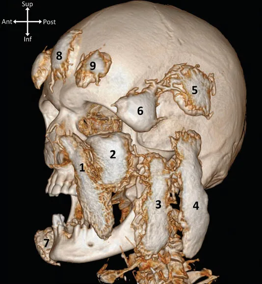 Fig 1. Superficial fat compartments of the upper face on 3D contrast-CT: 8 = central forehead, 9 = lateral forehead, 5 = superior temporal, 6 = inferior temporal. (Schenck, 2018, p. 3).
> Sources: Schenck 2018 [2].
Fig 1. Superficial fat compartments of the upper face on 3D contrast-CT: 8 = central forehead, 9 = lateral forehead, 5 = superior temporal, 6 = inferior temporal. (Schenck, 2018, p. 3).
> Sources: Schenck 2018 [2].
Temple, resolved (ten layers, Cotofana 2019) [1]: the five SCALP layers continue into the temple but change name past the superior temporal septum: the galea becomes the superficial temporal fascia (which carries the anterior and posterior branches of the superficial temporal artery), the periosteum becomes the deep temporal fascia.
 1  Skin
 2  Subcutaneous fat  (superficial temporal compartments; superficial technique deposits HERE, above the STA)
 3  Superficial temporal fascia  (= SMAS continuation; holds aSTA + pSTA)
 4  Loose CT: UPPER temporal compartment (no relevant NV) + LOWER temporal compartment
       (frontal branch of facial nerve [motor], zygomaticotemporal branch [sensory], sentinel vein)
 5  Superficial lamina of deep temporal fascia
 6  Superficial temporal fat pad + proximal sentinel vein (medial zygomaticotemporal vein)
 7  Deep lamina of deep temporal fascia
 8  Deep temporal fat pad (temporal extension of the buccal fat pad of Bichat)
 9  Temporalis muscle
10  Periosteum  (anterior + posterior deep temporal arteries run superficial to it)
The deep temporal fascia splits into superficial and deep laminae 2-5 cm cranial to the zygomatic arch, enclosing the superficial temporal fat pad and the proximal sentinel vein [1]. Deep-technique filler must sit on bone, close to the temporal crest, to avoid the deep temporal arteries [1]. The ten-layer temporal stack, with the temporalis, the deep temporal fascia laminae, the superficial temporal fat pad and the lower temporal compartment, is shown in Fig 2.
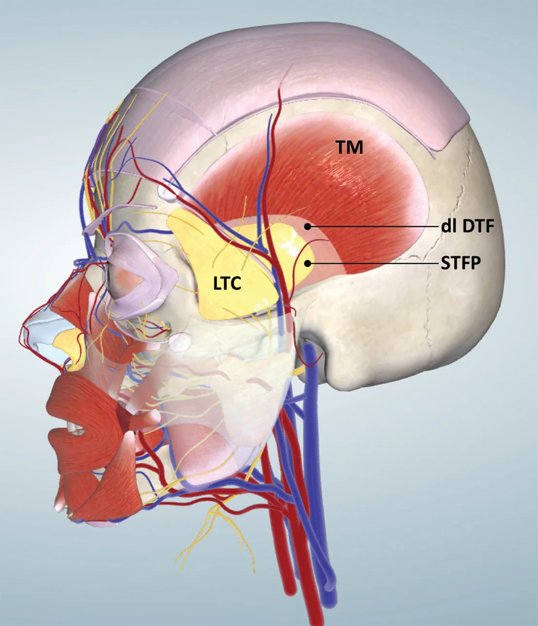 Fig 2. Ten-layer temporal anatomy: temporalis, deep temporal fascia laminae, superficial temporal fat pad and lower temporal compartment. (Cotofana, 2019, p. 8).
> Sources: Cotofana 2019 [1].
Fig 2. Ten-layer temporal anatomy: temporalis, deep temporal fascia laminae, superficial temporal fat pad and lower temporal compartment. (Cotofana, 2019, p. 8).
> Sources: Cotofana 2019 [1].
Glabella: thin skin directly over the muscle group (procerus in the midline; paired corrugator supercilii and depressor supercilii), scant subcutaneous fat, and a discrete glabellar fat body deep to the muscles [13]. The subfrontal fat carrying SO/STr converges here (Fig 3), and the supratrochlear and dorsal nasal arteries are terminal branches anastomosing directly with the ophthalmic artery, which is the anatomical basis of the region's blindness risk (D1.3, D1.10).
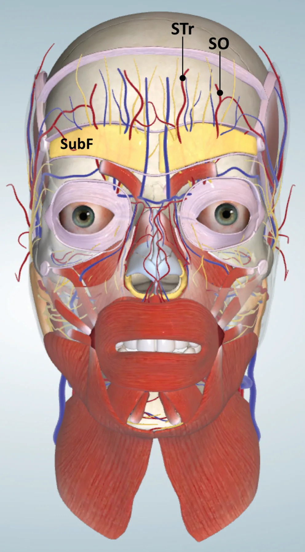 Fig 3. Subfrontal fat and the supratrochlear/supraorbital vessels it contains. (Cotofana, 2019, p. 5).
> Sources: Cotofana 2019 [1].
Fig 3. Subfrontal fat and the supratrochlear/supraorbital vessels it contains. (Cotofana, 2019, p. 5).
> Sources: Cotofana 2019 [1].
Surface landmarks the injector actually uses: the supraorbital notch/foramen (palpable at the medial third of the rim, marking the SO bundle), the temporal crest (the ridge that separates forehead from temple and the safe deep-temple deposit line), the temporal fusion line / superior temporal septum (the upper boundary of the temporal compartments), the zygomatic arch (the deep temporal fascia splits 2-5 cm above it), the frontozygomatic area (where the sentinel vein pierces), and the hairline (the upper limit of the treatable forehead). Marking these before injecting converts the layered anatomy above into a set of do-not-cross lines on the skin.
Two deep periorbital fat bodies that govern the brow and glabella: the ROOF (retro-orbicularis oculi fat) is the brow's deep cushion; its atrophy and descent are a main driver of brow heaviness and upper-lid folding with age, so a deep supraorbital deposit at the medial/central brow works by restoring the ROOF platform, not by filling a line [1][13]. The glabellar fat body lies deep to the procerus/corrugator knot; it is not a filler target (the terminal ophthalmic vessels converge here), which is another reason glabellar volume is handled by toxin and time, not product (D1.7). Neither is a place for a routine bolus.
Muscle balance (the functional one-liner): the frontalis is the only brow elevator; it has no inferior bony insertion and opposes corrugator, procerus, depressor supercilii and orbicularis. Everything on the forehead is the balance of those two forces, which is why weakening the elevator without addressing the depressors drops the brow (D1.8) [14].
Layer count and skin behaviour differ across the three subregions, which is why one rule cannot cover them:
| Subregion | Layers | Skin | Safe-plane logic |
|---|---|---|---|
| Forehead (paramedian) | up to 8 | thin centrally, thicker at hairline | plane inverts with height (SO/StR deep at rim → subcutaneous high) |
| Temple | 10 | thin, mobile | deep on crest OR subcutaneous above STA; mid-plane is the trap |
| Glabella | few (thin skin directly over muscle) | very thin, adherent | intradermal microdroplet only, if ever; terminal ophthalmic vessels |
Skin thickness governs product choice: the thin glabellar and central-forehead skin shows a superficial deposit (Tyndall, beading) and tolerates only the smallest, least-hydrophilic volumes, whereas the thicker temporal skin over a deep bony deposit hides product well.
Fat compartment summary of the region:
| Compartment | Layer | Clinical role |
|---|---|---|
| Superficial central + lateral forehead (SCFC, SLFC) | 2 | superficial diffuse volume; thin over frontalis |
| Subfrontal fat | 5 | vessel corridor (SO/STr); not a filler target |
| Deep central + lateral forehead (DCFC, DLFC) | 7 | deep structural deposit |
| ROOF | deep periorbital | brow support; atrophies with age |
| Superficial superior + inferior temporal | 2 | subcutaneous temple cannula plane |
| Upper temporal compartment | 4 | safe (no relevant NV) |
| Lower temporal compartment | 4 | frontal branch VII, sentinel vein: avoid |
| Superficial temporal fat pad | 6 | deep temple target with sentinel vein nearby |
| Deep temporal / buccal fat extension | 8 | deep volume; near deep temporal arteries |
Muscles of the region, with vector (the toxin map is a force map):
| Muscle | Origin → insertion | Action | Toxin role |
|---|---|---|---|
| Frontalis | galea aponeurotica → skin of brow/forehead | only brow elevator; folds horizontal lines | treat high, spare the lower band; never without depressors |
| Procerus | nasal bone/upper lateral cartilage → glabellar skin | pulls the medial brow down, folds the horizontal glabellar line | central point of the 5-point glabella map |
| Corrugator supercilii | superomedial orbital rim → mid-brow dermis | draws the brow medially and down, folds the vertical frown lines | two points per side; lateral point ≥1 cm above rim |
| Depressor supercilii | medial orbital rim → medial brow | depresses the medial brow | co-treated for a medial brow lift |
| Orbicularis oculi (orbital part) | medial orbital rim, encircles orbit | depressor of the brow (esp. the lateral tail) | lateral-tail point for the chemical brow lift |
The glabellar complex is a knot of these depressors around the procerus; the frontalis is their single antagonist, which is why the whole region reduces to one balance.
Frontalis shape and height variants: the frontalis is a paired muscle whose bellies can be unequal (asymmetric lateral lines are the rule, not a flaw), and its inferior extent and the presence of a central dehiscence vary; a high convex forehead, a narrow forehead, a low hairline and an asymmetric line pattern each demand a different point map, so the pivotal five-point frontalis template is a starting point, not a stencil (D1.8) [14][15]. The hairline and the lateral temporal crest bound the treatable zone.
Clinical use of the line of ligaments: the temporal-crest-to-mandible ligament line (temporal ligamentous adhesion, lateral orbital thickening, zygomatic and mandibular ligaments) separates a medial field where a deposit projects the overlying tissue from a lateral field where a deposit lifts more inferior tissue; the temporal deep deposit sits just lateral to this line, which is why a correctly placed temple deposit both fills the hollow and lends a lateral lift to the brow tail [1].
Classic pitfall: believing the deep-on-bone plane is safe throughout the forehead. It holds at the temporal crest and in the deep forehead compartments over frontal bone, but near the orbital rim the SO/STr vessels sit deep (subfrontal, layer 5) and only become subcutaneous higher up. The plane that is safe at the hairline is the dangerous one at the rim (D1.3).
D1.3 · Vessels, nerves and the danger zone
The one sentence that governs upper-third filler safety: the supratrochlear and supraorbital arteries emerge deep at the orbital rim (subfrontal fat, layer 5), then cross the inferior and middle frontal septa from deep to superficial as they ascend, becoming subcutaneous a few centimetres above the rim [1]. There is therefore no single safe plane for the whole forehead: deep-on-bone is safe high but dangerous low; subcutaneous is safe low but dangerous high (Fig 3, Fig 4).
Arterial map of the region:
| Vessel | Origin | Course / depth | Occlude it and… |
|---|---|---|---|
| Supratrochlear a. (STr) | ophthalmic (ICA) | exits ~1.7-2.2 cm from midline at/near the superomedial rim; deep at rim → subcutaneous high; terminal, direct ophthalmic anastomosis | retrograde embolus to central retinal a. → blindness; forehead/glabella skin necrosis [6][35] |
| Supraorbital a. (SO) | ophthalmic (ICA) | exits supraorbital notch/foramen lateral to STr; deep→superficial across the septa | same ophthalmic route; scalp/forehead necrosis [1][9] |
| Dorsal nasal a. | ophthalmic (ICA) | glabella/radix; terminal | direct ophthalmic route; the second half of "why not the glabella" [35] |
| Superficial temporal a. (STA) | external carotid | anterior + posterior branches within the superficial temporal fascia (layer 3); subcutaneous temple deposit is above it | temple skin necrosis, scalp; STA→ophthalmic anastomoses reported [8][36] |
| Deep temporal aa. (ant + post) | internal maxillary | run superficial to periosteum (layer 10) on temporalis | deep temple bleeding/haematoma; reason to keep deep deposits on the crest [1] |
| Middle temporal / sentinel vein | temporal venous system | large, thin-walled, valveless; sentinel vein pierces near the frontozygomatic area into the lower temporal compartment (layer 4/6) | venous embolism, pulmonary embolism reported; a temple-specific, non-arterial mechanism [1] |
Supratrochlear artery depths (Doppler, n=71) [6] measured for glabellar/forehead safety:
| Level | Epidermis-artery distance (EAD) | Artery-periosteum distance (APD) |
|---|---|---|
| Eyebrow | 1.8-5.9 mm | 0.7-3.7 mm |
| 1.5 cm above eyebrow | 1.8-5.1 mm | 0.6-3.8 mm |
EAD and APD rise with BMI; EAD rises with age; APD is greater in men. The operational reading: the artery is close to bone at the rim (APD as low as 0.6-0.7 mm), so "on periosteum = safe" fails there; the safest glabellar deposit is intradermal, just below the rhytide, not subcutaneous and not supraperiosteal [6].
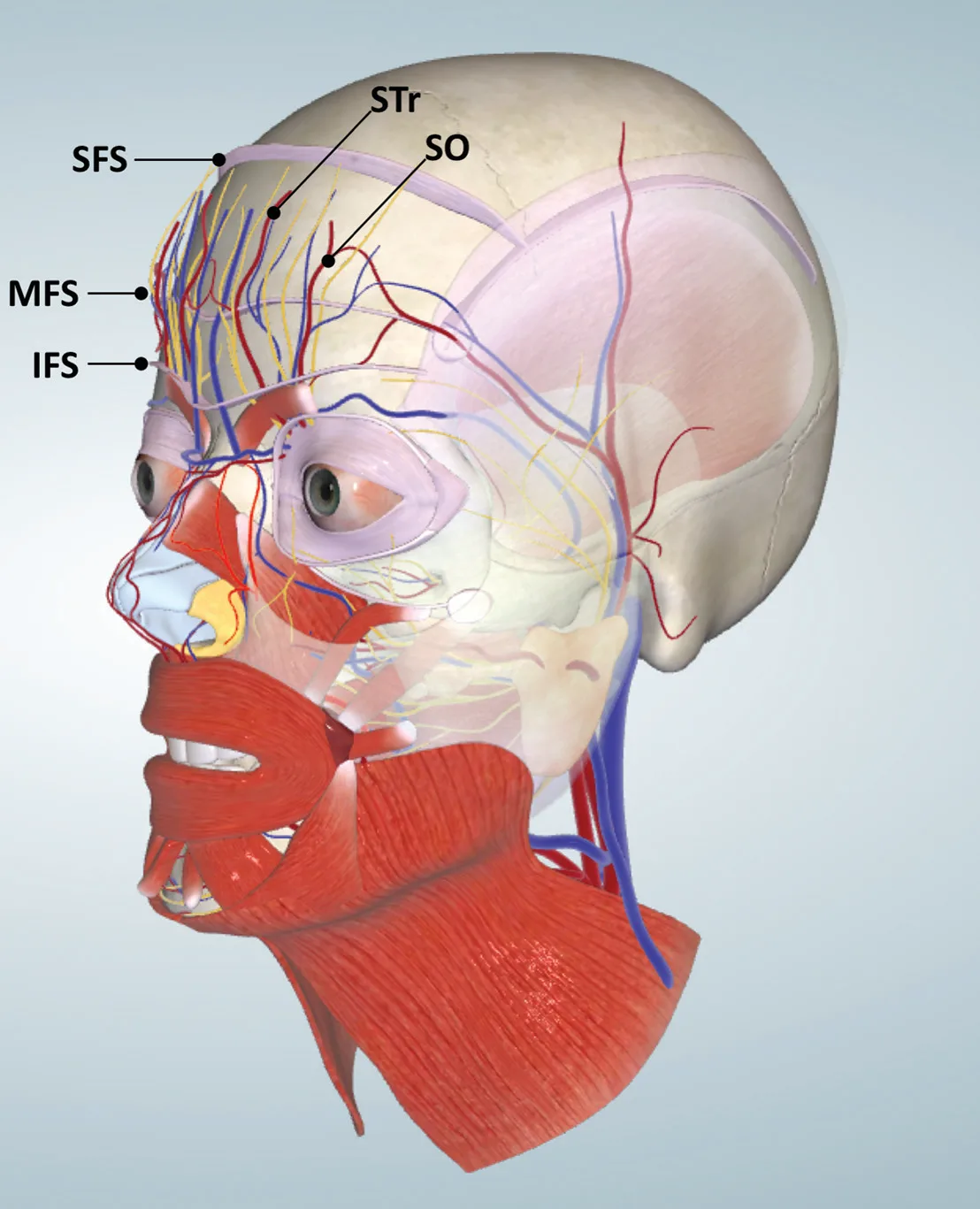 Fig 4. Supratrochlear/supraorbital vessels crossing the frontal septa from deep to superficial. (Cotofana, 2019, p. 6).
> Sources: Cotofana 2019 [1].
Fig 4. Supratrochlear/supraorbital vessels crossing the frontal septa from deep to superficial. (Cotofana, 2019, p. 6).
> Sources: Cotofana 2019 [1].
Venous anatomy of the temple (the reason a temple bolus is different from a cheek bolus): the temporal region drains through the middle temporal vein and the sentinel vein (medial zygomaticotemporal vein), large and thin-walled channels that, being valveless, offer no barrier to retrograde flow; a fast or high-volume bolus can therefore enter the venous system and embolise to the lungs [1]. The sentinel vein is often visible transcutaneously at the frontozygomatic area, and a visible vein at a planned entry is a direct instruction to move the point. This venous mechanism is independent of, and additional to, the arterial one, and it is the part of temple safety most often omitted from teaching.
Arterial territories, so you can read a necrosis pattern: the supratrochlear supplies a paramedian forehead strip and the medial brow; the supraorbital supplies the lateral forehead and anterior scalp; the superficial temporal branches supply the temporal skin and the lateral scalp; a necrosis that maps to one of these territories names the vessel that was hit and guides the flooding of hyaluronidase to that territory [8][9][31]. Skin mottling along a territory during injection is an occlusion until proven otherwise.
Sensory nerves (V1): the frontal nerve divides intra-orbitally into supraorbital (SON) and supratrochlear (STN). Cadaveric variants matter for both block anaesthesia and for predicting where a vessel travels with its nerve [7]:
- Two intra-orbital division patterns: variant I 42% (division in the distal half of the orbit), variant II 58% (division in the proximal half, giving a thick SON and a long thin STN) [7].
- SON splits into medial + lateral branches within the orbit in 27.8% of variant I versus 75% of variant II; a single passage over the supraorbital margin in ~80% of wet specimens and ~78% of skulls [7].
- The SON exits a true foramen in a variable minority and a notch in the majority; a foramen tethers the bundle and changes where a deep deposit meets it [1][7].
Arterial variability that changes the plan. The supratrochlear exits at roughly 1.7-2.2 cm from the midline but its exact position and its depth vary between people and sides; the supraorbital exits a notch in most and a true foramen in a minority, and a foramen tethers the bundle and fixes where a deep deposit meets it [1][7]. The superficial temporal artery has several documented branching patterns and can run unusually anteriorly, so the "safe" subcutaneous temple path is patient-specific, which is the practical argument for Doppler mapping before a high-risk temple deposit rather than trusting a textbook average [8][9].
Motor nerve (the temple's other danger): the frontal (temporal) branch of the facial nerve runs superficially in the temporal region, in the plane between SMAS (superficial temporal fascia) and the deep temporal fascia, within the sub-SMAS fat; accidental deep dissection or product bolus there risks frontalis paralysis and a true (not toxin) brow ptosis [5] (Fig 5).
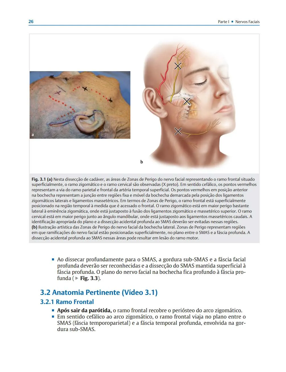 Fig 5. Frontal branch of the facial nerve in the temporal danger zone. (Rohrich, Facial Danger Zones, 2020, p. 33).
> Sources: Rohrich 2020 [5].
Fig 5. Frontal branch of the facial nerve in the temporal danger zone. (Rohrich, Facial Danger Zones, 2020, p. 33).
> Sources: Rohrich 2020 [5].
What gets occluded if you fail, by site (link to J2 — Vascular Occlusion & Emergency Response):
- Glabella / medial brow: STr or dorsal nasal → ophthalmic → central retinal artery. Vision loss is immediate, painful, and not reliably reversed by hyaluronidase even when the protocol is run in full [35][39].
- Central/high forehead: SO/STr subcutaneous branches → skin necrosis in a paramedian patch; ophthalmic retrograde still possible with high pressure.
- Temple, arterial: STA branches → temporal/scalp necrosis; ophthalmic anastomosis makes vision loss possible.
- Temple, venous: middle temporal / sentinel vein (valveless) → venous filler embolism with reported pulmonary embolism, a mechanism most temple courses omit [1].
The anastomotic trap (why a distal injection can blind). The external-carotid facial/angular system and the internal-carotid ophthalmic system meet at the medial canthus, glabella and dorsal nose; the supratrochlear, dorsal nasal and angular arteries form a continuous network with the ophthalmic artery, so a high-pressure bolus anywhere along it can drive filler retrograde into the ophthalmic artery and then anterograde into the central retinal artery once pressure is released [8][34][40]. This is why "the needle is far from the eye" is false reassurance in the glabella and medial brow.
Aspiration, honestly. A pre-injection aspiration can catch some intravascular needle positions but has a real false-negative rate: small terminal vessels, high-viscosity product and thin needles all defeat it, and a moving needle changes position after the aspiration [30][31]. It is a low-cost adjunct, not a clearance test; the safeguards that actually lower risk are plane, micro-volume, low pressure, motion, cannula in high-risk broad planes, ultrasound, and hyaluronidase in the room [31][41].
Cannula-versus-needle vessel mechanics. A blunt cannula pushes vessels aside rather than piercing them and lowers the observed occlusion odds across broad subcutaneous planes, but a moving cannula can still enter a tethered or larger vessel, and a larger-gauge cannula resists rather than prevents cannulation; needle precision is preferable for a bone-contact deposit at the crest [31][32][33]. Selection is by target plane, not by dogma.
Ultrasound signs of an evolving occlusion. On Doppler, loss of the expected arterial signal in a territory, a hyperechoic intraluminal filler column, and a mottled dermal perfusion pattern flag an occlusion earlier than the eye does; a pre-mapped baseline makes the change detectable, which is the argument for scanning before and re-scanning on any pain or blanching [9].
Deep temporal arteries, restated for the injector. The anterior and posterior deep temporal arteries (internal maxillary branches) run superficial to the periosteum on temporalis, so a "deep on bone" temple deposit must sit on the crest and be small, not swept medially across the muscle where these vessels run [1].
Classic pitfall: aspirating, seeing no flash, and treating that as an all-clear at the glabella. A negative aspiration does not exclude an intra-arterial needle in a small terminal vessel; the safeguards that matter are plane (intradermal), volume (micro), pressure (low), and motion, plus hyaluronidase ready [6][35][41].
D1.4 · Aging of the region, in order
Aging is a layered process and the layers do not age in step. The upper third loses, roughly in this sequence: bone first, then deep fat and ligament, then superficial fat, then skin [3][10]; Fig 6 links these tissue-layer changes youthful-versus-aged as one chained process. Reading the sequence backwards is the classic error: chasing a static forehead line (a skin/muscle sign) while the actual deficit is skeletal and deep-fat volume.
1 · Bone (the scaffold moves first). The frontal bone and orbital rim remodel lifelong [3][10][11]. Documented changes: protrusion of the glabella, expansion of the supraorbital ridges, lateral translation and widening of the orbital aperture, and a decrease in the glabellar, orbital, maxillary and pyriform angles with age [3][12]. The frontal bone thins and the soft tissue over it is thin to begin with, so frontal hollowing and a more skeletal upper face follow [1][11]. The widening orbital aperture undermines brow and periorbital support from below.
2 · Deep fat (structural deflation). The deep forehead compartments (DCFC, DLFC) and the deep temporal fat deflate; the temple, already thin in the upper temporal compartment, hollows and the temporal crest and superficial temporal vessels become visible [1]. Deep-fat loss removes projection, so the brow and lateral canthus lose their platform.
3 · Ligament (the hammock slackens). The retaining ligaments are biomechanically stiff (zygomatic stiffest, then orbital retaining, then mandibular) and may not intrinsically degrade much; what changes is their bony origin, shifted by skeletal remodelling, so the ligament's line of pull alters and the compartments it suspends descend [3]. The orbicularis retaining ligament tilts and ROOF support fails, feeding brow and upper-lid descent.
4 · Superficial fat (descent and thinning). Superficial forehead and temporal compartments thin; the ROOF descends into the frontal areolar plane, manifesting as brow heaviness/ptosis with upper-lid skin folding [13]. Loss here is why the lateral brow tail falls even when the muscle is intact (Fig 7). Fig 9 shows the superficial fat progressively losing volume and sliding inferiorly across three aging stages.
5 · Muscle (dynamic to static). Facial muscles undergo sarcopenia: they lengthen, resting tone rises toward maximal-contracture tone, and amplitude shortens [3]. Clinically, dynamic lines convert to static, etched lines, and the frontalis becomes chronically overactive, both cosmetically (deep horizontal lines) and compensatorily (holding ptotic lids up, D1.6). Fig 8 maps the glabellar wrinkling from procerus/corrugator/depressor supercilii action alongside the periorbital ROOF sag in the aged half.
6 · Skin (the envelope). Photoaging, dermal collagen/elastin loss and elastosis etch the lines the muscle folds, so a line that was purely dynamic at 30 is partly dermal at 55: toxin alone will not erase it (D1.7 skin lane).
What is lost, and what it demands:
| Layer | Upper-third change | Order | Correct modality |
|---|---|---|---|
| Bone | glabellar protrusion, supraorbital ridge expansion, orbital aperture widening, frontal thinning | 1st | deep structural filler / defer; not toxin |
| Deep fat | deep forehead + temporal deflation → temple hollow, skeletal forehead | 2nd | supraperiosteal/deep HA, CaHA, PLLA, fat |
| Ligament | origin shifted by bone → altered pull, ROOF/brow descent | 3rd | lateral support (temple/cheek), energy, surgery |
| Superficial fat | forehead/temporal thinning, ROOF descent → brow ptosis | 4th | subcutaneous support; toxin depressors for brow |
| Muscle | sarcopenia, dynamic→static lines, frontalis overactivity | 5th | toxin (balance elevator/depressors) |
| Skin | elastosis, etched lines | 6th | skinboosters, laser, peel, microneedling |
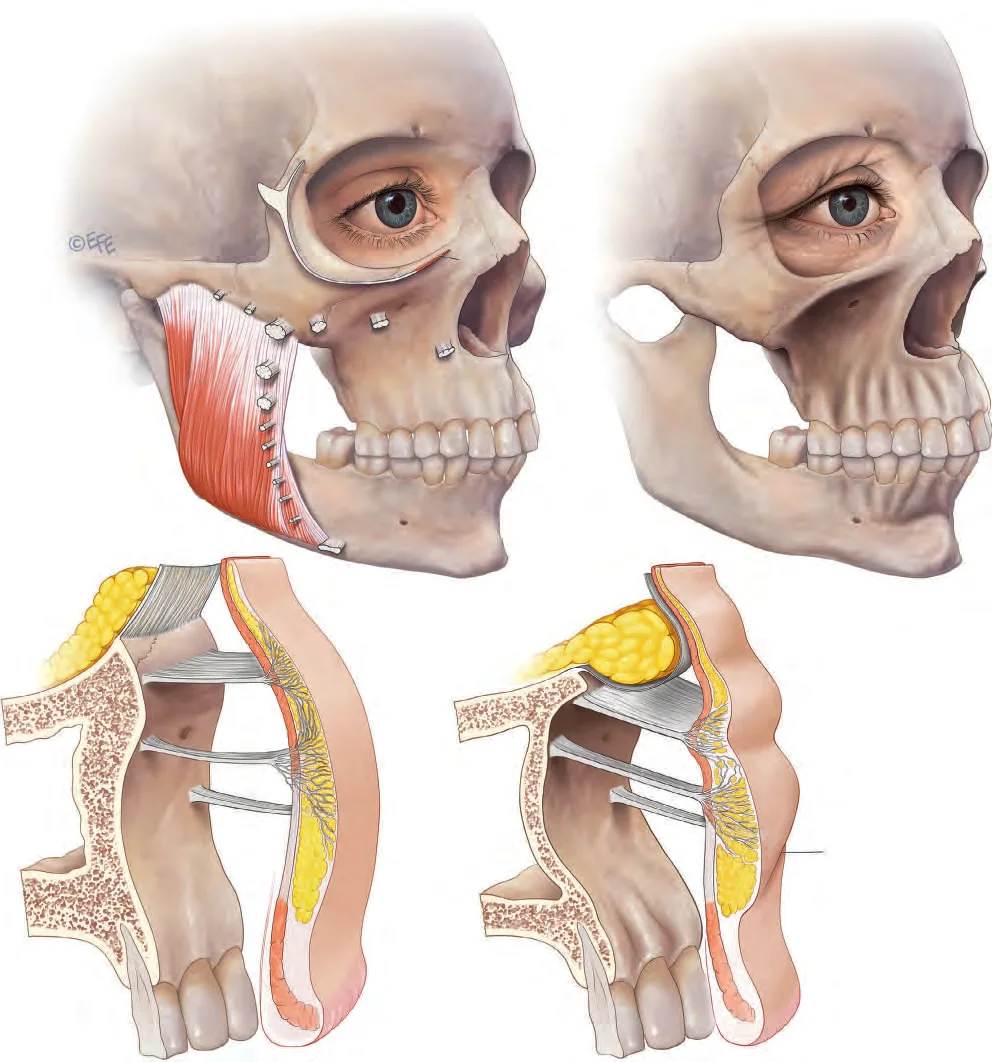 Fig 6. Linking of aging changes through the tissue layers, youthful vs aged. (Standring, Gray's Anatomy, 2015, p. 960).
> Sources: Standring 2016 [4].
Fig 6. Linking of aging changes through the tissue layers, youthful vs aged. (Standring, Gray's Anatomy, 2015, p. 960).
> Sources: Standring 2016 [4].
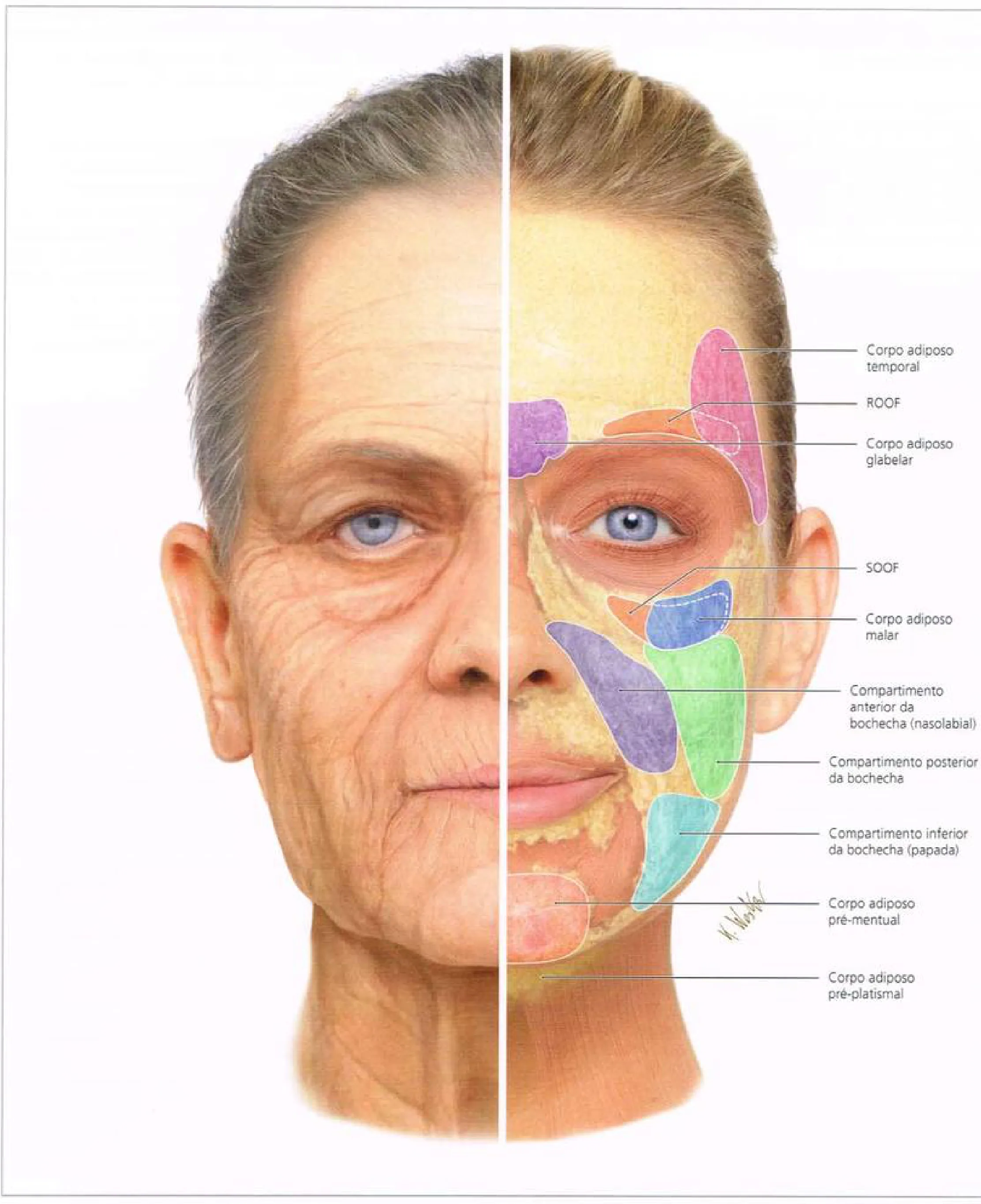 Fig 7. Upper-face fat compartments, youthful versus aged (temporal fat, ROOF, glabellar fat, SOOF). (Radlansky, Atlas Ilustrado de Anatomia Clínica da Face, p. 58).
> Sources: Radlansky [13].
Fig 7. Upper-face fat compartments, youthful versus aged (temporal fat, ROOF, glabellar fat, SOOF). (Radlansky, Atlas Ilustrado de Anatomia Clínica da Face, p. 58).
> Sources: Radlansky [13].
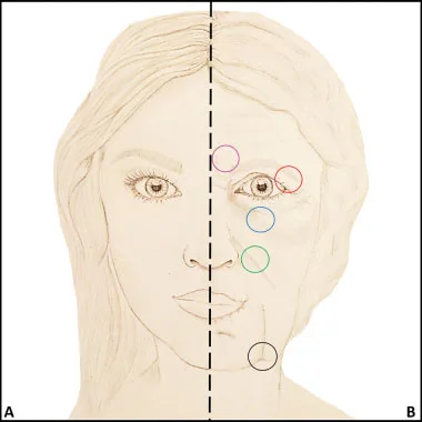 Fig 8. Anatomy of the aging face, glabellar and periorbital changes. (Cotofana, 2016, p. 5).
> Sources: Cotofana 2016 [3].
Fig 8. Anatomy of the aging face, glabellar and periorbital changes. (Cotofana, 2016, p. 5).
> Sources: Cotofana 2016 [3].
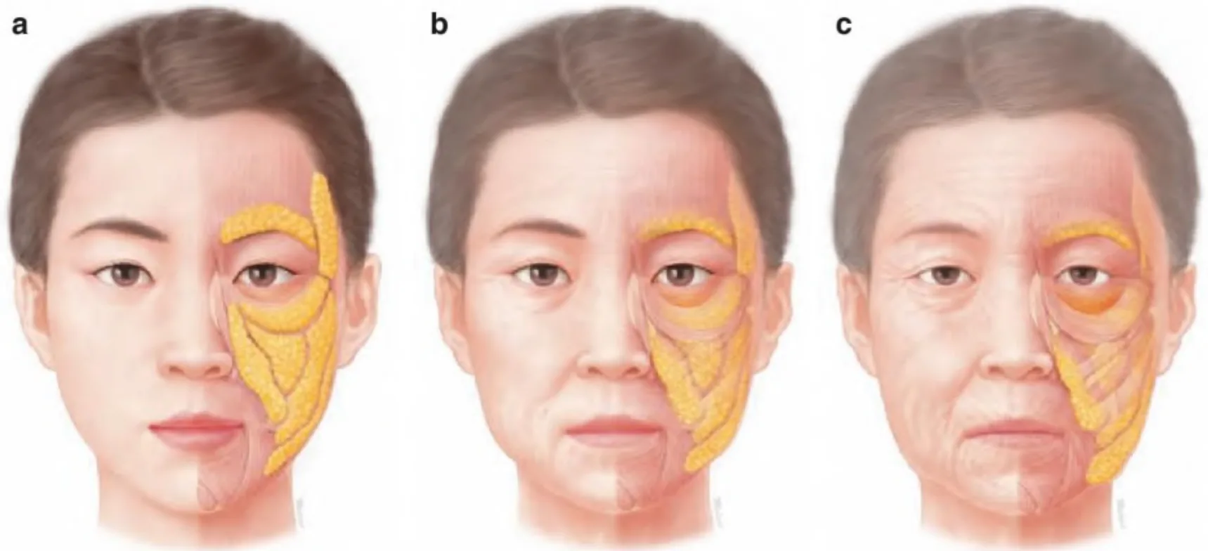 Fig 9. Progressive volume loss and descent of the superficial fat compartments with aging. (Kim, Clinical Anatomy of the Face, 2016, p. 59).
> Sources: Kim 2016 [MEDLIB]; corpus figure caption mislabelled it a "bone resorption a/b/c views", so it is captioned here from the opened image, not the corpus text.
Fig 9. Progressive volume loss and descent of the superficial fat compartments with aging. (Kim, Clinical Anatomy of the Face, 2016, p. 59).
> Sources: Kim 2016 [MEDLIB]; corpus figure caption mislabelled it a "bone resorption a/b/c views", so it is captioned here from the opened image, not the corpus text.
Skeletal angles, measured. Population studies quantify the skeleton-first change: the glabellar, orbital, maxillary and pyriform angles decrease with age, and the maxilla, pyriform aperture and infraorbital rim regress, driving the anterior orbital rim forward and undermining periorbital support; the orbital aperture widens, especially superomedially and inferolaterally [3][10][12]. Asian-population data show the same direction with a less marked orbital/maxillary change and a more marked pyriform change than in Caucasian series, so the direction of aging is shared but the magnitude is population-specific [3]. Facial-bone aging updates reinforce that the skeleton is the moving foundation, not a fixed frame [11].
The deflation-versus-descent debate, resolved for practice. Two schools describe upper-third aging: deflation (fat and bone volume loss) and descent/attenuation (ligament fatigue and gravity). The evidence supports both operating together: bone remodelling shifts ligament origins, which lets the deflating compartments descend, so the clinical answer is not "volume vs lift" but sequence-restore the skeletal/deep-fat platform, then address descent that remains [3]. This is why volume in the temple can lift a brow tail (platform) while volume in the central forehead cannot correct a ptotic brow (descent).
Sex and the aging upper third. Men start with a lower, flatter brow, a heavier glabellar complex and thicker skin; they lose bone and deep fat on the same schedule but the descent reads differently, and over-lifting during correction feminises the gaze [16]. Women show earlier brow-tail descent as the lateral platform (temple/lateral cheek) empties. Both are treated by the same order (bone/deep fat first), with a sex-specific brow-shape target (D1.6, D1.8).
Clinical translation of the aging order (why sequence beats modality-choice): because the platform (bone, deep fat) fails before the surface (superficial fat, skin), restoring the surface without the platform gives a heavy, over-filled result that still looks aged; and because muscle converts dynamic lines to static ones over decades, a 30-year-old frown is toxin-responsive while the same line at 55 is part dermal and needs the skin lane too. The order therefore reads as a treatment sequence: skeletal/deep-fat support and toxin first, superficial support and skin lane second, which is also the safest order because the deep, high-yield temple step is done before any central-forehead or glabellar consideration [3][10]. A patient treated against the aging order (skin first, structure last) spends more, looks less natural, and is exposed to the higher-risk central sites unnecessarily.
Temporal aging in detail. The upper temporal compartment is nearly avascular and thin from the start, so it hollows early and predictably; the lower temporal compartment and the deep/superficial temporal fat pads deflate later, and the temporalis itself can atrophy, together exposing the temporal crest and the superficial temporal vessels, which both worsens the aesthetic and raises the injection risk (visible vessels are cannulable vessels) [1].
Classic pitfall: offering forehead filler for a "flat forehead" that is really an aged skeleton plus temporal deflation. Restore the temple and orbital rim first; the central forehead step often disappears, and the central forehead is the higher-risk site [1].
D1.5 · Assessment: measure, photograph, animate, scan
Measure (at rest and at maximal animation):
| Parameter | How | Why it decides |
|---|---|---|
| Brow position vs bony rim | relate the brow to the supraorbital rim, per side | male brow sits at/near the rim, female above; elevating a male brow feminises the gaze [16] |
| Brow-tail height | mark peak and tail, compare sides | tail descent is fat/ligament, not muscle; predicts who needs lateral support not toxin [1] |
| Margin-reflex distance (MRD1) | pupil light reflex to upper-lid margin | separates true blepharoptosis (surgical) from brow ptosis [20] |
| Frontalis compensation | observe the resting forehead unobserved; ask to open eyes without using the brows | a constantly raised resting brow marks a frontalis crutch, a relative contraindication to frontalis toxin [14] |
| Interbrow / glabellar contraction pattern | frown; classify V, U, converging arrows, omega, inverted omega | historically used to tailor points; 2026 multicentre data show a standardized 5-point algorithm works across patterns [17] |
| Temple depth grade | inspection + palpation; visible vessels/arch | grades the deficit and the risk (visible vessels are cannulable vessels) |
| Forehead line severity | dynamic + static, per band, note asymmetry | lateral frontalis bellies are often unequal; treat asymmetrically [14] |
| Lid tone (snap/distraction test) | before any pretarsal or low periorbital work | poor tone contraindicates lid-margin techniques |
Temple deficit grading (region-specific):
| Grade | Finding | Implication |
|---|---|---|
| Mild | subtle concavity, contour still continuous brow-to-arch | little product; question whether the risk is worth it |
| Moderate | clear depression, temporal crest visible, marked transition to brow tail | clearest indication, best aesthetic yield |
| Severe | skeletonised temple, visible vessels, prominent zygomatic arch | high volume over sessions; ⚠ maximal vascular risk, the vessels you see are the ones you can cannulate |
Photograph: standardized, same distance/lighting/background, at rest and at maximal animation (brow elevation, frown, forceful eye closure), frontal, oblique and profile. Without the animation frame there is no honest before/after and no way to document baseline asymmetry, which must be recorded before treatment, not discovered after [14].
Animate (the dynamic exam, in order): elevate brows (frontalis strength, line pattern, asymmetry) → frown (glabellar complex, corrugator/procerus/depressor supercilii vectors) → gentle then forceful eye closure (orbicularis, lateral tail) → then the two decisive manoeuvres: watch the resting forehead when the patient is not aware, and ask them to open the eyes without lifting the brows. A patient who cannot is compensating (D1.6).
Scan (Doppler ultrasound, before high-risk filler): duplex Doppler maps the supratrochlear and supraorbital arteries and the superficial temporal branches, and identifies the sentinel/middle temporal vein before temple work [6][9]. High-risk artery mapping for the forehead has been formalised on Doppler and shifts the entry point and plane per patient [9]. Ultrasound also confirms deposit plane in real time and detects an early occlusion. It does not make the glabella safe; it lowers, not abolishes, the risk, and the default (avoid glabellar filler) stands [6][35].
Validated severity scales (use them to grade and to track):
| Scale | What it grades | Use |
|---|---|---|
| Glabellar Line Severity Scale (GLSS) | glabellar frown, 0-3, at rest and max frown | the standard glabellar endpoint; the 2026 pattern study used it [17] |
| Forehead / dynamic line scales | horizontal forehead lines, at rest and max elevation | separates dynamic from etched, drives toxin vs skin lane |
| Allergan Temple Hollowing Scale (ATHS) | temple concavity grade | temple filler endpoint; the PLLA RCT used it (96.5% ≥1-grade at month 6) [28] |
| Merz/GAIS global aesthetic improvement | overall change | patient-reported endpoint across upper-third studies [17][28] |
Standardised photography protocol: fixed camera-to-subject distance, identical lighting and plain background, neutral head position (Frankfort plane), and matched frames at rest and at maximal animation (brow elevation, frown, forceful closure) in frontal, oblique and profile. Repeat every session for honest before/after and to catch baseline asymmetry; store with the record, not the phone.
Doppler ultrasound protocol (before high-risk filler, and to teach the plane): identify and mark the supratrochlear and supraorbital arteries at and above the rim, note the epidermis-artery depth (which varies with BMI, age and sex) [6]; map the anterior/posterior superficial temporal artery branches and the sentinel/middle temporal vein before temple work [1][9]; confirm the deposit plane in real time; and rescan if pain or blanching appears to catch an early occlusion. Ultrasound lowers, it does not abolish, the risk, and it does not license glabellar filler [6][9].
Brow-position measurement, with the caveat: the "ideal" brow position is an aesthetic convention that has shifted across decades and differs between populations and sexes; measure and record the patient's actual brow-to-rim relationship and inter-side difference as a descriptive baseline, not as a target to force (cross-ref B5 — Schools of Facial Beautification — Competing Philosophies, B6 — Ethnic, Racial & Cultural Considerations in Face & Body Reshaping).
Baseline record before the first treatment (what the note must contain): brow position per side relative to the rim; MRD1 per side; the frown pattern; frontalis compensation (present/absent, with the two manoeuvres documented); temple grade; forehead line severity at rest and on animation, noting asymmetric bellies; lid tone; any pre-existing ptosis or asymmetry the patient has not noticed; and the standardized photo set. Documenting a pre-existing asymmetry before treatment converts a later complaint into an expected finding rather than an alleged complication.
Structured exam order (repeatable): inspect at rest → elevate brows → frown → gentle then forceful eye closure → watch the resting forehead unobserved → open eyes without brows → palpate the temple and the brow-supporting compartments → Doppler if high-risk filler is planned. Running the same order every time is what catches the compensator and the baseline asymmetry that a freehand look misses.
Classic pitfall: measuring only at rest. The line the patient hates may vanish on animation (pure dermal, treat skin) or only appear on animation (pure dynamic, treat muscle), and the brow that looks symmetric at rest may be markedly asymmetric on elevation. A rest-only exam mis-assigns the modality.
D1.6 · Goal and patient selection: who benefits, who does not, who is referred
Who benefits:
| Profile | Finding | First move |
|---|---|---|
| Dynamic frown/forehead lines, good dermis | lines only on animation | toxin, excellent yield [14][18] |
| Moderate temple hollow, good skin | clear concavity, crest visible | temple filler (best-yielding upper-third filler) [1][24][28] |
| Etched static lines, damaged dermis | lines visible at rest | toxin plus skin lane (skinbooster/laser/peel), staged [3] |
| Skeletal forehead / orbital-rim loss | frontal hollowing, rim step | deep structural filler or defer; not toxin |
| Ptotic brow tail, muscle intact | tail descent, fat/ligament loss | toxin depressors then lateral support ladder [1][21] |
Who does not benefit (and why offering treatment harms):
- Frontalis compensator. A patient with dermatochalasis or true ptosis who keeps the brows elevated to see is using frontalis as a crutch; weakening it closes the visual field. This is a strong relative contraindication to frontalis toxin, and it must be actively sought, not asked about, because patients do not volunteer it [14].
- True blepharoptosis / dermatochalasis with lid-on-lash overhang. Neither toxin nor filler removes skin or lifts the lid; offering it produces a disappointing result and erodes trust. This is surgical (see referral) [20].
- Volume-loss mistaken for a line. A skeletonised forehead treated as a wrinkle problem gets over-toxined and under-corrected.
- Unrealistic expectation / body dysmorphic features, or a patient who wants a "frozen" upper face. The frozen forehead, dropped brow and dissatisfied patient is a selection failure, not a dosing one.
Who is referred:
| Sign | Referral |
|---|---|
| Upper-lid skin hanging over the lash line | oculoplastics / blepharoplasty |
| True blepharoptosis with visual-field loss | oculoplastic surgery |
| Marked frontal/brow descent beyond injectable range | brow-lift surgery |
| Neuromuscular disease (myasthenia gravis, Eaton-Lambert, ALS) | neurology; toxin is contraindicated [see J8 — Contraindications & Special Populations] |
Sex-specific selection [16]: the male brow is lower (at or near the rim), flatter, thicker, with a less lateral apex; elevating and arching it feminises the gaze, one of the most common and least-discussed unwanted effects of upper-third treatment in men. Men also have a heavier, thicker frontalis and glabellar complex and generally need higher toxin doses than the female templates imply; under-dosing a man reproduces the frown in weeks [16][23]. Brow shape in men is a decision, not a default inherited from the female pattern.
Toxin-relevant contraindications to screen (detail in J8 — Contraindications & Special Populations): neuromuscular disease (absolute); aminoglycosides and other agents that potentiate neuromuscular blockade; pregnancy and lactation; active infection at the site; prior hypersensitivity or suspected toxin resistance.
Referral thresholds, made concrete: upper-lid skin resting on or overhanging the lash line (functional dermatochalasis) is blepharoplasty, not filler or toxin; a low upper-lid margin with reduced MRD1 and a normal brow is true blepharoptosis for oculoplastic surgery; a brow that sits well below the rim with skin redundancy beyond what a chemical brow lift can move is a brow-lift candidate; and a "tired eyes" complaint that is actually visual-field-driven compensation is an ophthalmology referral before any toxin. Offering an injectable across these thresholds produces a predictable disappointment and is the fastest way to lose a patient's trust in the region [20]. The honest sentence, said early, is that some upper-third problems are surgical.
Selection logic that de-risks the whole region: when volume need is uncertain, treat toxin first in its own session and reassess at 2 weeks. Toxin changes the surface anatomy (a relaxed frontalis drops the compensatory elevation; a relaxed glabella lets a static line soften over 2-3 months), and the volume decision made after that is smaller, safer and more accurate than the one made on day zero [14].
Special populations and situational cautions:
| Situation | Handling |
|---|---|
| Pregnancy / lactation | defer toxin and elective filler [see J8 — Contraindications & Special Populations] |
| Prior permanent/semi-permanent filler in the region | map it (ultrasound); avoid layering reversible over unknown permanent product; irreversible-in-max-risk-zone caution compounds |
| Anticoagulants / antiplatelets | higher bruising/haematoma risk; do not stop without the prescriber; fine needle, compression, consider deferring temple bolus |
| Immunosuppression / active infection | defer filler; biofilm and infection risk |
| Dry eye, poor lid tone, prior blepharoplasty | avoid lower-periorbital and lid-margin techniques; the compensator screen matters more |
| Neuromuscular disease, aminoglycosides | toxin contraindicated / potentiated J8 — Contraindications & Special Populations |
| Keloid / post-inflammatory hyperpigmentation tendency | caution with the skin lane (laser/microneedling), test and stage |
Psychological selection and expectation: screen for body-dysmorphic features and the patient who wants a "frozen" or radically altered upper face; the glabella carries a documented research line as an adjunct in depression (randomized trials exist) but this is not an approved psychiatric indication and is not offered as treatment [C]. What is usable today is reframing the goal: in the glabella the aim is to change a negative expression (frown), not to erase a line, which improves realistic satisfaction (cross-ref B2 — Patient Psychology & Selection).
The consultation as the decision point (and its economics): the upper-third consultation decides more than any gesture that follows it, because assigning the four problems correctly (dynamic, etched, volume, descent) and screening the compensator determines whether the patient gets a low-risk, high-satisfaction toxin result or an over-filled, higher-risk one. Treating toxin-first and reassessing at 2 weeks also spends the least product: a temple/rim step deferred until the relaxed face is seen is often smaller than the one planned on day zero, and the central-forehead step frequently disappears. The disciplined pathway is cheaper for the patient and safer, which aligns the incentive with the anatomy [1][14].
Consent specifics for the region: name the blindness risk explicitly (not a generic formula) before any upper-third filler and state it can be permanent; list off-label areas; state the expected duration and the possibility of asymmetry and a 2-week retouch; confirm hyaluronidase availability for HA; and record that eyelid-skin excess or true ptosis was discussed as surgical when present [20][35].
Timing: avoid treating immediately before a major event (bruising, the 2-week toxin settling window, and the possibility of a retouch); schedule toxin at least 2 weeks ahead of a deadline.
Classic pitfall: treating the modality the patient names ("fill my forehead lines") instead of the problem the exam finds (dynamic lines + skeletal deflation + a compensating frontalis). Decide by problem, not by request.
D1.7 · Technique: the full regional grid
This block closes with the whole grid of options for the region, not one technique. Every row carries school, plane and evidence. A row without an alternative is an unclosed region.
Product axis (what, by rheology and behaviour):
| Product | Behaviour | Where in the upper third | Reversible | Evidence |
|---|---|---|---|---|
| HA, high G′ / high cohesivity | firm structural projection on bone | deep temple (supraperiosteal), deep forehead compartment | yes (hyaluronidase) | [24][30] [C] |
| HA, low G′ / low cohesivity | soft, spreads, superficial | subcutaneous temple, intradermal glabellar microdroplet | yes | [6][30] [C] |
| CaHA, standard | volumiser + biostimulation, subdermal/supraperiosteal | temple (deep) | ⚠ no antidote | [MEDLIB] [C] (cross-ref A4 — Bioestimuladores: ciencia de materiales) |
| CaHA, hyperdilute | biostimulation > volume, diffuse subdermal | thin/skeletonised temple, skin trophism | ⚠ no | [MEDLIB] [C] |
| PLLA | gradual collagen-mediated volume over weeks-months | temple (RCT-supported) | ⚠ no | [28] [B] |
| PCL (polycaprolactone) | collagen stimulation, longer scaffold | temple volume, off-label upper third | ⚠ no | [MEDLIB] [C] |
| PMMA | permanent microsphere implant | ⚠ not advised upper third (irreversible in max-risk zone) | no | [MODELO] |
| Autologous fat | living graft, large-volume temple | temple, brow platform | partial | [29] [C] |
| Polynucleotides / PN-PDRN | dermal regeneration, no volume | etched glabellar/forehead lines (intradermal) | n/a | [MEDLIB] [C] (cross-ref F2 — PRP, Polynucleotides (PN-PDRN) & Exosomes) |
| Skin booster (low-crosslink HA) | hydration/quality, no lift | etched forehead lines | yes | [MEDLIB] [C] |
| Toxin | weakens, does not fill | see D1.8 | n/a (wanes) | [14] [A] |
Rheology logic, not averaged numbers: higher G′ and cohesivity buy projection on bone (deep temple/forehead); lower G′ and cohesivity buy soft, superficial correction (glabellar microdroplet). Specific G′ values vary by product and are not averaged here; see A6 — Mapa comparativo de rellenos y equivalencia entre marcas. The same rule governs CaHA dilution: more dilution = more biostimulation, less volume; less dilution = more volume, more superficial-nodule risk [MEDLIB].
Instrument axis:
| Tool | Typical calibre/length | Use in region | Note |
|---|---|---|---|
| Needle 30-32 G, short | glabellar intradermal microdroplet; toxin | precise, superficial | in a terminal-vessel field, precision does not equal safety [6] |
| Needle 27 G, on bone | deep temple supraperiosteal on the crest | bone-contact deposit | keep contact, aspirate, low pressure [1] |
| Cannula 22-25 G, 50-70 mm | subcutaneous temple; forehead from lateral entry | broad-plane spread, fewer occlusion events | not absolute protection; a moving cannula still enters vessels [31][32][33] |
| Ultrasound guidance | any high-risk deposit | maps STr/SO/STA, confirms plane, catches early VO | lowers risk, does not abolish it [6][9] |
Plane axis (region-specific meaning): supraperiosteal (deep temple on crest, deep forehead compartment) · deep fat (subfrontal is a vessel corridor, not a target) · SMAS/superficial temporal fascia (holds the STA, avoid) · superficial fat (subcutaneous temple/forehead cannula plane) · subdermal (hyperdilute CaHA, biostimulation) · intradermal (glabellar microdroplet, etched lines).
Movement axis: bolus (deep temple on bone, small) · microbolus (deep forehead compartment) · retrograde linear threading (subcutaneous forehead/temple with cannula) · fan (subcutaneous temple from one entry) · cross-hatch (diffuse superficial temple) · serial puncture / microdroplet (glabellar intradermal, etched lines) · tower/column (deep temple, layered on bone). Never a large fast bolus in the temple (valveless middle temporal vein) [1].
Temple, point by point (the best-yielding, highest-risk upper-third filler):
- Deep supraperiosteal (on bone at the crest). Entry: single point ~1 cm above the zygomatic arch and behind the temporal crest line. Direction: needle perpendicular to firm bone contact, aspirate, deposit a small bolus (tower) on periosteum, keeping medial to avoid the deep temporal arteries which run superficial to periosteum [1]. Product: high-G′ HA or CaHA. Volume: small per point (order of 0.2-0.5 mL), do not chase the whole hollow from one deep bolus. Retouch at 2-4 weeks.
- Subcutaneous (superficial, above the STA). Entry: lateral, cannula 22-25 G, plane in layer 2, above the superficial temporal fascia that carries the STA [1]. Movement: retrograde fan/cross-hatch, low pressure, in motion. Product: low-to-mid G′ HA or hyperdilute CaHA for trophism. Avoid the sentinel vein path; if a visible vein sits at the entry, move the point.
- Interfascial / subfascial (between the deep temporal fascia laminae). A contained compartment reached by cannula; reliable plane identification is the whole game, and ultrasound changes it from a blind to a guided technique [24][25]. Penetration-force studies distinguish the fascial planes and support a deliberate, felt entry rather than a guessed one [25].
- PLLA temple (RCT-supported): a randomized, no-treatment-control, evaluator-blinded, multicentre trial (n=174, 2:1) gave 2-3 bilateral temporal sessions with 12-month follow-up; at month 6, 96.5% of treated vs 0% of controls achieved at least one-grade improvement on the temple hollowing scale [28]. This fills the region's prior evidence gap for a biostimulator with controlled data at the temple; it remains irreversible, which the max-risk zone weighs heavily.
Consensus: the temple is treated either deep on bone at the crest or subcutaneously above the STA; the mid-plane is the trap because it loses bone contact without clearing the vascular compartment [1][24].
Discrepancy (temple planes, both with rationale, not averaged): - A - deep supraperiosteal / submuscular focal augmentation: product on bone, below the superficial vessels, stable; risk is the deep temporal arteries and the need to keep bone contact [1]. - B - interfascial/subfascial cannula augmentation: contained compartment, broad distribution; risk is plane misidentification without ultrasound [24][25]. - C - superficial temporal fat-pad / subcutaneous augmentation: avoids the deep compartment; risk is the STA and surface irregularity if too superficial [1][26]. - Decide by: deficit severity, skin thickness, operator ultrasound skill, and reversibility need. Deep bolus for a moderate hollow with good skin; superficial cannula (or hyperdilute biostimulator) for the thin, skeletonised temple.
Discrepancy (needle vs cannula, region-wide): - A - cannula lowers observed vascular-occlusion odds and distributes product across broad planes, and is preferred in high-risk areas by adverse-event reviews [31]. - B - needle or cannula can place a precise supraperiosteal deposit when angulation and plane are controlled, and cannula is not absolute protection: a moving cannula still enters vessels, and selection is by target plane, not dogma [32][33]. A single-entry-point technique for forehead and temple reduces punctures and vessel encounters [27]. - Decide by: plane (bone-contact deposit favours a short needle; broad subcutaneous spread favours a cannula) and calibre (a larger cannula resists, not prevents, cannulation).
Forehead, point by point (rarely indicated; the answer is usually toxin, skin or nothing):
- Legitimate indications: skeletonised forehead in weight loss or lipoatrophy, a transverse or post-traumatic depression, and a marked bony step. Outside these, do not fill the central forehead.
- Entry and tool: cannula from a lateral entry point, never a perpendicular midline needle, because the SO/STr territory sits medial-paramedian and the plane inverts with height (D1.3, Fig 4).
- Product and volume: low-hydrophilic, low volume; forehead oedema is visible and persistent. Deposit in the deep central forehead compartment or subcutaneously depending on the deficit, not in the subfrontal vessel corridor.
- Sequence rules: treat the temple and orbital rim first; the central-forehead step often disappears. Never fill forehead and glabella in the same session (the highest-risk combination of the region). Consent naming blindness explicitly; hyaluronidase in the room.
Forehead, plane detail: the two defensible deposits are a deep central-forehead-compartment microbolus on periosteum (high on the frontal bone, above where SO/StR are still deep) and a subcutaneous retrograde thread from a lateral entry (high forehead, where the vessels have become superficial and can be pushed aside by a cannula). The dangerous middle is a needle in the subfrontal fat near the rim, exactly where the vessels run. Correcting a transverse depression uses low-volume retrograde threading tangential to the line, not a bolus into it; a bony step is filled on periosteum where the bone allows. Because forehead oedema is visible and slow to clear, the ceiling is reached at "flat", not "full" [1][6][27].
Glabella filler, treat vs avoid (the region's sharpest controversy):
Consensus: everyone agrees the glabella is, with the nose, the highest published blindness-incidence site, because the supratrochlear and dorsal nasal arteries are terminal and anastomose directly with the ophthalmic artery, and that retinal-embolic blindness is not reliably reversed by hyaluronidase [6][35][39].
Discrepancy (both schools, with rationale, never averaged): - A - treat selected static lines with minimal, low-pressure intradermal aliquots of low-crosslink product, just below the rhytide, for the residual etched line that survives toxin [6]. - B - avoid glabellar filler and prefer neuromodulator or non-filler correction because the consequence of vascular entry is catastrophic and the benefit is small; this atlas adopts B as default. - Decide by: there is no clinical variable that makes the glabella low-risk; the decision is a risk-tolerance one, and the default is avoidance.
Glabellar static-line ladder (what to do instead): (1) toxin and wait: a large part of the static line improves on its own over 2-3 months once the muscle stops folding it, the most underused first step; (2) repeat the toxin cycle, the improvement is cumulative between sessions; (3) dermis: microneedling, fractional radiofrequency, fractional laser, medium peel; (4) skinbooster or polynucleotides intradermal, very superficial, microdroplets; (5) only if all else fails, minimal low-crosslink product, intradermal just below the rhytide, microdroplets, never a bolus, never deep, never midline with a perpendicular needle, with specific consent and hyaluronidase in the room [6][35].
Why the glabellar static line improves without filler: the etched vertical line is folded thousands of times a day by the corrugators; once the muscle is relaxed, the dermis is no longer creased and remodels, so a substantial part of a static line softens over 2-3 months on toxin alone, and further over repeated cycles. Filling that line while the muscle keeps folding it works against the deposit and, in the glabella, buys a small cosmetic gain at the region's highest vascular price. The disciplined sequence (toxin, time, dermis, and product last if ever) is not caution for its own sake; it is the highest-benefit-to-risk path for a low-benefit target [6][35].
Brow tail ladder (the tail does not fall alone): the tail rests on the temple and lateral cheek; when those compartments empty, it descends even with a perfect muscle [1].
- Toxin to depressors (lateral orbicularis tail, depressor supercilii, lateral corrugator), preserving frontalis (D1.8) [21].
- Lateral structural support in the temple and lateral cheek (see C2 — MD Codes Systematic Approach for the temple points), not a brow bolus.
- Energy for genuine skin laxity (focused ultrasound, RF).
- Threads, with their complication rate, when descent exceeds the above.
- Surgery (brow lift) when descent is beyond injectable range.
MD Codes and named techniques: the temple deep and superficial deposit points are codified in the MD Codes system (exact codes in C2 — MD Codes Systematic Approach); the single-entry-point forehead/temple technique is a named low-puncture approach [27]. Where a named school does the opposite (superficial temporal fat-pad augmentation vs deep supraperiosteal), both are printed above with their reason, not merged.
Non-injectable alternatives (when the correct answer is NOT an injectable):
| Modality | Upper-third indication | When it beats injection |
|---|---|---|
| Focused ultrasound / RF (energy) | genuine skin laxity, brow-tail heaviness | descent is laxity, not volume; injecting adds weight |
| Fractional / non-ablative laser, peel | etched static forehead/glabellar lines, texture | the deficit is dermal, not muscular or volumetric (G3 — Non-Ablative & Fractional Lasers) |
| Threads | moderate brow-tail descent | between energy and surgery on the ladder |
| Brow-lift surgery | frontal/brow descent beyond injectable range | no injectable lifts a ptotic brow envelope |
| Blepharoplasty | upper-lid skin excess over the lash line | no injectable removes skin |
| Skinboosters / polynucleotides | etched lines with damaged dermis | dermal regeneration, not volume (F1 — Mesotherapy, Skin Boosters & Microneedling, F2 — PRP, Polynucleotides (PN-PDRN) & Exosomes) |
Products and plans to avoid in the upper third (with the reason, so it is a rule not a taboo): PMMA and other permanent implants (irreversible in a mobile, thin-skinned, maximal-vascular-risk zone); a large single glabellar or forehead bolus with a needle (terminal ophthalmic vessels, high embolic pressure); a mid-plane temple deposit (loses bone contact without clearing the vascular compartment); forehead and glabella filler in one session (compounded blindness risk); and any HA deposit without hyaluronidase stocked. Each is banned for a mechanism, not by convention [1][35].
Volume ceilings: per point (deep temple bolus small, order 0.2-0.5 mL, keep bone contact) [1][24]; per region (temple corrected over sessions, not in one pass; forehead low-volume by design); per session (never forehead + glabella together); overfill ceiling: the temple looks convex/masculinised and the forehead oedematous before the true anatomical need is exceeded, so stop at flat, not full.
Gauge and tool by plane (region):
| Target and plane | Tool | Rationale |
|---|---|---|
| Deep temple, supraperiosteal on crest | 27 G short needle, bone contact | precise single deposit; aspirate; keep on the crest away from deep temporal arteries |
| Subcutaneous temple | 22-25 G cannula, 50-70 mm, lateral entry | broad spread above the STA; blunt tip pushes vessels aside; retrograde fan/cross-hatch |
| Subcutaneous / deep forehead | 25 G cannula from lateral entry | avoids the midline SO/STr corridor; retrograde threading |
| Glabellar dermis (if ever) | 30-32 G needle | intradermal microdroplet just below the rhytide only |
| Interfascial temple | cannula, ideally ultrasound-guided | plane identification is the whole safety margin [24][25] |
Core injection-safety principles applied to the region (the physics, not the brand): small aliquots per pass; low injection pressure (a slow plunger keeps a mis-placed deposit small and lets you feel resistance); constant motion so no single point loads a vessel; inject on withdrawal (retrograde) so the tip is moving away from a vessel it might have entered; aspirate as a low-cost adjunct while knowing its false-negative rate; ultrasound for high-risk deposits; and hyaluronidase stocked for any HA. None of these individually clears a vessel; together they define the margin [31][41].
Other and adjunct products, for completeness: agarose and dextran-based fillers exist but have scant upper-third-specific data and no reversal, so they are not upper-third defaults [MODELO]; collagen stimulators are covered above; a skin booster or polynucleotide is a quality, not a volume, tool and belongs in the etched-line lane, not the temple hollow (cross-ref F1 — Mesotherapy, Skin Boosters & Microneedling, F2 — PRP, Polynucleotides (PN-PDRN) & Exosomes).
Biostimulator and permanent-product detail (temple): - PLLA: collagen-mediated, gradual over weeks-to-months, delivered in 2-3 sessions; the temple RCT gives controlled 12-month efficacy (96.5% ≥1-grade at month 6) [28]. Irreversible; reconstitution and settling time discipline matter, and nodule risk rises with under-dilution or too-superficial placement. - PCL (polycaprolactone): longer-lasting collagen scaffold, off-label in the temple; same irreversibility caveat and the same max-risk-zone caution [MEDLIB]. - CaHA (standard and hyperdilute): immediate volume plus biostimulation; hyperdilution shifts the effect toward trophism for a thin, skeletonised temple; no antidote, so plane and dilution must be right the first time [MEDLIB]. - PMMA: permanent microsphere implant; not advised in the upper third, because an irreversible permanent product in a maximal-vascular-risk, mobile, thin-skinned zone has an unfavourable risk-benefit [MODELO]. - Autologous fat: a living graft for large temple deficits and the brow platform, with its own harvest/processing and partial-take variability; useful when the deficit is large and the patient accepts a surgical-adjacent procedure [29].
Autologous fat, procedural notes: harvested, processed and injected in small aliquots on retrograde withdrawal, deep and subcutaneous, avoiding the venous plane; over-correction is planned for partial resorption, and the same valveless-vein caution applies [29].
MD Codes and threading patterns: the temple deep and superficial deposit points are codified (exact codes in C2 — MD Codes Systematic Approach); superficial temple correction uses retrograde linear threading and cross-hatch from a lateral entry, deep correction uses a single small bolus/tower on the crest, and the forehead uses retrograde threading from a lateral entry, never a midline perpendicular push. Always inject on withdrawal (retrograde), low pressure, in motion, and re-confirm the plane by feel or ultrasound before each pass.
Temple product-selection algorithm (reversibility-weighted): moderate hollow, good skin, wants reversibility → HA (deep on crest or subcutaneous cannula). Thin, skeletonised temple, poor trophism, accepts irreversibility → hyperdilute CaHA or PLLA for biostimulation. Large deficit, accepts a surgical-adjacent step → fat. Never PMMA. When two options tie, the reversible one wins in this zone [28][29].
Classic pitfall: using an irreversible product (CaHA, PLLA, PMMA, fat) in the temple by default for its biostimulation, in a maximal-vascular-risk zone. Default to a reversible product in the highest-risk zones unless there is a compelling reason; if an irreversible biostimulator is chosen, it must be correct dilution, correct plane, cannula technique, and full disclosure that there is no reversal [MEDLIB].
D1.8 · Toxin of the region: muscle, dose, points, safety distances, antagonist
Three rules govern every number below. (1) Units are not interchangeable across brands: the ona:abo ratio published runs ⚠ 1:2.5 to 1:3 by scenario, printed with context and never averaged; all figures are in onabotulinumtoxinA units unless stated [see A7 — Mapa comparativo de neurotoxinas y equivalencia entre marcas]. (2) The approved cosmetic indications are glabella, forehead (with glabella) and crow's feet; the brow-shaping and Mephisto corrections are established off-label uses [14]. (3) Dose the function, not the resting wrinkle; the bad toxin result is the wrong muscle or a broken elevator/depressor balance, rarely raw overdose.
Regional grid (dose per point, points, plane, safety distance, antagonist to spare):
| Target | Muscles | Dose (ona) | Points | Plane | Safety distance | Antagonist NOT to touch |
|---|---|---|---|---|---|---|
Glabellar complex [A] |
corrugator supercilii, procerus, depressor supercilii, medial orbicularis | 20 U total, 5 × 4 U (2 per corrugator, 1 procerus) | 5 | intramuscular, needle up | lateral corrugator point ≥1 cm above the bony rim; below → septal diffusion to levator → lid ptosis | do not over-weaken so an unopposed frontalis over-elevates a residual band [14][18] |
Frontalis [A] |
frontalis (sole elevator) | 10-20 U, 4-6 × 2-4 U; label pairs it with glabella (20 + 20) | 4-8 | intramuscular/superficial (thin muscle) | keep points ≥2 cm above the orbital rim; the lower band drops the brow | spare the lower/lateral frontalis in brow-dependent patients; co-treat depressors always [14][15] |
| Chemical brow lift | lateral orbicularis tail, depressor supercilii, lateral corrugator | 1-2 U per side (tail) | 1-2/side | superficial, subdermal | stay at the lateral tail, do not creep into central frontalis | spare the frontalis (it must keep elevating) [21] |
| Mephisto / Spock correction | lateral frontalis fibres above the peak | 1-2 U | 1-2 | superficial | above the point of maximal elevation only | keep the medial elevation intact [14] |
Brand equivalence (region, not averaged):
| Brand (glabella label) | Dose/points | Note |
|---|---|---|
| onabotulinumtoxinA | 20 U, 5 points of 4 U | reference units of this chapter |
| abobotulinumtoxinA (Azzalure) | 50 U Speywood, 5 points of 10 U | ⚠ Speywood units, not convertible by a fixed rule |
| incobotulinumtoxinA (Bocouture) | 20 U, 5 points | glabella + forehead data [18] |
| prabotulinumtoxinA (Nuceiva) | 20 U, 5 points | [23] male glabellar data |
| letibotulinumtoxinA (Letybo) | 20 U, 5 points |
Discrepancy (glabellar point map): five-point algorithm vs contraction-pattern tailoring. - A - standardized anatomy-based five-point corrugator/procerus algorithm. A 2026 prospective multicentre study (n=125, five centres) classified patients by surface contraction pattern (V, U, converging arrows, omega, inverted omega) and gave everyone the same standardized 5-point technique; GLSS improved across patterns, so outcomes did not differ by surface pattern [17]. - B - contraction-pattern-tailored dosing (V/U/omega maps), the older school that adapts points to the visible pattern. - Decide by: the 2026 data support the 5-point default; pattern reading still refines an asymmetric or atypical frown, but is no longer required for a good standard outcome [17].
Discrepancy (frontalis): fixed template vs tailored map. - A - fixed symmetric point map with a standard lower-border margin, reproducible and teachable. - B - dynamic individualized map that preserves lower or lateral frontalis when brow elevation is compensatory and adapts to forehead shape, height, tone and sex; the SAMCEP consensus explicitly moves away from templates toward dynamic assessment [14], and the Lines-and-Dots technique aligns the grid to the supraorbital/supratrochlear nerve pathways with a predictive dose model [15]. - Decide by: the five-point-style glabella template is a safe floor; the frontalis is where tailoring matters most, because the compensator and the asymmetric belly both punish a template [14][15].
Chemical brow lift, mechanism and limits: selectively weakening the depressors (lateral orbicularis tail, depressor supercilii, lateral corrugator) at small doses while sparing the frontalis lets the elevator win; the effect is modest in millimetres, notable in perception, reversible, cheap and low-risk, and it precedes threads and brow filler on the ladder [21][22]. Chemomodulation combined with myomodulation (toxin plus HA lateral support) extends the effect where descent has a volume component [21].
Onset, duration, reassessment: onset over days, full effect by ~2 weeks, so reassess and retouch at 2 weeks, not at 7 days; retouching early is the usual cause of over-dosing. Duration is on the order of months and is dose-dependent [14].
Sex: men need higher frontalis and glabellar doses (heavier muscle), and the brow must be kept low and flat; elevating it feminises the gaze [16][23].
Dose adjustment by patient (region): heavier musculature (many men, some women) needs higher frontalis and glabellar doses and reproduces the frown quickly if under-dosed [16][23]; a very expressive or first-time patient is started at the lower end with micro-dosing and a 2-week review to titrate up; brow-shape and line-pattern asymmetry are treated asymmetrically, not to a symmetric template; and population differences in brow convention and frontalis strength are respected as targets, not corrected toward one ideal (cross-ref B6 — Ethnic, Racial & Cultural Considerations in Face & Body Reshaping). The through-line is that the template is a floor to individualise from, not a prescription [14][15].
Complication link: lid ptosis from glabellar diffusion to the levator is managed symptomatically with apraclonidine (alpha-agonist, contracts Müller's muscle, raises the lid ~1-2 mm) while it wanes; neither apraclonidine nor phenylephrine reverses the toxin, they buy time [20] (see J4 — Botulinum Toxin Complications).
Glabella, step by step: mark with the patient frowning, seated; pinch and locate each corrugator belly (do not deduce it from a drawing); enter intramuscular, needle angled up and away from the orbit; place 2 points per corrugator (medial belly and a lateral point ≥1 cm above the bony rim) and 1 in the procerus at the radix; keep the volume small and the concentration adequate to limit diffusion to the levator; compress briefly. Reassess at 2 weeks for a residual band, retouch then.
Frontalis, step by step: mark the horizontal lines with the patient elevating the brows; map to the individual belly pattern (treat asymmetric bellies asymmetrically); keep every point ≥2 cm above the orbital rim and off the lower band; use 4-6 points at 2-4 U [14][15]; in a brow-dependent or lateral-line patient, drop the lateral and lower doses to preserve tail elevation; always pair with the depressor/glabella treatment so the elevator is not left unopposed. The single deep low-forehead crease is usually a compensation fold: leave it (D1.6).
Chemical brow lift, step by step: identify the lateral orbicularis tail at the brow's lateral end; place 1-2 U superficially per side [21]; add the depressor supercilii and the lateral corrugator if a medial lift is wanted; spare the frontalis, which must keep elevating; expect millimetres of lift and a larger perceptual change. If the tail is heavy from volume loss, add lateral support (temple/cheek), not more toxin (D1.7 ladder).
Reconstitution and dilution (region logic, not a fixed table): reconstitute with preservative-free (or, per practice, bacteriostatic) saline without shaking; diffusion is proportional to injected volume, not only to dose, so in the glabella a lower dilution (smaller volume per unit) limits spread toward the levator and the lid, while a higher dilution suits the broad, thin frontalis and the microdroplet skin-quality technique [14]. Record the exact dilution, brand and lot per session; a result that cannot be reproduced cannot be audited. Onset is over days with full effect at ~2 weeks and duration on the order of 3-4 months in the upper face, longer with repeated cycles [14][18].
Onset, duration and interval (region): onset over 2-4 days, full effect at ~2 weeks (the reason retouch waits until then), duration on the order of 3-4 months in the upper face and often longer with repeated cycles as the muscle deconditions; do not re-treat inside the interval to "top up", which drives dose creep and the frozen look. Between-session dose is adjusted from the observed result, not escalated by default [14][18].
Resistance and immunogenicity (brief): repeated high total doses at short intervals raise the theoretical risk of neutralising antibodies and secondary non-response; the mitigations are the lowest effective dose, the longest acceptable interval and a low-protein-load formulation where relevant. A true non-responder is rare and is worked up (technique, storage, dilution, brand switch) before it is called resistance [14].
Whole-upper-third mapping systems: proprietary maps treat the forehead, glabellar complex and lateral orbital region as one functional unit rather than three separate areas; the Merz One21 map for incobotulinumtoxinA places 21 points of 1 U (21 U total) across the upper third [45], trading many small-dose points for few large ones to reduce per-point diffusion and the treated-versus-untreated step. It is a manufacturer methodology ([C]/[D], no independent PMID), useful as a reproducible, teachable, auditable template on top of the elevator-versus-depressor principle, but no fixed map replaces the dynamic exam (full comparison in C3 — Botulinum Toxin - Full Technique Map §C3.8).
Full-face balance (why the upper third is never dosed in isolation): the frontalis-versus-depressors equilibrium is the local case of a face-wide elevator-depressor system; over-weakening the glabellar complex can unmask a compensatory frontalis band, and over-weakening the frontalis unmasks the depressors, so the map is a system of forces, not isolated points [14]. The trend across recent practice is more points at smaller per-point doses (microdosing), which reduces per-point diffusion and the "frozen" step between treated and untreated zones, at the cost of shorter duration and a managed expectation.
Microbotox / intradermal toxin for upper-third skin quality (status stated plainly): very dilute toxin in multiple intradermal micropapules is used on the forehead (and glabella dermis) to soften fine superficial lines, reduce sebum and pore appearance and add a light tightening, targeting the most superficial muscle fibres inserting into the dermis and the cholinergic-innervated glands. The sweat/sebaceous-gland mechanism is well established (it is the hyperhidrosis mechanism); the superficial muscle-tone effect is plausible but far less proven. The indexed primary literature under this technique's own name is thin, so it is [D]/[MODELO] here and the consent must say so; the logic (lower dose per point, more points, intradermal plane) is carried, not a specific dilution table (dilutions vary widely between authors and are not averaged) [MEDLIB, cross-ref C3 — Botulinum Toxin - Full Technique Map §C3.9].
Off-label and consent: the approved cosmetic indications are glabella, forehead (with glabella) and crow's feet; brow shaping, Mephisto correction, microbotox and any temple-adjacent use are off-label, which changes the consent, the documentation and the burden of proof; mark it region by region and record it [14][19].
Classic pitfall: treating the frontalis without treating the depressors, or without exploring compensation. The frontalis is the sole elevator; weaken it alone and the brow drops; weaken it in a compensator and the visual field closes. Co-treat depressors, keep points high, and screen the crutch first [14].
D1.9 · Combination and sequence: what goes before, what after, what interval
Ordering principle: in the upper third, toxin comes first because it changes the surface anatomy the volume decision depends on. Relaxing the frontalis drops the compensatory brow elevation; relaxing the glabellar complex lets a static line soften over 2-3 months; both reduce the dynamic displacement that would otherwise migrate or crease a filler [14][3]. Filling first, then toxining, treats an anatomy that is about to change.
Sequence within an upper-third plan:
| Step | Modality | Timing | Reason |
|---|---|---|---|
| 1 | Toxin (glabella, frontalis, brow depressors) | day 0 | sets muscle balance; reveals true volume need |
| 2 | Reassess | +2 weeks | full toxin effect; decide volume against the relaxed face |
| 3 | Temple / structural filler | +2 weeks (or same day if plan is clear) | restore the platform the brow tail rests on |
| 4 | Skin lane for etched lines (skinbooster, PN, laser, peel, microneedling) | staged after filler settles | dermis is treated last, over the now-relaxed muscle [3] |
Same session vs staged: toxin and filler can be done in one visit when the plan is clear and the areas differ; when volume need is uncertain, stage (toxin first, reassess at 2 weeks). Never combine forehead and glabella filler in the same session (highest-risk vascular combination). Do the toxin before the filler within a combined visit, so the injection field is not distorted by fresh filler oedema.
Interval rules by pair (cross-ref L2 — Treatment Sequencing & Combination Protocols):
| Pair | Order | Interval | Reason |
|---|---|---|---|
| Toxin → filler | toxin first | same day or +2 wk | reveal volume need; avoid dynamic displacement |
| Toxin → energy (RF/US/laser) | toxin first | energy after ~2 wk, or before toxin | heat and post-energy massage can diffuse fresh toxin; avoid same-day heat over a just-toxined muscle |
| Filler → energy | filler first (HA) | energy after filler settles (days-weeks) | high-energy devices may affect fresh HA; time it |
| Toxin → skinbooster/PN | toxin first | days-weeks | treat the dermis over a relaxed muscle so the etched line stops re-folding [3] |
| Peel/laser → filler | resurfacing first if inflammatory | separate visits | avoid injecting through inflamed/compromised skin |
Skin-quality and regenerative lane, sequenced. Etched forehead and glabellar lines that survive toxin are a dermal problem, so the skin lane (skinboosters, polynucleotides, fractional laser, medium peel, microneedling with or without radiofrequency) is layered after the muscle is relaxed and, for filler patients, after the volume has integrated; running these in their own spaced series avoids stacking heat or needling trauma on fresh toxin or filler (cross-ref F1 — Mesotherapy, Skin Boosters & Microneedling, F2 — PRP, Polynucleotides (PN-PDRN) & Exosomes, F3 — Peels & Chemical Exfoliation, G3 — Non-Ablative & Fractional Lasers). The order is deliberate: toxin stops the folding, volume restores the platform, and only then does the dermal lane remodel a line that is no longer being creased a hundred times a day.
Post-toxin instructions that shape sequencing: no rubbing or massage of the treated area for a few hours, no lying flat immediately, avoid intense heat and vigorous exercise the same day. These constraints are why an energy treatment is not stacked on the same day as toxin over the same muscle.
Combination across regions: the upper third is planned with the periorbital region (D2 — Periorbital Region) and the midface, because the brow tail rests on temple and lateral cheek; a temple/cheek support step often does more for a falling tail than anything done at the brow itself [1]. In a full-face plan (link L2 — Treatment Sequencing & Combination Protocols), the upper third is usually addressed early (toxin) with structural volume sequenced against the midface plan.
Upper third within the periorbital and midface plan: the crow's-feet orbicularis (D2) shares the lateral canthal field with the brow-tail depressor point, so glabella, frontalis, brow tail and crow's feet are usually mapped in one toxin session as a single balanced system, while the volume steps (temple, lateral cheek, rim) are staged against the midface plan because the brow tail rests on the temple and lateral cheek platform (cross-ref D2 — Periorbital Region, L2 — Treatment Sequencing & Combination Protocols). Treating the upper third in isolation from the lateral platform is why some brow-tail results disappoint: the muscle was addressed, the platform was not.
When to combine in one visit vs stage, decided: combine when the plan is clear, the areas differ and the patient prefers one visit; stage when volume need is uncertain (toxin first, reassess at 2 weeks), when treating a first-time patient (see the response before adding), or when the plan involves forehead and glabella filler (never the same session). Energy and threads are always separate visits from fresh toxin over the same muscle.
Documentation per session (region-specific): area, muscle or point, brand, lot, dilution, units or millilitres, plane and tool. Without recorded dilution and lot, a result cannot be reproduced and a complication cannot be investigated.
Device- and product-specific timing rationale:
| Combination | Preferred spacing | Why |
|---|---|---|
| Toxin then focused ultrasound / RF | energy at ~2 weeks, or before toxin | intense heat plus post-treatment manipulation can diffuse fresh toxin; the muscle is also a moving target until the toxin settles |
| HA filler then energy | filler placed first, energy after it integrates | high-energy devices can theoretically affect fresh HA; time by days-to-weeks |
| Biostimulator (PLLA/CaHA) then anything | stage; slow onset over weeks | the volume result is not visible on day zero, so downstream volume decisions wait [28] |
| Toxin then skinbooster / polynucleotides | toxin first, dermis after | treat the dermis over a relaxed muscle so the etched line stops re-folding [3] |
| PDO threads (brow tail) then filler/toxin | threads after the toxin/volume base | the ladder is toxin-depressors then support then energy then threads then surgery, so threads sit late [1] |
| PRP / PN with microneedling | own series, spaced weeks | regenerative dermal lane for etched lines; adjunct, not a volume substitute (cross-ref F2 — PRP, Polynucleotides (PN-PDRN) & Exosomes) |
What not to combine (the region's specific incompatibilities): never forehead + glabella filler in one session; never high-energy heat or vigorous massage over the same muscle on the same day as toxin; do not layer a reversible filler over an unmapped permanent product; and do not run an aggressive resurfacing lane through skin that will be injected the same day. Each incompatibility maps to a mechanism (compounded vascular risk, toxin diffusion, unknown-product interaction, compromised-barrier injection) rather than a blanket rule [1][14].
Sample staged upper-third plan (uncertain volume need): Visit 1, toxin to glabella + frontalis (balanced with depressors) + a chemical brow lift if indicated. Visit 2 (+2 weeks), reassess the relaxed face; if the temple/rim deficit persists, treat the temple (deep on crest or subcutaneous cannula) and reassess the central forehead, which often no longer needs anything. Visit 3 (staged), skin lane for any residual etched lines (skinbooster, PN, laser or peel). Energy or threads for genuine laxity are their own visits, spaced from fresh toxin. This order spends the least product, exposes the patient to the least vascular risk, and makes each decision against a stable baseline [14][3].
Classic pitfall: stacking energy or aggressive massage on the same day as forehead/glabella toxin. Heat and mechanical spread can diffuse the toxin beyond the target and drop a brow or a lid; separate them, and put toxin first with a heat-free interval.
D1.10 · Region-specific complications
These are the complications that happen here and largely nowhere else, not the generic bruise/swelling list (link J1 — General Complications & Prevention through J8 — Contraindications & Special Populations).
1 · Filler-induced vision loss (the defining risk of the region). The glabella and forehead, with the nose, carry the highest published blindness incidence from filler, because the supratrochlear, supraorbital and dorsal nasal arteries are terminal ophthalmic-system branches; retrograde embolism reaches the central retinal artery [35][39][31]. The temple adds an ophthalmic route via STA anastomoses [8][36]. Presentation: immediate pain, vision loss, sometimes ophthalmoplegia or skin mottling along the vessel. Hyaluronidase does not reliably reverse retinal-artery occlusion even when the full protocol is run [35][39]. A 2026 UK multidisciplinary consensus formalised the emergency pathway: immediate measures, rapid access to ophthalmology/oculoplastics, improved consent, and awareness [35] (execute via J2 — Vascular Occlusion & Emergency Response and J3 — Hyaluronidase - Pharmacology & Clinical Protocols).
2 · Skin necrosis (arterial, non-ocular). A paramedian forehead or glabellar patch along the SO/STr territory, or a temporal patch along the STA. Mechanism is intravascular embolus or extravascular compression [31][40]. Management is prompt hyaluronidase flooding of the territory (HA only), warmth, and the occlusion protocol. Hyaluronidase dosing is debated: a 2024 meta-analysis (15 studies, 223 patients) found pooled complete scar resolution 77.8%, with high-dose (>500 IU) 69.6% vs low-dose (≤500 IU) 88.1% (p=0.18, not significant, low certainty) [38]; the pragmatic reading is that repeated adequate dosing matters more than a single very high dose, and 2024-2026 guidance still centres hyaluronidase as first line [37][36].
3 · Venous filler embolism / pulmonary embolism (temple-specific). The middle temporal vein is large, thin-walled and valveless; a fast or high-volume temple bolus can embolise venously, with pulmonary embolism reported [1]. This non-arterial mechanism is why the temple rule is small volume, low pressure, in motion, and why a visible sentinel vein at the entry means move the point. Experimental models confirm both anterograde and retrograde embolic routes for filler [34][40].
4 · Brow ptosis vs eyelid ptosis (distinguish, they are different problems):
| Brow ptosis | Eyelid (lid) ptosis | |
|---|---|---|
| What falls | the brow | the upper lid margin |
| Cause after toxin | frontalis weakened (esp. lower/lateral band) | septal diffusion to the levator from a too-low glabellar point |
| Sign | heaviness, tired gaze, brow below rim | reduced MRD1, evident asymmetry |
| Prevention | keep points high, co-treat depressors, screen the compensator | lateral corrugator point ≥1 cm above the rim, small volume, adequate concentration |
| Management | compensatory depressor toxin, wait | apraclonidine drops (raise lid ~1-2 mm), wait for waning |
Detail in J4 — Botulinum Toxin Complications [20][14].
5 · Mephisto / Spock brow. Over-elevated lateral tail from treating central frontalis and leaving the lateral belly free; corrected with 1-2 U lateral frontalis above the peak, prevented by even distribution [14].
6 · Temple contour and product complications. Overfilled temple looks convex and masculinised; superficial HA can show a Tyndall/bluish hue; irreversible products (CaHA, PLLA, PMMA, fat) give nodules or irregularity that cannot be dissolved, a heavy penalty in a high-risk zone. Dizziness and pain after temporal augmentation with HA is reported and usually self-limited, but flags proximity to the sensory nerves and vessels [42].
7 · Forehead-specific soft complications. Visible, persistent oedema (the forehead does not hide fluid), beading/lumpiness from too-superficial or too-hydrophilic product, and Tyndall over thin skin. Prevention: low-hydrophilic product, low volume, correct plane.
8 · Toxin soft effects of the region. Post-injection headache (common, transient), transient asymmetry, and the unmasking of a latent ptosis in a frontalis compensator (the reason the compensation exam is mandatory, D1.6); aesthetic-toxin adverse effects across the face are catalogued in recent reviews [14][43][44].
Region complication matrix:
| Complication | Mechanism | Site | Prevention | Management |
|---|---|---|---|---|
| Vision loss | retrograde arterial embolus to ophthalmic/retinal | glabella, forehead, temple | avoid glabella; intradermal micro; US map; low pressure | emergency pathway, ophthalmology; hyaluronidase (retinal not reliably reversed) [35][39] |
| Skin necrosis | embolus/compression | paramedian forehead, glabella, temple | plane, volume, motion, cannula in high-risk | flood territory with hyaluronidase, warmth, protocol [37][38] |
| Venous/pulmonary embolism | valveless middle temporal vein | temple | small volume, low pressure, avoid sentinel vein | supportive, occlusion protocol [1][40] |
| Lid ptosis | levator diffusion | glabella | ≥1 cm above rim, small volume | apraclonidine, wait [20] |
| Brow ptosis | frontalis over-weakening | forehead | keep points high, co-treat depressors | depressor toxin, wait [14] |
| Nodule/irregularity | irreversible product, wrong plane | temple | reversible product default, correct plane | limited if non-HA; excise/steroid per type [MEDLIB] |
8b · Asymmetry (the common, benign, manageable one). Some post-toxin asymmetry is expected because the frontalis bellies and the brow platforms are rarely equal; it is assessed and corrected at the 2-week review, not day 7, with a small balancing dose (for example 1-2 U to the higher lateral frontalis). Pre-existing asymmetry documented at baseline is the reason this reads as an expected finding rather than a complication, which is why the baseline photo and note matter (D1.5) [14]. A filler asymmetry (one temple flatter) is corrected by topping up the deficient side, not by adding to both.
9 · Bruising and haematoma (region-dense, but usually minor). The periorbital and lateral-temporal skin is vascular and superficial, so ecchymosis is common; brief compression after each point, a fine needle, and avoiding the visible superficial veins reduce it. A tense temporal haematoma is rare but flags a vein or STA branch; compress and observe [30][31].
10 · Infection and biofilm (delayed, product-related). Any filler can seed a low-grade biofilm that presents weeks-to-months later as a tender, fluctuant or inflamed nodule; asepsis, no injection through infected or inflamed skin, and treating an inflamed nodule as possible biofilm (culture-guided antibiotics, avoid incising first) are the operating rules [30][43] (cross-ref J5 — Biofilm, Sterilization & Asepsis).
11 · Delayed-onset nodules and granuloma. Late nodules occur with all fillers and are harder to resolve with irreversible products; HA nodules can be dissolved, whereas CaHA/PLLA/PMMA/fat nodules in the temple may need intralesional steroid, time or excision, which is the operational reason the max-risk zones default to reversible product [28][30]. A superficial HA deposit over thin forehead/glabellar skin can show a bluish Tyndall hue, corrected with hyaluronidase.
12 · Toxin soft complications, managed. Post-injection headache is common and transient; transient asymmetry is corrected at the 2-week review (not at day 7); an over-elevated lateral brow is the Mephisto/Spock, corrected with 1-2 U lateral frontalis [14][43].
Nodule and Tyndall management, by product: an HA nodule or a bluish Tyndall over thin forehead/glabellar skin is dissolved with hyaluronidase, titrated to the deposit; a non-HA nodule (CaHA, PLLA, PCL, PMMA, fat) cannot be dissolved and is managed by watchful waiting, intralesional steroid (with or without 5-FU per practice) for an inflammatory nodule, or excision for a persistent discrete lesion, which is the standing argument against irreversible product in the temple [28][30]. An inflamed late nodule is treated as possible biofilm first (culture-guided antibiotics, avoid incising blindly), not squeezed [30][43].
Headache and transient effects: post-injection headache is common after glabellar/frontalis toxin and settles within days; a persistent or severe headache after temple filler warrants re-examination for haematoma or an evolving vascular event rather than routine analgesia [42][43].
Vascular-occlusion recognition and first response (region summary; full protocol in J2 — Vascular Occlusion & Emergency Response and J3 — Hyaluronidase - Pharmacology & Clinical Protocols):
| Step | Action |
|---|---|
| Recognise | disproportionate pain, blanching/mottling along a vessel, or any visual change (pain, blurring, field loss) during or after injection [31][35] |
| Stop | halt injection immediately |
| Vision involved | treat as an emergency, arrange immediate ophthalmology/oculoplastics; run the protocol but counsel that retinal occlusion is often irreversible [35][39] |
| Skin/soft-tissue | flood the territory with hyaluronidase (HA only), warmth, gentle massage; re-dose adequately rather than relying on one very high dose [37][38] |
| Adjuncts | consider aspirin and the wider protocol per J2 — Vascular Occlusion & Emergency Response; document time-course and product/lot [41] |
Aftercare and follow-up (region-specific):
| Item | Instruction |
|---|---|
| Post-toxin, first hours | do not rub or massage the treated area, do not lie flat, avoid intense heat and vigorous exercise the same day (limits unwanted diffusion) |
| Post-filler, first days | avoid extreme heat, strenuous exercise and dental/facial procedures briefly; sleep head-elevated to limit forehead oedema |
| Bruise/oedema care | cool compress, arnica per preference; the forehead holds oedema visibly, so warn about a few days of fullness |
| Follow-up | routine review at 2 weeks (toxin retouch, filler asymmetry); photograph again |
Warning signs the patient must report immediately (home VO recognition): severe or increasing pain out of proportion, skin that turns white, dusky or mottled, and above all any visual symptom (pain in or around the eye, blurred vision, double vision, loss of vision). Any visual change is an emergency and the patient is told, in writing, to contact the clinic and attend immediately; this instruction is part of the consent, not an afterthought [35][41].
Complication timeline (when each one shows up): immediate/intra-procedure = vascular occlusion (pain, blanching, visual change), which is the emergency; hours-to-days = bruising, oedema, headache, early ptosis onset (toxin), and the tail of an untreated occlusion (necrosis); 1-2 weeks = toxin effect complete, so brow/lid ptosis, Mephisto and asymmetry are judged and corrected here; weeks-to-months = delayed nodules, biofilm, Tyndall, and the biostimulator's gradual result; long term = repeated masseter-style atrophy is not a temple concern, but chronic over-elevation avoidance and product longevity are. Matching the complaint to the timeline tells you whether you are looking at an emergency, an expected settling phenomenon, or a late product problem [31][35][43].
Prevention checklist that actually lowers the region's event rate: avoid glabellar filler; treat the temple/rim before the central forehead; choose the plane by height and by ultrasound, not by a fixed rule; cannula in broad high-risk planes and a controlled short needle for bone-contact deposits; small aliquots, low pressure, constant motion, inject on withdrawal; hyaluronidase stocked and dosed adequately; and never combine forehead and glabella filler in one session [1][31][41].
Classic pitfall: running the vascular-occlusion protocol as if hyaluronidase guarantees rescue in the glabella. It does not for retinal-artery occlusion; the real safety margin is prevention (avoid glabellar filler, intradermal-only if ever, ultrasound, low pressure), and consent must state that vision loss can be permanent [35][39].
Coverage vs UPO
UPO master material is corpus [D] (slide-level, never_sufficient_alone) and is the lane that ages fastest; where the chapter rests on it, the number is corroborated against label or primary literature. UPO's genuine strength for this region is the T10 complications module (ultrasound-directed detection, acute ischaemia, chronic complications), which is recent (2026-dated presentations) and aligns with the external consensus lane; its toxin and filler technique modules (T8) are sound but slide-level, so the atlas supplies the anatomy, the danger-zone depth data and the controlled-trial evidence UPO slides do not carry.
| UPO topic (M2 Tratamientos Faciales) | Status in this chapter | What the atlas adds |
|---|---|---|
| T4 Valoración facial / Understanding Facial Aging | Covered (D1.4, D1.5) | layer-by-layer order of aging (bone→deep fat→ligament→superficial fat→muscle→skin) [3][10]; measured brow/MRD/compensation exam; Doppler mapping protocol [9] |
| T8.1 Materiales de relleno | Covered (D1.7) | region-specific product grid by rheology; CaHA-hyperdilute/PLLA/fat rows; irreversible-in-max-risk-zone default rule [24][28] |
| T8.2 Toxina botulínica (upper face) | Covered (D1.8) | brand non-equivalence (ona:abo 1:2.5-1:3, not averaged); 5-point vs pattern-tailored controversy [17]; compensation screen [14] |
| T8.3 Mesoterapia / microneedling | Covered as skin lane (D1.7) | positioned as the dermal lane for etched lines; cross-ref F1 — Mesotherapy, Skin Boosters & Microneedling |
| T6 Peelings; T9.5 Endolifting | Covered as non-injectable alternatives (D1.7) | when the correct answer is energy/resurfacing not injection; cross-ref G3 — Non-Ablative & Fractional Lasers |
| T10 Complicaciones rellenos (isquemia, ecografía dirigida, 2026) | Covered (D1.3, D1.10) | region VO map with ophthalmic routes; hyaluronidase high vs low-dose debate [38]; venous/pulmonary embolism from the valveless middle temporal vein [1] |
Rows UPO does not cover (atlas-only for this region):
| Atlas topic | Where | Source |
|---|---|---|
| Eight-layer forehead / ten-layer temple with the vessel plane change | D1.2, D1.3 | Cotofana 2019 [1] |
| Supratrochlear artery Doppler depths (EAD 1.8-5.9 mm, APD 0.6-3.8 mm) | D1.3 | [6] |
| Supraorbital/supratrochlear nerve variants and frequencies | D1.3 | [7] |
| Middle temporal vein valveless → venous/pulmonary embolism | D1.3, D1.10 | [1] |
| Temple plane schools (deep vs interfascial vs superficial) | D1.7 | [1][24][25] |
| PLLA temple RCT (n=174, 96.5% at month 6) | D1.7 | [28] |
| Glabella treat-vs-avoid editorial default (avoid) | D1.2, D1.7 | [6][35] |
| Chemical brow lift mechanism + brow-tail ladder | D1.7, D1.8 | [21] |
| Male-specific brow/dosing (don't feminise) | D1.6, D1.8 | [16][23] |
| Gauge-and-tool selection by plane; aspiration false-negative honesty | D1.3, D1.7 | [31][32][33] |
| Hyaluronidase high vs low-dose debate and territory flooding | D1.10 | [37][38] |
| Staged multi-visit plan and device/product interval rules | D1.9 | [3][14] |
| Home vascular-occlusion recognition and written warning-sign consent | D1.10 | [35][41] |
| Reconstitution/dilution logic (diffusion ∝ volume) and immunogenicity | D1.8 | [14] |
Self-assessment
- In the forehead, why is the deep-on-bone plane not uniformly safe near the orbital rim?
Answer
The supraorbital and supratrochlear arteries sit deep (subfrontal fat, layer 5) at the rim and only become subcutaneous higher up, crossing the frontal septa deep-to-superficial; near the rim the deep plane holds the artery (APD as low as 0.6-0.7 mm) [1][6].- Glabellar toxin: where must the lateral corrugator point sit and why?
Answer
At least 1 cm above the bony supraorbital rim; below it the toxin diffuses through the septum to the levator palpebrae and causes lid ptosis [14][18].- What are the two defensible temple planes, and what is the trap between them?
Answer
Deep supraperiosteal on bone at the temporal crest, or subcutaneous above the superficial temporal artery; the mid-plane is the trap because it loses bone contact without clearing the vascular compartment [1][24].- Why is the temple's danger not only arterial?
Answer
The middle temporal vein is large, thin-walled and valveless, so a fast/high-volume bolus can embolise venously, with pulmonary embolism reported [1].- What is the order in which the upper third ages?
Answer
Bone first (glabellar protrusion, supraorbital ridge expansion, orbital aperture widening), then deep fat and ligament, then superficial fat, then muscle (dynamic→static), then skin [3][10].- Frontalis is the only brow elevator: what does that dictate for toxin?
Answer
Weakening it without co-treating the depressors drops the brow; keep points ≥2 cm above the rim and always treat elevator and depressors together [14].- Do glabellar surface contraction patterns change the outcome of a standard 5-point toxin?
Answer
A 2026 multicentre study (n=125) found the standardized 5-point algorithm improved GLSS across V/U/converging/omega/inverted-omega patterns, so outcomes did not differ by surface pattern [17].- Does hyaluronidase reliably reverse glabellar filler blindness?
Answer
No; retinal-artery occlusion is not reliably reversed even when the full protocol is run, which is why avoidance is the default and consent must state permanence [35][39].- What distinguishes brow ptosis from eyelid ptosis after toxin, and how is lid ptosis managed?
Answer
Brow ptosis = frontalis weakened, the brow falls; lid ptosis = levator diffusion, MRD1 drops. Lid ptosis is managed symptomatically with apraclonidine (raises the lid ~1-2 mm) while it wanes [20][14].- Why treat the temple and orbital rim before the central forehead?
Answer
Restoring the temple/rim platform often makes the central-forehead step disappear, and the central forehead is the higher-risk site [1].What's new and trends
Dated changes in the last cycle, using only references already cited in this chapter:
| Year | Change | Maturity | Reference |
|---|---|---|---|
| 2023 | Consensus moves upper-face toxin from fixed templates to dynamic, tailored assessment | clinically actionable now | SAMCEP [14] |
| 2023 | Temple fascial planes distinguished by penetration force, supporting a felt (not guessed) interfascial entry | promising but not validated | [25] |
| 2024 | Lines-and-Dots forehead technique aligns the toxin grid to the supraorbital/supratrochlear nerve pathways with a predictive dose model | promising but not validated | [15] |
| 2024 | Doppler high-risk artery mapping for the forehead formalised | clinically actionable now | [9] |
| 2024 | Hyaluronidase for necrosis meta-analysis: pooled resolution 77.8%; high vs low dose not significantly different | clinically actionable now | [38] |
| 2025-2026 | Interfascial temple filler technique detailed anatomically | promising but not validated | [24][26] |
| 2026 | PLLA temple RCT (n=174): 96.5% ≥1-grade improvement at month 6 vs 0% control | promising but not validated | [28] |
| 2026 | Glabellar contraction patterns do not change standard 5-point outcomes (multicentre, n=125) | clinically actionable now | [17] |
| 2026 | UK multidisciplinary consensus on filler-induced vision loss (pathway, consent, awareness) | clinically actionable now | [35] |
| 2026 | Embolism-mechanism models (anterograde/retrograde) inform prevention but are not a protocol | preclinical/speculative | [34][40] |
| 2026 | Marketing framing of "cannula = absolute protection" | unsupported commercial claim | [32][33] |
| 2026 | Facial-bone aging updates reinforce skeleton-first sequencing | preclinical/speculative | [11] |
Maturity of each change (so recency is not mistaken for readiness): clinically actionable now = the 2023 tailored consensus [14], the 2024 hyaluronidase dosing evidence [38], the 2026 glabellar 5-point outcome [17] and the 2026 vision-loss pathway [35], all directly usable at the chair. Promising but not validated = the 2026 PLLA temple RCT (single controlled trial, irreversible product) [28], the interfascial temple technique [24] and the LADs nerve-pathway model [15], useful but not yet standard of care. Preclinical/speculative = the embolism mechanism models [34][40], informative for prevention but not a protocol. Unsupported commercial claim = any brand-driven "safer cannula" absolutism, which the 2025-2026 literature explicitly rejects ("not absolute protection") [32][33]. Recency alone does not move a row up this ladder.
What did not change, and why the older references still hold. The terminal ophthalmic anatomy of the supratrochlear/dorsal nasal arteries and the resulting blindness risk are anatomical constants, so the avoid-glabellar-filler default and the "hyaluronidase does not reliably rescue the retina" caution stand unchanged [1][35][39]. The physiology that the frontalis is the sole brow elevator and must be balanced against the depressors is unchanged, as are the ≥1 cm above rim (corrugator) and ≥2 cm above rim (frontalis lower border) safety distances and the approved glabellar dose of 20 U ona in five points [14][18]. The five-layer SCALP framework and the compartment map from 2016-2019 remain the anatomical basis; 2026 work refines, it does not overturn, them [1][3]. The needle-versus-cannula question also did not resolve into a winner: 2025-2026 work reframed it as plane-dependent selection and explicitly stated that a cannula is not absolute protection, which is a refinement of, not a reversal of, the earlier "cannula in high-risk areas" guidance [31][32][33]. And the toxin core numbers (glabella 20 U in five points, the ≥1 cm and ≥2 cm distances, the frontalis-plus-depressors rule) are unchanged from the pivotal era; what changed is the move from a fixed template toward tailoring and micro-dosing on top of that stable floor [14][15][17]. Where the corpus leans on UPO master slides, those are the fastest-ageing lane and are corroborated here against label and 2022-2026 literature rather than cited alone.
Unexplored directions (AI speculation)
> Speculative section. The items below are AI-generated research directions, not clinical recommendations. Each is tagged [IA-ESPEC] and states an anchor already cited in this chapter, a proposal, its expected effect, the main confounder, and what would settle it. None contains a dose, a product choice or a protocol a reader could act on.
[IA-ESPEC]Anchor: the PLLA temple RCT shows a biostimulator gives controlled 12-month temple correction [28], while venous-embolism risk is tied to the valveless middle temporal vein and bolus behaviour [1]. Proposal: a diffuse subdermal biostimulator instead of a deep HA bolus at the temple. Expected effect: fewer venous embolic events by avoiding the venous plane. Confounder: irreversibility of the biostimulator and operator plane control could offset any venous-safety gain. What would settle it: a comparative trial recording venous and ischaemic event rates by product and plane at the temple.[IA-ESPEC]Anchor: supratrochlear artery depth varies with BMI, age and sex on Doppler [6]. Proposal: a per-patient Doppler-derived depth threshold defining how superficial a glabellar deposit must stay. Expected effect: fewer occlusion and necrosis events than freehand placement. Confounder: operator ultrasound skill and vessel motion during injection. What would settle it: a prospective study comparing Doppler-thresholded versus freehand deposits on occlusion/necrosis rates.[IA-ESPEC]Anchor: a fixed 5-point algorithm equals pattern-tailored dosing on outcomes [17], and a nerve-pathway grid predicts dose [15]. Proposal: a nerve-pathway grid as the default frontalis map. Expected effect: equal outcomes with fewer total units and longer inter-treatment intervals. Confounder: individual frontalis-belly asymmetry independent of nerve pathway. What would settle it: a head-to-head trial of the two mapping systems on units used, duration and satisfaction.[IA-ESPEC]Anchor: the frontalis compensator is a strong relative contraindication missed because patients do not report it [14]. Proposal: a quantified "lid-open-without-brow" test scored before treatment. Expected effect: prediction of post-toxin brow drop before it happens. Confounder: day-to-day variability in a patient's compensatory effort. What would settle it: a cohort correlating the pre-treatment metric with measured post-toxin brow position.[IA-ESPEC]Anchor: the sentinel/middle temporal vein is the temple's venous hazard and is mappable on ultrasound [1][9]. Proposal: routine pre-temple sentinel-vein ultrasound mapping. Expected effect: lower venous complication rates. Confounder: venous variability and the small absolute event rate needing large samples. What would settle it: a registry comparing ultrasound-mapped versus freehand temple procedures on venous events.[IA-ESPEC]Anchor: aging follows a fixed order (bone→deep fat→ligament→superficial fat→muscle→skin) and each layer is separately imageable [3][10]. Proposal: a layer-resolved imaging score assigning each patient a personalised treatment order. Expected effect: more natural results with less product than a modality menu. Confounder: imaging cost and inter-rater scoring reliability. What would settle it: a prospective cohort testing imaging-ordered versus complaint-led treatment on naturalness and product economy.[IA-ESPEC]Anchor: the male brow is lower and flatter and over-elevation feminises the gaze [16]. Proposal: a sex-specific frontalis dose-and-point model for men. Expected effect: less feminisation and less under-dosing than a female template applied to men. Confounder: within-sex variation in brow preference and frontalis strength. What would settle it: a comparative study of a male-specific map versus a standard template on brow position and satisfaction in men.
§Safety
Region red lines, each with its mechanism (numbered sources in References):
- Glabella filler is avoided by default. Terminal ophthalmic anastomoses (supratrochlear, dorsal nasal) give the shortest arterial route to the retina; benefit is small, and blindness is not reliably reversible [6][35][39].
- No single safe plane in the forehead. SO/STr are deep at the rim and subcutaneous higher up; choose the plane by height and, ideally, by ultrasound [1][6].
- Never fill forehead and glabella in the same session (the highest-risk vascular combination of the region) [35].
- Temple: contact bone at the crest or stay subcutaneous above the STA; never mid-plane; small volume, low pressure, in motion (valveless middle temporal vein, deep temporal arteries) [1].
- Hyaluronidase in the room before any upper-third HA filler; dose adequately and repeat rather than relying on one very high dose [37][38].
- Toxin distances: lateral corrugator ≥1 cm above the bony rim; frontalis points ≥2 cm above the rim; co-treat depressors with frontalis [14][18].
- Screen the frontalis compensator before frontalis toxin; weakening a crutch closes the visual field [14].
- Brand units are not interconvertible (ona:abo 1:2.5-1:3, not averaged); redose from zero per brand [MEDLIB, see A7 — Mapa comparativo de neurotoxinas y equivalencia entre marcas].
- Irreversible products (CaHA, PLLA, PMMA, fat) are not the default in the highest-risk zones; a nodule or occlusion there cannot be dissolved [28].
- Consent names blindness explicitly and states it can be permanent; eyelid-skin excess and true blepharoptosis are surgical, not injectable [20][35].
- Manage vascular occlusion by the protocol immediately (J2 — Vascular Occlusion & Emergency Response, J3 — Hyaluronidase - Pharmacology & Clinical Protocols); manage lid ptosis symptomatically with apraclonidine while it wanes [20].
- Neuromuscular disease is a contraindication to toxin; screen for it and for potentiating drugs (J8 — Contraindications & Special Populations).
- A visible vein at the temple entry means move the point; the middle temporal/sentinel vein is valveless and a bolus into it can embolise to the lungs [1].
- Aspiration is an adjunct, not a clearance test; rely on plane, micro-volume, low pressure, motion and retrograde injection, not on a negative aspiration [30][31].
- The patient must be told, in writing, to report any visual symptom immediately; any visual change after upper-third filler is an emergency [35][41].
- Do not re-treat toxin inside the interval to "top up"; wait for the 2-week effect and dose from the observed result to avoid dose creep and the frozen look [14].
- Ultrasound before high-risk filler lowers but never abolishes risk; it does not license glabellar filler [6][9].
References
[B]Cotofana S, et al. Anatomy of the Facial Fat Compartments and their Relevance in Aesthetic Surgery. J Dtsch Dermatol Ges 2019. DOI 10.1111/ddg.13737[B]Schenck TL, et al. Functional Anatomy of the Facial Superficial Fat Compartments. Plast Reconstr Surg 2018;141(6). DOI 10.1097/PRS.0000000000004364[C]Cotofana S, et al. The Anatomy of the Aging Face: A Review. Facial Plast Surg 2016;32:253-260. DOI 10.1055/s-0036-1582234[C]Standring S (ed). Gray's Anatomy: The Anatomical Basis of Clinical Practice. 41st ed. Elsevier; 2016. ISBN 9780702052309[C]Rohrich RJ, et al. Facial Danger Zones (Zonas Faciais de Perigo). Thieme/DiLivros; 2020. ISBN 9786555720044[B]Evaluation of supratrochlear artery depth and course variations by Doppler ultrasonography along the glabellar frown lines for safer filler injections. 2022. PMID 36066329[B]Anatomical Variations of the Supraorbital and Supratrochlear Nerves: Their Intraorbital Course and Relation to the Supraorbital Margin. 2019. PMID 31301129[C]The Superficial Temporal Artery: Anatomy and Clinical Significance in the Era of Facial Surgery and Aesthetic Medicine. 2022. PMID 36799316[B]Mapping High-Risk Arteries Using Doppler Ultrasound for Forehead filler injections. 2024. PMID 39082612[B]Insight into age-related changes of the human facial skeleton. 2023. PMID 37996537[C]Facial bone aging: An update and literature review. 2026. PMID 41716329[B]Morphological and Morphometric Analysis of the Orbital Aperture. 2021. PMID 34659952[C]Radlansky RJ, van der Zypen E. Atlas Ilustrado de Anatomia Clínica da Face. ISBN 9788578890773[A]SAMCEP Society consensus on the treatment of upper facial lines with botulinum neurotoxin type A: a tailored approach. 2023. PMID 37408173[B]Optimizing Botulinum Toxin A Administration for Forehead Wrinkles: the Lines and Dots (LADs) Technique and a Predictive Dosage Model. 2024. PMID 38393186[C]Neuromodulators in Men. 2024. PMID 39196837[B]Advancing the Evidence on Glabellar Contraction Patterns: a Prospective Multicenter Study. 2026. PMID 42130059[B]IncobotulinumtoxinA for the Treatment of Glabella and Forehead lines. 2023. PMID 36987400[C]Avoiding Complications on the Upper Face Treatment With Botulinum Toxin. 2022. PMID 34341857[C]Botulinum toxin-induced blepharoptosis: anatomy, etiology, prevention and management. 2021. PMID 34378298[C]Eyebrow elevation using chemomodulation and myomodulation (toxin plus HA). 2025. PMID 41288638[B]A Novel OnabotulinumtoxinA Treatment Technique to Obtain Predictable brow effects. 2020. PMID 33149650[B]Efficacy, Patient-Reported Outcomes, and Safety in Male Subjects treated for glabellar lines. 2020. PMID 31343446[B]Filler Injection Between the Superficial and Deep Temporal Fascia. 2026. PMID 41466484[B]Comparative study of the penetration force between the superficial and deep temporal fascia. 2023. PMID 36592538[C]Leveraging anatomy to improve safety and efficacy in temple augmentation. 2025. PMID 41079298[C]Singular entry point technique for forehead and temple filler. 2024. PMID 38837504[B]Efficacy and Safety of a Poly-L-Lactic Acid Filler for Temporal Augmentation: a Randomized, No-treatment Control, Evaluator-blinded, Multicenter Study. 2026. PMID 41637104[C]Autologous fat grafting for cosmetic temporal augmentation. 2024. PMID 39371685[C]Facial fillers: relevant anatomy, injection techniques, and complications. 2023. PMID 37780674[C]Adverse Effects Associated with Dermal Filler Treatments: Part II Vascular Complication. 2024. PMID 39061692[C]Needles Versus Cannulas: Advantages, Disadvantages, and Selection. 2026. PMID 41457906[C]Cannula is safer than needle in filler injection? 2025. PMID 41541903[B]An Experimental Model Exhibiting Anterograde and Retrograde filler embolism. 2023. PMID 37711726[A]Consensus Guidelines for the Management of Tissue Filler-induced Vision Loss in the United Kingdom. 2026. PMID 41490283[A]Guide for Managing Vascular Occlusion Caused by Fillers with hyaluronidase. 2026. PMID 40770496[C]Hyaluronidase for Dermal Filler Complications: Review of Applications. 2024. PMID 38231537[B]Conventional High-Dose vs Low-Dose Hyaluronidase for Skin Necrosis after Hyaluronic Acid Fillers: a Systematic Review and Pilot Meta-Analysis. 2024. PMID 39214904[B]Asymptomatic Stroke After Hyaluronic Acid Filler Injection. 2021. PMID 33351073[C]Mechanisms of fat and soft tissue filler embolism. 2025. PMID 41631193[C]The 10-Point Plan 2021: Updated Concepts for Improved Procedural Safety in facial filler injections. 2021. PMID 34276222[D]Dizziness and Pain After Temporal Augmentation With Hyaluronic Acid. 2023. PMID 37336475[C]Management of Complications Following Botulinum Toxin Facial treatment. 2026. PMID 41640941[C]Adverse effects of the aesthetic use of botulinum toxin. 2024. PMID 39616095[D]Merz Aesthetics. One21 upper-face treatment map for incobotulinumtoxinA (manufacturer methodology; no independent indexed identifier; cross-ref C3.8).
Verification: 2026-08-24. Corpus lane [MEDLIB]: 10 subchapter retrieval runs on disk (evaluation/runs/D1.1-D1.10.jsonl; D1.3 re-run solo after a first exit=143 under engine contention). Anatomy from Cotofana 2019 [1], Schenck 2018 [2], Cotofana 2016 [3], Standring [4], Radlansky [13], Rohrich [5]; UPO master material treated as [D], corroborated against label/primary literature. External lane: 39 PMIDs verified via PubMed/Europe PMC (2019-2026), covering the chronically thin dose/contraindication facets and the corpus-acquisition gap (PLLA temple RCT [28], 2026). Salvage: prior D1.es (D1.1, D1.2) and C3.es (C3.1, C3.8) reconciled into docs/salvage/D1.prev.md; no prior fact dropped (glabella 20 U/5 points, ona:abo 1:2.5-1:3 not averaged, ≥1 cm corrugator distance, One21 21×1 U, apraclonidine 1-2 mm all carried forward). Figures: 9 opened and captioned from the image, not the corpus caption; the Kim p59 corpus caption was mislabelled ("bone resorption a/b/c views") and was re-captioned from the opened figure as three-stage superficial-fat aging progression. ⚠ flags: no single safe forehead plane; glabellar blindness incidence and irreversibility; valveless middle temporal vein and venous/pulmonary embolism; irreversibility of CaHA/PLLA/PMMA/fat in the temple; feminisation of the male brow; surgical limit of eyelid-skin excess. No new subchapter added (the 10-block region template covered the region without a homeless theme).
Navigate
Prev: — · Next: D2 — Periorbital Region · Domain: D — Region-by-Region - Face & Head