B1 · Facial Assessment & Aesthetic Analysis
> Currency and provenance — 47 references · median 2017, range 1986-2026, 13 % from 2022 on · provenance: verified external 34 % (16) · MEDLIB corpus 66 % (31, of which 4 from the UPO master's) · 5 flagged [D] never_sufficient_alone.
Domain: B — Patient Assessment & Consultation · The consultation starts here. A1 — Anatomía facial y regional tells you what lies underneath; this chapter tells you what to look at, in what order, and what to write down before anything is touched. B5 — Schools of Facial Beautification — Competing Philosophies compares the philosophies; here is the method common to all of them, plus the paperwork, the metrics and the Spanish law that govern the encounter.
> Tags: [A] guideline/consensus/normative instrument with year · [B] primary literature with PMID/DOI · [C] monograph · [D] slide or opinion, never sufficient alone · [MEDLIB] own corpus · [MODELO] structure, never a figure · ⚠ disputed or population-bound number.
The idea that governs the whole chapter: the canons are not treatment targets. They are a language to describe what you see and to communicate it. The target is always this patient's face, improved on its own structure — never the approximation of one face to a template. Whoever mistakes the canon for the goal manufactures homogeneous faces, and that homogeneity is exactly what the patient fears. A correct assessment yields a list ordered by impact, not a list of defects: if the patient walks out with more problems than they walked in with, the assessment failed even when every observation was true.
Subchapters
- [x] B1.1 — In 30 seconds
- [x] B1.2 — Applicable Spanish and EU regulation
- [x] B1.3 — The assessment consultation, step by step
- [x] B1.4 — Templates and documents
- [x] B1.5 — Frequent errors and their cost
- [x] B1.6 — Metrics and reference values
- [x] B1.7 — The Spanish particularity
- [x] B1.8 — Organizational alternatives
- [x] B1.9 — Instrumentation and imaging devices (added over the 8-block template; rationale in the Verification footer)
B1.1 · In 30 seconds
The consultation in one screen — do these in this order, every time.
| # | Step | The one thing that must happen | Red line |
|---|---|---|---|
| 1 | Listen, no exam yet | Record the patient's words verbatim: "what would you change?" then "what does your face tell you when you look at it?" | Do not start measuring while they talk |
| 2 | Ask by perception | tired / sad / angry / saggy — the message the face sends, not the defect list [1] | Do not offer a menu |
| 3 | Ask what to keep | "what would you keep about your face, no matter what?" | This is the question that prevents over-treatment and westernisation |
| 4 | History | prior product (date, zone), permanent fillers (ask actively), autoimmunity, anticoagulation, herpes, isotretinoin, prior surgery, event date [33] | A permanent filler changes the entire plan |
| 5 | Photograph 5 views + dynamics | frontal · oblique 45° R · oblique 45° L · profile 90° R · profile 90° L, then smile and maximal animation [32] | No photo before touching → no defence against a pre-existing-asymmetry claim |
| 6 | Static exam | thirds · fifths · symmetry · Ogee in 3/4 · jawline · chin projection [2] | — |
| 7 | Dynamic exam | smile, maximal effort, brow elevation, commissure animation | Many findings exist only in movement |
| 8 | Separate the three ageing processes | deflation vs descent vs skin change — supine test + digital lift [12] | Treating descent with volume is the most expensive diagnostic error in the field |
| 9 | Palpate | skin thickness, laxity, old nodules, pulses, bony contour | — |
| 10 | Show 2–3 findings, not 10 | mirror + dynamic demonstration [5] | Ten defects shown is consultation-room iatrogenesis |
| 11 | Write a phased plan | today / 3 months / 12–24 months, with prices [3] | — |
| 12 | Document pre-existing asymmetry explicitly, marked on the photo | The asymmetry claim is the single most common complaint | |
| 13 | Consent + reflection period | health-data consent (Ley 41/2002 [35], RGPD [36]) | Photography is health data; teaching/marketing use needs a separate, revocable consent |
Reference values you will reach for (full grid in B1.6): vertical thirds ideally equal; lower third split subnasale→stomion 1/3, stomion→menton 2/3; face = 5 eye-widths wide; nasofrontal angle 115–130°; nasolabial 90–100° ♂ / 95–110° ♀ ⚠; mentocervical 80–95°; Ricketts E-line — lower lip ~2 mm, upper lip ~4 mm behind the nasal-tip–pogonion line ⚠; phi ≈ 1.618 (a discipline of restraint, not a validated predictor of attractiveness [34]).
Legal red lines (Spain, full text in B1.2 and B1.7): the act is performed by a physician, in a health centre authorised for unit U.48 (RD 1277/2003 [41]); the clinical photograph is a special-category health datum (Art. 9 RGPD [36], LOPDGDD [37]); the informed-consent document and the reflection period are mandatory, not courtesy (Ley 41/2002 [35]); any before/after you publish is advertising and is bound by RD 1907/1996 [40].
The five red lines, condensed: (1) photograph the five views and mark any asymmetry before touching [32]; (2) ask directly about permanent fillers [11]; (3) never treat descent with volume — run the supine and digital-lift tests first [12]; (4) show two or three findings, not ten [5]; (5) a physician, in an authorised U.48 centre, with act-specific consent and a reflection period — verify your community's rule (Cataluña restricts toxin/laser to physicians) [41][45]. A simulation, if shown, is guidance in writing and archived, never a promise [3].
Common pitfalls: the "quick opinion" with no photograph and no written note. It feels efficient and it is the origin of most litigation — because six weeks later the only record of the baseline is the patient's memory, and their memory is that the asymmetry is your fault [32].
B1.2 · The applicable law — Spanish and EU regulation you operate under
Assessment is a regulated medical act before it is a clinical one. Five bodies of law touch the valuation encounter directly, and each one converts into a concrete obligation at the chair. The table is the operative summary; the prose under it gives the article and the reason.
| Instrument | What it governs | Your obligation at the chair |
|---|---|---|
| Ley 41/2002 (autonomía del paciente) [35] | information, informed consent, clinical history | veracious, comprehensible information on diagnosis, alternatives, risks and benefits; consent (verbal or written, revocable at any time); a clinical history that is the centre's property but which the patient may access and copy |
| RGPD (UE) 2016/679 + LOPDGDD 3/2018 [36][37] | personal data, special-category health and biometric data | the clinical photograph is a special category (Art. 9 RGPD); processing is prohibited by default and needs explicit consent; a separate legal basis for teaching/marketing use; a data-protection impact assessment when photographs feed biometric identification or large databases |
| Reglamento (UE) 2017/745 (MDR) + RD 192/2023 [38][39] | medical devices | a skin-analysis or 3D-imaging device with a medical purpose is a medical device, CE-marked and classified per Annex VIII; AEMPS is the competent authority in Spain |
| RD 1907/1996 (publicidad sanitaria) + Ley 34/1988 + Ley 14/1986 [40][42][43] | advertising and commercial promotion of health services | before/after images and claims must be truthful and verifiable (Art. 7 RD 1907/1996); the general prohibitions of Art. 4 bind you; the autonomic health authority controls it (Art. 27 Ley 14/1986) |
| RD 1277/2003 (autorización de centros) [41] | health-centre authorisation | non-surgical aesthetic acts are unit U.48; surgical ones are U.47; prior administrative authorisation is required to install and operate (Art. 29.1 Ley 14/1986) |
| Ley 44/2003 (LOPS) [47] + RD 2026 modifying RD 1277/2003 [44] | who may perform the act | a physician with the adequate qualification and competence; the 2026 reform restricts procedures to appropriately trained specialists (in force 1 July 2026) |
Consensus across every one of these instruments: the patient must receive real information, consent to a specifically named act and its specifically named risks, have their data treated as sensitive, and be treated by a qualified physician in an authorised centre. None of that is discretionary and none of it is satisfied by a signature on a generic form at the reception desk.
Ley 41/2002 — consent and the record. The law reconstructs the doctor–patient relationship around autonomy [35]. Article 4 grants the right to information that is adequate, truthful and comprehensible on the state of health, diagnosis, prognosis and the treatment alternatives with their risks and benefits. Article 8 requires free consent after that information; Article 10 lists the limits and consent by representation; Article 11 sets the conditions for written consent. For aesthetic medicine the operative reading is that this is satisfaction (not curative) medicine, so Spanish jurisprudence has consistently raised the information duty here above that of curative medicine: the more elective the act, the more exhaustive the disclosure, and the higher the standard of proof that it happened. Articles 14–15 define the clinical history — property of the centre, accessible to the patient — and Article 12 covers advance directives. The consult note, the five-view photograph and the signed, act-specific consent are the three documents that discharge this law; their absence is not a formatting lapse but a legal exposure.
RGPD + LOPDGDD — the photograph is sensitive data. Health data are a special category under Article 9 RGPD, processing prohibited by default and lawful only on an enumerated basis, of which explicit consent is the practical one in a private clinic [36]. The RGPD created two further special categories — genetic and biometric data [37]. A clinical facial photograph is at minimum a health datum; when it is subjected to technical processing for the purpose of uniquely identifying a person it becomes a biometric datum, which is why facial-recognition-grade image banks and 3D morphometric databases carry the heavier duty and, in practice, a data-protection impact assessment. The clinical consequence is a two-consent structure that recurs through this chapter: one consent for the care record, a separate and independently revocable consent for any teaching, congress or marketing use. LOPDGDD Articles 8–9 supply the Spanish habilitation on top of the Regulation.
MDR + RD 192/2023 — the analysis device. The MDR has applied directly across the EU since 26 May 2021, replacing the old device directives [38]. A device is a medical device by its intended purpose: a camera-based skin-analysis system or 3D imaging rig marketed for diagnosis, monitoring or treatment planning falls in scope, must be CE-marked, and is classified through the Annex VIII rules. RD 192/2023, in force since 22 March 2023, is the Spanish adaptation and names AEMPS as the competent authority; it replaced RD 1591/2009 [39]. The practical filter: a VISIA-type unit sold as a cosmetic complexion analyser may sit outside the medical-device regime, whereas the same hardware sold with a diagnostic or treatment-planning claim is inside it — and your marketing copy is what decides which.
RD 1907/1996 + Ley 34/1988 — before/after is advertising. The moment a before/after leaves the record and enters a website or a social post, it is commercial promotion of a health service and is bound by the advertising rules [40][42]. RD 1907/1996 requires truthfulness and technical/scientific backing (Art. 7) and prohibits a defined set of health claims (Art. 4); the Sociedad Española de Medicina Estética singles out Article 7 as the operative one for the specialty. The general principles of the Ley General de Publicidad and Articles 27 and 102 of the Ley General de Sanidad apply on top [43]. The clinical translation: standardised photography is not only a diagnostic instrument, it is the only lawful basis for a truthful before/after — a result fabricated by changing the light or the lens (see B1.4) is, here, also an unlawful advertisement.
RD 1277/2003 — the unit and the centre. Annex II defines U.48 as the unit in which a physician performs non-surgical treatments with facial or bodily aesthetic purpose, and U.47 as cosmetic surgery performed by a plastic-surgery or other qualified surgical specialist [41]. Article 29.1 of the Ley General de Sanidad requires prior administrative authorisation of the centre. The reading that matters at the point of care: an infiltration or a device treatment is a sanitary act that may only be carried out in an authorised health centre, under a physician — a distinction that the 2026 reform and several autonomic norms tightened (B1.7).
Discrepancy that changes the gesture — who may inject. National law (LOPS [47]) requires a qualified professional; the open, litigated question is whether a nurse may perform a hyaluronic-acid infiltration under medical indication, on which Spanish court rulings diverge, and several autonomous communities (Cataluña) restrict botulinum toxin and laser to physicians outright. Decide by jurisdiction, not by preference: verify the current rule of your autonomous community and clinic authorisation before delegating any injectable or energy act — the answer is not uniform across Spain and it is moving (B1.7).
Who may perform the act — LOPS and the 2026 reform. The Ley de ordenación de las profesiones sanitarias frames who is a health professional and within what competence [47]; on top of it, the state has moved to restrict aesthetic procedures to appropriately trained specialists through a real decreto reforming RD 1277/2003, approved 24 March 2026 and in force 1 July 2026, after a 2024 ministerial order attempting the same was struck down by the Audiencia Nacional for using the wrong instrument [44]. The practical reading for the assessment: the person who assesses and plans an injectable or energy act is a physician, the centre is an authorised U.48, and — from mid-2026 — the competence requirement is explicit across all sanitary care, so the note records who performed the act and under what qualification (B1.7).
Consent by representation and the elective standard. Articles 9 and 10 of Ley 41/2002 govern consent by representation and its limits, which is where minors and incapacity are handled; for elective aesthetic acts the disclosure standard is the higher, satisfaction-medicine one, and Spanish courts have repeatedly turned on whether the specific risk that materialised was named in the specific consent [35]. Article 11 requires written consent for procedures that carry appreciable risk, which every injectable and energy act does. The clinical history must be retained under Article 17 and the autonomic extensions, and the patient's right to access and copy it (Articles 14–18) is exercised routinely in aesthetic disputes — another reason the standardised photograph and the dated note are the physician's own protection, not only the patient's right.
Device classification, in practice. Under Annex VIII of the MDR, most stand-alone diagnostic imaging and analysis software and hardware fall into class I or IIa by the applicable rules, and software that drives a treatment decision can climb higher; the operator's duty is to confirm CE marking, the class, and that the intended purpose on the label matches the use [38][39]. Where a device is sold as a non-medical cosmetic analyser, the treatment claim must not be made in the clinic's own marketing, because the claim itself would pull the device — and the clinic — into the medical-device and advertising regimes at once (B1.9).
Advertising, the concrete prohibitions. Beyond truthfulness (Art. 7), RD 1907/1996 Article 4 prohibits a defined set of health claims, and the general advertising and consumer law applies on top [40][42]; a before/after that omits the standardisation, that implies a guaranteed or typical result, or that uses a testimonial as clinical proof is exposed under these rules. Sound assessment practice and lawful marketing therefore converge on the same artefact: a standardised, consented, truthfully-captioned image (B1.4). Running an aesthetic practice competently means treating the consent, the record, the device status and the advertising as one compliance surface, not four afterthoughts [26].
What the consent document must actually contain. Discharging Ley 41/2002 and the RGPD together means the document, not the conversation, carries these elements: the named procedure and the named material; the specific, foreseeable risks including vascular occlusion and, for periocular work, blindness; the realistic alternatives, including no treatment; the expected course and recovery; the separate data blocks for care-record storage versus teaching/marketing use, the latter revocable; and the date and signatures [35][36]. For a satisfaction-medicine act the courts weigh whether the risk that materialised was named, so a generic risk paragraph is weaker than an enumerated, act-specific one [35]. The consent references sit in the assessment record beside the baseline photograph, so the three artefacts — note, image, consent — are retrievable together (B1.4).
The compliance surface is one, not four. For the physician running the assessment, Ley 41/2002, the RGPD/LOPDGDD, the MDR/RD 192/2023 and the advertising rules are not four separate obligations but one surface met by one disciplined workflow: a standardised, consented, dated baseline; a device whose label matches its use; and a truthful, standardised before/after that is simultaneously the diagnostic record and the only lawful advertisement [26][40]. A practice that treats them as one surface passes an inspection and defends a complaint on the same paperwork; one that treats them as afterthoughts fails both on the same gap.
Common pitfalls: treating consent as a one-time signature. The consent that protects you is act-specific, risk-specific, dated, and paired with a photograph and a note — and it is revocable, so a patient who withdraws consent for image use after a good result is exercising a right, not breaching a contract. A generic "I consent to aesthetic treatment" form discharges none of Ley 41/2002, the RGPD, or the advertising rules [35][36][40].
B1.3 · The assessment consultation, step by step
The procedure, as a schematic. Below the ladder are the three techniques that make it diagnostic rather than descriptive: separating the three ageing processes, asking by emotional perception (MD Codes), and phasing the plan over time (AART/HIT).
| Phase | Action | Instrument / manoeuvre | Output |
|---|---|---|---|
| Open | listen without examining | open questions, verbatim notes | the patient's own words and priority |
| ask by perception | tired / sad / angry / saggy [1] | a message to correct, not a defect list | |
| ask what to keep | direct question | the preservation constraint | |
| History | targeted medical history | prior product, permanent filler, autoimmunity, anticoagulation, herpes, isotretinoin, event date [33] | contraindications and timing |
| Capture | photograph | 5 views + dynamics, standardised (B1.4) [32] | the legal and diagnostic baseline |
| Static | measure and read | thirds, fifths, symmetry, Ogee in 3/4, jawline, chin, E-line [2] | the descriptive map |
| Dynamic | animate | smile, maximal effort, brow elevation | movement-only findings |
| Differentiate | the three processes | supine test, digital lift, skin pinch [12] | deflation vs descent vs skin |
| Palpate | contour and tissue | skin thickness, laxity, nodules, pulses, bone | the tactile layer |
| Communicate | show 2–3 findings | mirror, dynamic demonstration [5] | shared, prioritised problem list |
| Plan | write phases | today / 3 months / 12–24 months, priced [3] | a document, not a promise |
The three ageing processes — the diagnosis that governs everything
Three independent processes coexist on every ageing face, and each has a different treatment. Separating them in the assessment is the difference between a plan and a guess.
| Process | What happens | The tool that fits |
|---|---|---|
| Structural / deflation | bone resorption (orbit, maxilla, pyriform, mandible) and deep fat-compartment atrophy [12][13] | deep supportive filler, biostimulation |
| Positional / descent | descent of superficial fat, ligamentous laxity, pseudoptosis | threads, energy, surgery. Filler here only adds weight |
| Cutaneous | elastosis, loss of collagen/HA, dyschromia, texture | skinbooster, peel, laser, topicals |
The most expensive diagnostic error in all of aesthetic medicine is treating the second process with the tool of the first: filling a face whose problem is descent and laxity. The result is the over-filled face the patient never asked for and that must later be dissolved [11]. The literature that maps the deep fat compartments and the ligamentous retaining system underpins this reading — mid-facial ageing is compartmental deflation plus descent, not a uniform loss [12].
How to distinguish them in 30 seconds of exam: - Supine test: ask the patient to lie back. What corrects lying down is descent (gravity); what persists is volume loss. - Digital lift in the mirror: lift the cheek soft tissue with two fingers. If the groove corrects, it is descent; if the groove stays, there is a true deficit. - Skin pinch: elasticity and recoil report the cutaneous component.
Ask by perception — the MD Codes attribute method
The change that reshaped more consultations than any point-coding scheme is the change of question. Instead of "what bothers you?" — which produces a defect list and an anxious patient — the physician asks about the perception the face transmits [1].
| Attribute | What the patient says | Typical corrective target |
|---|---|---|
| Tired | "I look tired even when I sleep" | periorbital region, tear trough, temple, medial cheek |
| Sad | "I look serious or switched-off" | oral commissure, marionette line, brow tail, chin |
| Angry | "people think I'm in a bad mood" | glabella, brow tail, frown — toxin first |
| Saggy | "my face is falling" | deep support, mandible, jawline, threads/energy |
The staged, coded plan of Fig 1 shows this vocabulary applied point by point. It works because it reframes the visit from defect correction to message correction, and that does three things: it shortens the list, it prioritises, and it gives a success criterion the patient can verify ("do you look less tired?") instead of a millimetric one. The eight structural points of the coding scheme (Ck1–Ck5 malar, C1–C3 chin, Jw1–Jw3 mandible and the rest) are a vocabulary of location, [D]+[C]: a proprietary, highly teachable, commercially-originated system of great practical use and no evidence level [1][27]. It is printed here as an organising method, never as the backing for a number.
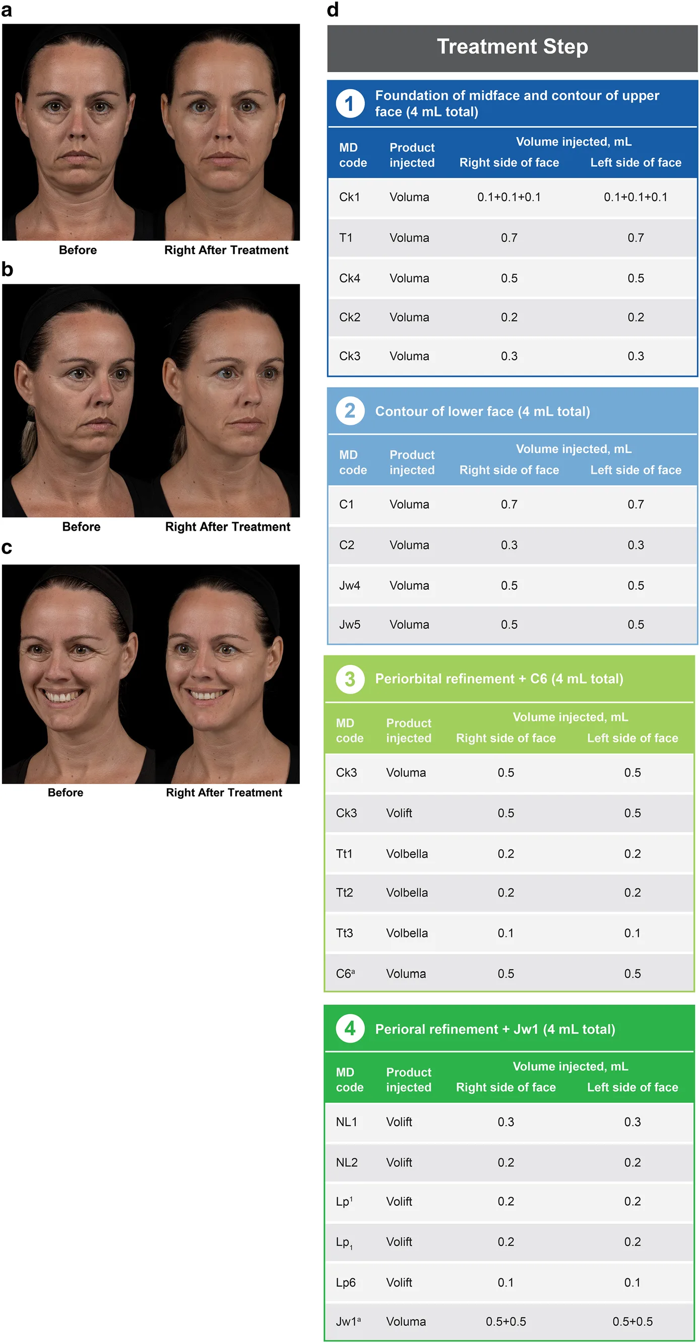 Fig 1. A saggy mid-face treated by the MD Codes method: frontal, oblique and smiling before/after (a–c) beside the coded plan (d), which lists the point (Ck1, T1, C1, Jw1…), the product and the volume per side. The point-and-volume table is a planning vocabulary, not a dose recommendation — the assessment (B1.3) decides which points, not the table. — (de Maio, 2020, p. 15) [1]
> Sources: de Maio 2020 [1]
Fig 1. A saggy mid-face treated by the MD Codes method: frontal, oblique and smiling before/after (a–c) beside the coded plan (d), which lists the point (Ck1, T1, C1, Jw1…), the product and the volume per side. The point-and-volume table is a planning vocabulary, not a dose recommendation — the assessment (B1.3) decides which points, not the table. — (de Maio, 2020, p. 15) [1]
> Sources: de Maio 2020 [1] [MEDLIB]
The MD Codes attribute is dose-responsive, which is a communication tool, not a target. Fig 2 shows the same face at successive total volumes; the "tired/saggy" reading softens progressively. The point of showing it to a patient is to make the incremental, phased nature of the plan concrete — not to commit to a millilitre count on day one.
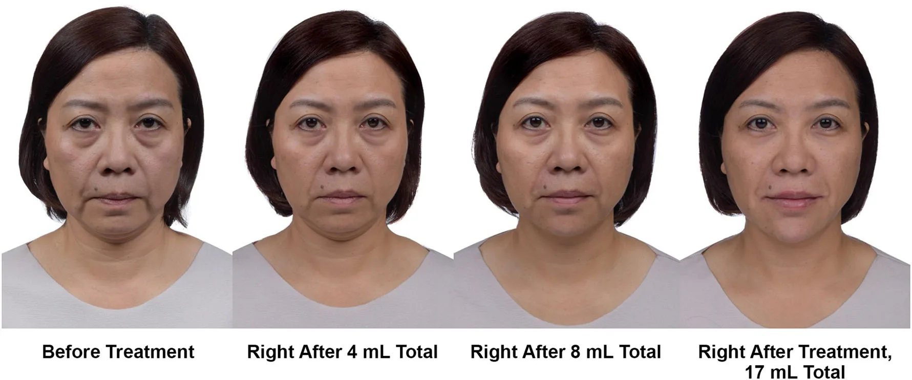 Fig 2. The same patient photographed before treatment and after successive increments of total volume: the perceived "tired/saggy" attribute lifts progressively as structure is restored. Read as a demonstration of phasing and of the attribute method, not as a volume prescription. — (de Maio, 2020, p. 12) [1]
> Sources: de Maio 2020 [1]
Fig 2. The same patient photographed before treatment and after successive increments of total volume: the perceived "tired/saggy" attribute lifts progressively as structure is restored. Read as a demonstration of phasing and of the attribute method, not as a volume prescription. — (de Maio, 2020, p. 12) [1]
> Sources: de Maio 2020 [1] [MEDLIB]
Phase the plan over time — AART / HIT
A structurally similar method of different corporate origin adds the time axis. Anatomy → Assessment → Regimen → Therapy; the Holistic Individualised Treatment variant writes the plan across 12–24 months [3]. What it adds over point-coding is the forced discipline of a phased plan with budget and calendar — precisely where consultations that only treat what the patient points at fall down. What it shares is that it is a commercial teaching method [D], useful and not evidence.
The practical synthesis this atlas recommends keeps three things from both: 1. Ask by perception, not by defect (MD Codes). 2. Write a phased plan with a 12–24-month horizon (AART/HIT). 3. Commit to no points until the three-process differentiation of this block is done — that is the real diagnosis.
The modern preprocedural consultation literature converges on the same content from the safety side: patient-, product- and procedure-related risk factors, standardised photography, act-specific and adverse-event-specific consent, and recognition that ethnicity, gender and social-media exposure shape the request [33].
The static exam, zone by zone
The static exam is a fixed sweep, top to bottom, so nothing is missed and the note is comparable across visits [17]:
| Zone | What to read | Finding that changes the plan |
|---|---|---|
| Brow | height, shape, tail position, asymmetry | brow ptosis mimics excess upper-lid skin; lifting the tail feminises (B1.6) |
| Upper eyelid | dermatochalasis, true ptosis, sulcus hollowing | ptosis is surgical, not a filler/toxin target |
| Tear trough / lid–cheek junction | groove depth, negative vector, festoons | a negative vector (globe anterior to the malar) makes tear-trough filler prone to malar oedema |
| Midface / malar | projection, Ogee, submalar hollow | deflation vs descent decided here (supine test) [12] |
| Nose | dorsum, tip, base width, naso-labial angle | base width read against intercanthal (B1.6) |
| Perioral / lips | vermilion, Cupid's bow, philtrum, upper-lip length, rhytids | lower-third 1/3–2/3 ratio broken by over-filling |
| Jawline / jowl | definition, pre-jowl sulcus, gonial angle | jowl is descent + pre-jowl deflation, not one problem |
| Chin | projection to the vertical, microgenia vs retrognathia | chin projection is often the highest-yield single manoeuvre [24] |
| Submental / neck | mentocervical angle, platysmal bands, submental fat | reads the lower boundary of any lifting plan |
The dynamic exam reveals what the static one cannot: frontalis raises the brow and prints horizontal lines; the glabellar complex (corrugator, procerus) prints the "angry" signature; orbicularis prints crow's feet; the depressor anguli oris pulls the "sad" commissure down; mentalis dimples the chin; platysma bands stand out on effort. Many findings — a gummy smile, a depressor-driven downturned mouth, asymmetric animation — exist only in movement and are missed entirely by a static-only assessment [3].
Palpation closes the exam: skin thickness and sebaceous quality by zone (thin periorbital vs thick nasal), laxity and recoil, old nodules or filler, the bony landmarks (orbital rim, pyriform aperture, zygoma, mandibular angle and pogonion), and an awareness of the vessels — the facial and angular arteries — that the danger-zone anatomy locates [14]. Prior product is palpated as well as asked for: a permanent or minimally-degradable filler is often felt before it is admitted, and ultrasound resolves the doubt where available [10][11].
The structural (foundation) reading
The structural school (Liew and others) reads the face from the deep support outward: bone and deep fat first, the profile before the front, the surface last [11]. It fits the three-process model directly — a face that has lost its skeletal and deep-compartment support is restored by rebuilding the foundation, and treating the surface of an unsupported face chases the descent. Skeletal deficiency that is beyond filler (a truly retruded chin, a deficient pyriform) is where implants and, in the surgical lane, bony work belong [24]. The assessment does not choose the treatment; it produces the ordered problem list — foundation, then position, then surface — that the treatment plan follows.
The AART regimen, in detail
Written across 12–24 months, the regimen sequences the three processes rather than treating them together [3][23]: skin quality first or in parallel (biostimulation, skinboosters, microneedling, topicals) so the envelope improves while the plan runs; deep structural support next (deep filler to the compartments and bony contact points identified at assessment); refinement last (superficial lines, lips, fine contour). Review points at 3, 6, 12 and 24 months re-photograph and re-measure, so the plan is a living document with verifiable milestones rather than a one-visit sale. The skin-quality lane is where the regenerative material — PRP, polynucleotides, biostimulators — is placed on the calendar [22][23].
History, timing and the preservation question
The targeted history is not a form to tick but a set of answers that each change the plan [33]. Prior product by zone and date establishes the substrate; the permanent-filler question is asked directly because patients do not volunteer it and a permanent or minimally-degradable product changes layering risk and the entire plan [11]. Autoimmune disease, a history of hypersensitivity, and recent or planned dental work bear on filler safety; anticoagulation and antiplatelet use bear on bruising; a herpes-simplex history bears on peri-oral and injectable work; recent isotretinoin bears on resurfacing and on wound healing; keloid and HPI history bear on energy and peel selection (B1.7). Timing matters as its own variable: a patient with an event in two weeks is planned differently from one with a 12–24-month horizon, because the phased plan (B1.4) sequences the acts across time and an imminent event compresses or defers that sequence.
The preservation question, mechanically. Asked literally — what would you keep about your face, no matter what? — before any proposal, it does three jobs at once: it surfaces the feature the patient values and would resent losing, it constrains the plan against westernisation and over-treatment (B1.7), and it converts an unspoken expectation into a documented one that the consent and the plan can honour [34]. A consultation that skips it plans against an assumed ideal rather than the patient's actual one, which is the root of the "I don't look like myself" complaint.
Common pitfalls: starting the consult at the mirror. Examining before listening produces a physician-led defect list, inverts the priority order, and misses the preservation constraint entirely — the patient then spends the encounter defending features you have already flagged for treatment [1].
B1.4 · Templates and documents
Four documents carry the encounter: the standardised photograph, the assessment record, the two-part consent, and the phased plan. Each is a template you fill the same way every time, because the value of all four comes from reproducibility.
Document 1 — clinical photography: the protocol.
Standardised photography is a diagnostic instrument and a legal document, not a keepsake. Without a baseline image there is no way to prove that an asymmetry pre-existed, and the pre-existing asymmetry is the most common complaint of all.
The five mandatory views (three of them in Fig 3): frontal · oblique 45° right · oblique 45° left · profile 90° right · profile 90° left. Add dynamics (maximal contraction and smile) whenever toxin is on the plan.
| Variable to fix and never touch again | Set to | Why |
|---|---|---|
| Focal length / distance | ≥ 85 mm equivalent, constant | a phone at 30 cm widens the nose — that is the lens, not a finding [32] |
| Background | plain, neutral blue or grey | isolates the subject, stabilises exposure |
| Lighting | fixed, diffuse, bilateral | never top-light the "before" and front-light the "after" — the commonest way to fabricate a result |
| Head position | Frankfort plane horizontal (infra-orbital rim–tragus) | head tilt alone changes apparent chin projection |
| Face | no make-up, hair back, no glasses, no jewellery | removes confounders |
| Expression | neutral in the five static views; dynamics separate | consistency across visits |
The standardisation is the whole point: even small variations in angle, distance or light change the image drastically and destroy its clinical and medico-legal value [32]. A grid or crosshair focusing screen (Fig 4) makes the alignment repeatable across months and across operators.
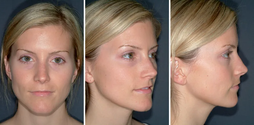 Fig 3. Standardised positioning: the same subject in frontal, oblique 45° and profile 90° views under fixed distance and lighting. Three of the five mandatory views; add the mirror-image oblique and profile to complete the set. — (Nouri, 2018, p. 518) [7]
> Sources: Nouri 2018 [7]
Fig 3. Standardised positioning: the same subject in frontal, oblique 45° and profile 90° views under fixed distance and lighting. Three of the five mandatory views; add the mirror-image oblique and profile to complete the set. — (Nouri, 2018, p. 518) [7]
> Sources: Nouri 2018 [7] [MEDLIB]
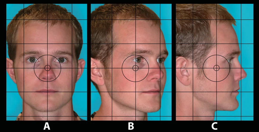 Fig 4. A grid pattern with a central crosshair superimposed on frontal, oblique and profile views: the grid documents head position and the Frankfort plane, so the same alignment is reproducible at the follow-up visit and between operators. — (Naini, 2011, p. 102) [2]
> Sources: Naini 2011 [2]
Fig 4. A grid pattern with a central crosshair superimposed on frontal, oblique and profile views: the grid documents head position and the Frankfort plane, so the same alignment is reproducible at the follow-up visit and between operators. — (Naini, 2011, p. 102) [2]
> Sources: Naini 2011 [2] [MEDLIB]
Consent and data protection for the image. The clinical photograph is health data (B1.2). The template requires two separate consents: one for care/record use, one for teaching, congress or advertising use — and the second is independently revocable [35][36]. The physician who publishes a before/after must be able to show the second consent and the truthfulness backing that the advertising rules demand (B1.2) [40].
Document 2 — the assessment record: what the note must contain.
The note is the spine of Ley 41/2002 compliance and of every future comparison. A complete assessment record carries:
- The patient's verbatim motive and their perception attribute (tired/sad/angry/saggy).
- The preservation constraint ("what to keep").
- Relevant history, with permanent filler answered explicitly (yes/no/unknown), anticoagulation, autoimmunity, herpes, isotretinoin, prior product by zone and date.
- The three-process reading (deflation / descent / skin) with the supine and digital-lift results.
- Static metrics actually measured (thirds, fifths, angles, E-line) and the dynamic findings.
- Pre-existing asymmetry, marked on the photograph — the single most protective line in the record.
- The 2–3 findings shown to the patient and the ones deliberately not raised.
- The phased plan and the consent references.
Document 3 — the two-part consent: template logic.
A generic "I consent to aesthetic treatment" discharges nothing. The consent template is:
| Part | Content | Governing rule |
|---|---|---|
| Act-specific care consent | the named procedure, the named material, the named risks (including vascular occlusion and, for periocular filler, blindness), alternatives, and the option of no treatment | Ley 41/2002 Arts. 4, 8, 10, 11 [35] |
| Data / image consent | separate blocks for care-record storage vs teaching/marketing use, the second revocable, retention period stated | Art. 9 RGPD, LOPDGDD [36][37] |
The elective nature of aesthetic medicine raises the disclosure standard: the more discretionary the act, the more exhaustive and the better-documented the information must be. Specific consent for both the adverse event and its treatment is now standard practice given the rise in vascular complications [33].
Document 4 — the phased plan: the deliverable that prevents over-treatment.
The plan is written, dated, priced and split into today / 3 months / 12–24 months [3]. Writing it in phases is what stops the consultation from treating everything the patient points at in one sitting; it also gives the patient a document to think about during the reflection period (B1.7). A simulation, if shown, is annexed to the plan with the mandatory line in writing: this image is guidance, not a guaranteed result — and it is stored in the record, because a simulation shown to a patient becomes, in their memory, a commitment [3].
Document 5 — the perception questionnaire: the intake instrument.
A short intake sheet that asks the patient to rank the four attributes (tired/sad/angry/saggy) and to write the one feature they would keep converts the MD Codes method into a repeatable document [1]. It anchors the consultation to the patient's own priority and produces a verifiable success criterion for the follow-up.
Video and dynamic documentation. When toxin is on the plan, static images are insufficient: a short standardised clip of maximal frontalis, glabellar and orbicularis contraction and of the smile records the dynamic findings that the treatment targets and against which the result is judged [29]. The same standardisation rules apply — fixed distance, fixed light, neutral background — because a dynamic "before/after" fabricated by changing capture conditions fails both the clinical and the advertising test (B1.2) [40].
Retention, access and the two-consent audit. The record is retained under Article 17 of Ley 41/2002 and the autonomic extensions, and the patient may access and copy it; in an aesthetic dispute this access is exercised routinely, so the note, the dated baseline and the signed act-specific consent are the physician's defence as much as the patient's right [35]. The image consent is audited separately: the care-use block and the teaching/marketing block are stored so that a later withdrawal of marketing consent can be honoured without touching the care record [36][37].
The record spine, expanded. A complete note is reconstructable by another clinician from these fields alone: the verbatim motive and perception attribute; the preservation constraint; the structured history with the explicit permanent-filler answer; the three-process reading with the supine/digital-lift results; the measured static metrics and the dynamic findings; the palpation notes; the marked pre-existing asymmetry; the two-to-three findings shown and those withheld; the phased plan with prices and review points; and the consent references [26]. A note missing any of these leaves a gap that a complaint, weeks later, will find.
Simulation governance. If a Vectra-style simulation or an AI morph is shown, it is annexed to the plan with the written line — this image is guidance, not a guaranteed result — dated and stored, because the image becomes a remembered commitment and, unstored, an undocumented promise [3]. Showing the full multi-parameter skin-analysis report at the same visit is the parallel error to avoid: one parameter, the one the plan treats, not the whole anxious printout [16].
Common pitfalls: letting the light or the lens drift between "before" and "after". A top-lit before and a front-lit after, or a 30 cm phone shot against an 85 mm clinic shot, manufactures a result that is not yours — and, once published, an advertisement that is not truthful (B1.2) [32][40].
B1.5 · Frequent errors and their cost
Every error in this table is an assessment error — it happens before a needle is loaded — and each has a cost that is clinical, legal, financial or reputational, often all four. The schematic lists the error, why it happens, its cost and the manoeuvre that prevents it.
| Error | Why it happens | Cost | Prevention |
|---|---|---|---|
| Treat the canon as the target | it gives a clear answer | homogenised, westernised faces; loss of the feature the patient valued | the canon is vocabulary; the target is this face [34] |
| Fill a face whose problem is descent | filler is the tool the operator owns | over-filled face, later dissolution, refund, distrust | supine test + digital lift before deciding [12] |
| Show ten defects | mistaken for rigour | anxious patient, longer list, consultation-room iatrogenesis | show 2–3, prioritised [5] |
| Drift light/distance between before and after | convenience | fabricated result; unlawful advertisement | fixed protocol; ≥85 mm, fixed light [32][40] |
| Not document pre-existing asymmetry | haste | the most common complaint, indefensible without a marked baseline | mark it on the photo and in the note [32] |
| Apply the female pattern to a man | automatism | feminising result the male patient perceives but cannot name | jaw and chin first; brow still; cheek lateral and low [5] |
| Apply the Caucasian canon to any face | the published canons are those | over-treatment of normal variants, HPI in skin of color | ask what to keep; know the normal variants [6][8] |
| Show the full VISIA report | it impresses | seven red parameters → seven anxieties → over-consumption | show only the parameter you will treat [16] |
| Promise the simulation | it sells | a memorised commitment you cannot meet; complaint | in writing: guidance not commitment; archive it [3] |
| Skip the permanent-filler question | the patient does not volunteer it | a plan built on a false substrate; delayed complications | ask directly and explicitly [33] |
| Consent as a single generic signature | speed | discharges neither Ley 41/2002 nor the RGPD nor the advertising rules | act-specific + data consent, dated [35][36] |
| No reflection period on an elective act | commercial pressure | invalidated consent, higher liability on satisfaction medicine | offer and document a reflection interval (B1.7) [35] |
The three costliest, expanded.
Descent treated as deflation. This is the error with the largest financial and reputational cost because it is self-reinforcing: filler placed into a descended, lax face adds weight, accelerates the descent, and reads as "more ageing", which invites more filler. The exit is expensive — hyaluronidase, a period of distortion, a refund, and a patient who tells others. The supine test and the digital lift cost thirty seconds and prevent all of it; the compartmental-ageing literature is the anatomical justification for reaching for support, energy or surgery instead of volume when the manoeuvre says descent [12][13][11].
The undocumented pre-existing asymmetry. Almost no face is symmetric, and the fifth view exists precisely to record it (B1.6, Fig 6). The cost of skipping it is entirely legal and it lands weeks later: a patient attributes a baseline asymmetry to the treatment, and with no marked baseline the clinician cannot rebut it. On satisfaction medicine the burden of proving adequate information and baseline documentation sits heavily on the physician (B1.2), so the missing photograph is not a gap in tidiness but a lost case [32][35].
The full skin-analysis report shown to the patient. A VISIA-style printout with several parameters in the red generates one anxiety per red bar and a patient who now has several reasons to consume [16]. The cost is a distorted plan (treating the report, not the face) and an erosion of trust when the "diagnoses" turn out to be relative percentiles, not disease. The rule is to show the single parameter that the agreed plan addresses, and to hold the rest (B1.9).
The generalisable lesson. Most of these are the same disease: substituting a tool the operator owns, or a template the operator knows, for a diagnosis of the individual face. The 2024 review that concludes there is no universal standard of beauty is the evidence-level statement of that lesson — physical attributes influence perceived attractiveness, but the golden ratio and the neoclassical canons do not define a target, and diversity of the normal is the rule, not the deviation [34].
Four more errors that start in the assessment.
Averaging a discrepant number. When two schools give different targets for the same angle, the wrong response is to split the difference. A nasolabial angle read against a male norm and a female norm does not average to a target; the reading is kept with its context (sex, ancestry, the patient's wish) and the decision is made on the clinical variable, not the mean [34]. Averaging a dose, a ratio or an angle manufactures a value that no school endorses.
Assessing statically only. A downturned mouth driven by the depressor anguli oris, a gummy smile, or asymmetric animation exist only in movement; a static-only assessment plans the wrong act — filler for a muscle problem, or nothing for a dynamic one [3]. The cost is a treatment that does not touch the patient's actual complaint.
Ignoring the negative vector before tear-trough filler. A negative-vector, festooned lower lid filled with hyaluronic acid predictably swells; the cost is months of malar oedema and, often, hyaluronidase and a refund. The vector is a thirty-second oblique read that reclassifies the case as unsuitable for the "simple" under-eye correction (B1.6).
Treating the device percentile as a diagnosis. A skin-analysis percentile ranks this face against a reference base; read as disease it invents pathology and inflates the treatment list [16]. The cost is a plan built on the machine's agenda rather than the patient's, and the trust lost when the "diagnoses" turn out to be relative numbers (B1.9).
The cost ledger, summed. The recurring costs are four: clinical (over- or mistreatment, complications, reversals), legal (indefensible complaints on satisfaction medicine, invalid consent), financial (refunds, hyaluronidase, lost repeat custom), and reputational (the westernised or over-filled result the patient shows others). Every one is cheaper to prevent at assessment than to remediate after treatment, and the prevention is the same short list — listen, photograph, differentiate the three processes, show two to three findings, write it down, consent it specifically [32][35].
Consultation-room iatrogenesis, named. Showing ten defects is not thoroughness; it is a harm with a mechanism. Each flagged item becomes a worry the patient did not arrive with, the list lengthens, the plan sprawls, and the patient leaves less content than they came — the exact failure state the chapter's opening defines [5]. The same mechanism runs the full skin-analysis report and the AI proportion score: more red flags generate more anxiety and more consumption, none of it indexed to the patient's actual request [16][34]. The corrective is a discipline of subtraction — surface the two or three findings that carry the patient's own priority, hold the rest, and let the phased plan (B1.4) reveal the remainder only if and when it becomes relevant.
Common pitfalls: the confident, fast opinion that skips the note and the photograph. It maximises throughput and it is the common root of the litigable errors above, because it removes the one artefact — a standardised, dated baseline with a signed, act-specific consent — that turns a complaint into a defensible record [32][35].
B1.6 · Metrics: what is measured and the reference value
The reference grid, at a glance. Every value below is a descriptor, derived from mostly young Caucasian reference populations, and is used as vocabulary — never as a norm to enforce. A large deviation from the population mean tends to be read as unattractive and the population mean as attractive; that, not any single ideal, is the defensible reading of the evidence [34].
| Metric | Reference value | Read in which view | Note |
|---|---|---|---|
| Vertical thirds | trichion–glabella–subnasale–menton, ideally equal | frontal | lower third changes most with age and responds most to treatment |
| Lower-third split | subnasale→stomion 1/3 · stomion→menton 2/3 | frontal / profile | governs lips and chin; broken by excess lip filler |
| Horizontal fifths | face = 5 eye-widths | frontal | central fifth (intercanthal) = one eye width |
| Intercanthal distance | ≈ one eye width | frontal | telecanthus vs true hypertelorism |
| Alar width | ≈ medial intercanthal distance | frontal | reference for alar base |
| Oral width | vertical from the medial limbus of each iris | frontal | commissure position |
| Nasofrontal angle | 115–130° | profile | glabella–nasion–dorsum |
| Nasolabial angle | 90–100° ♂ · 95–110° ♀ ⚠ | profile | sex-dependent, population-bound |
| Mentocervical angle | 80–95° | profile | submental contour, chin projection |
| Ricketts E-line | lower lip ~2 mm, upper lip ~4 mm behind the nasal-tip–pogonion line ⚠ | profile | varies markedly by ethnicity |
| Phi (φ) | ≈ 1.618 | any | a discipline of restraint, not a validated attractiveness predictor ⚠ [34] |
| Ogee curve | smooth malar-convex → submalar-concave → mandibular-convex S | oblique 45° | ageing flattens it; the reason the 45° view is mandatory |
> ⚠ All of these numbers come from mostly young, mostly Caucasian reference populations. Applied outside that frame they "diagnose" normal variants as anomalies. They are a language, not a standard [2][34].
Vertical thirds and the lower face
The vertical thirds — trichion→glabella, glabella→subnasale, subnasale→menton — are equal in the canon and almost never equal in reality [2]. The lower third is the one that changes most with age and responds most to treatment, and it subdivides again: subnasale→stomion 1/3, stomion→menton 2/3. That 1:2 ratio governs the lip–chin relationship and is the proportion most often broken by excessive lip filler; the profile thirds are shown in Fig 5 and the millimetric lower-third canon in Fig 6.
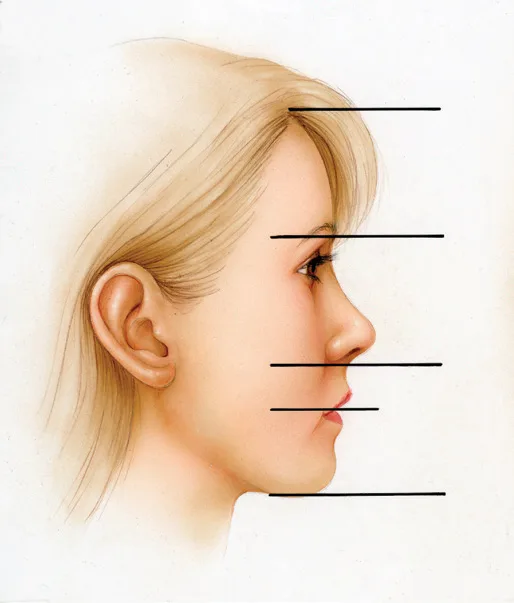 Fig 5. The horizontal reference lines of the vertical thirds on a profile: trichion, glabella, subnasale and menton. In the canon the three segments are equal; the lower third is the mobile one across the lifespan. — (Master Techniques in Facial Rejuvenation, 2018, p. 35) [3]
> Sources: Master Techniques in Facial Rejuvenation / Azizzadeh 2018 [3]
Fig 5. The horizontal reference lines of the vertical thirds on a profile: trichion, glabella, subnasale and menton. In the canon the three segments are equal; the lower third is the mobile one across the lifespan. — (Master Techniques in Facial Rejuvenation, 2018, p. 35) [3]
> Sources: Master Techniques in Facial Rejuvenation / Azizzadeh 2018 [3] [MEDLIB]
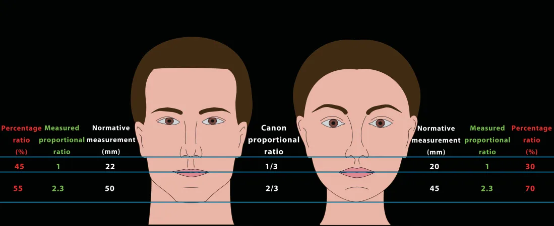 Fig 6. The lower-facial-height canon compared with proportional ratios in a male and a female schematic: the subnasale→stomion segment sits near the 1/3 and the stomion→menton near the 2/3 of the lower third, with the measured millimetric values shown beside the canonical ratio. The mismatch between "measured" and "canon" is the normal individual variation the assessment describes, not a defect to erase. — (Naini, 2011, p. 182) [2]
> Sources: Naini 2011 [2]
Fig 6. The lower-facial-height canon compared with proportional ratios in a male and a female schematic: the subnasale→stomion segment sits near the 1/3 and the stomion→menton near the 2/3 of the lower third, with the measured millimetric values shown beside the canonical ratio. The mismatch between "measured" and "canon" is the normal individual variation the assessment describes, not a defect to erase. — (Naini, 2011, p. 182) [2]
> Sources: Naini 2011 [2] [MEDLIB]
Horizontal fifths, intercanthal, alar and oral width
The face is read as five vertical eye-widths from helix to helix [4]. Three readings matter clinically: the intercanthal distance (the central fifth should equal one eye width), the alar width (ideally matching the medial intercanthal distance), and the oral width (a vertical dropped from the medial limbus of each iris). Symmetry is judged against the central midline, and the fifths make the asymmetry legible and photographable; the "five eyes" reading is shown in Fig 8.
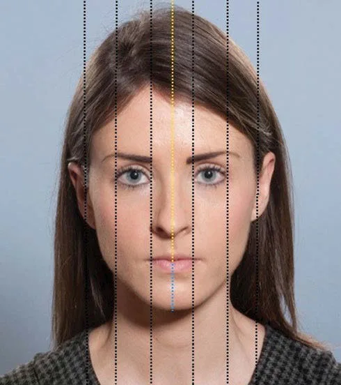 Fig 7. Frontal assessment of transverse proportions: six vertical lines divide the face into five equal eye-widths, with the orange midline exposing left–right asymmetry. This is how a pre-existing asymmetry is documented objectively — the manoeuvre B1.4 and B1.5 make mandatory. — (Farhadieh, 2015, p. 298) [4]
> Sources: Farhadieh 2015 [4]
Fig 7. Frontal assessment of transverse proportions: six vertical lines divide the face into five equal eye-widths, with the orange midline exposing left–right asymmetry. This is how a pre-existing asymmetry is documented objectively — the manoeuvre B1.4 and B1.5 make mandatory. — (Farhadieh, 2015, p. 298) [4]
> Sources: Farhadieh 2015 [4] [MEDLIB]
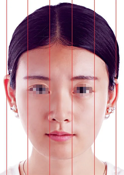 Fig 8. The five-eye-width division applied to a frontal photograph: the central fifth (intercanthal) should measure one eye width, and the medial iris lines locate oral width. The same grid reveals nasal base width against the intercanthal reference. — (Liu, 2021, p. 26) [5]
> Sources: Liu 2021 [5]
Fig 8. The five-eye-width division applied to a frontal photograph: the central fifth (intercanthal) should measure one eye width, and the medial iris lines locate oral width. The same grid reveals nasal base width against the intercanthal reference. — (Liu, 2021, p. 26) [5]
> Sources: Liu 2021 [5] [MEDLIB]
The neoclassical canons — descriptive, not prescriptive
The neoclassical canons come from Leonardo and Dürer (the proportion grid in Fig 9), and their value today is descriptive [2]. They fix the profile angles — nasofrontal 115–130°, nasolabial 90–100° in men and 95–110° in women ⚠, mentocervical 80–95° — and the Ricketts E-line, on which the lower lip sits ~2 mm and the upper lip ~4 mm behind the nasal-tip–pogonion line ⚠, a value that shifts markedly with ethnicity (B1.7). These are read on the profile and the oblique, which is why the 90° and 45° views are not optional.
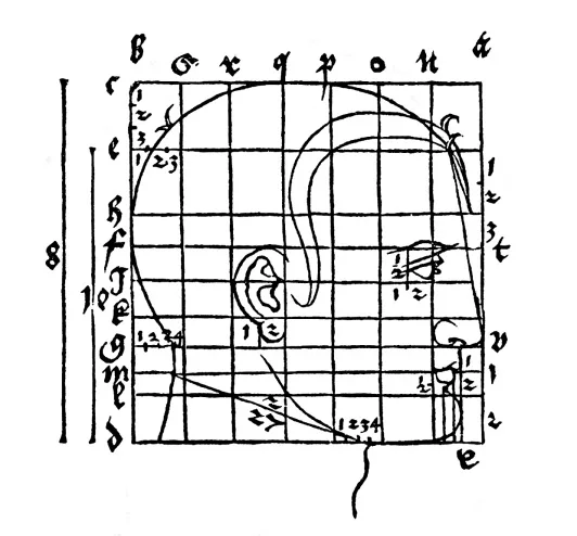 Fig 9. A Dürer proportion grid: the head mapped onto a fractional grid that fixes the relationships between features. The historical canons are a grid of relationships, and their modern use is to force you to look at ratios rather than points — not to prescribe a target. — (Naini, 2011, p. 173) [2]
> Sources: Naini 2011 [2]
Fig 9. A Dürer proportion grid: the head mapped onto a fractional grid that fixes the relationships between features. The historical canons are a grid of relationships, and their modern use is to force you to look at ratios rather than points — not to prescribe a target. — (Naini, 2011, p. 173) [2]
> Sources: Naini 2011 [2] [MEDLIB]
Phi and the Ogee — a discipline of restraint
There is a long tradition of mapping the face with masks built on the golden ratio (φ ≈ 1.618), and it underlies Swift's BeautiPHIcation school (B5 — Schools of Facial Beautification — Competing Philosophies). ⚠ The evidence that phi predicts perceived attractiveness is weak and heavily disputed [34]. What holds is the inverse: a large deviation from the mean reads as unpleasant and the population mean reads as attractive. Phi therefore works as a discipline of restraint — it forces you to look at relationships, not points — and that is its real use; the phi-point cheek construction in Fig 10 is the manoeuvre. The Ogee curve, the double S of the oblique profile (malar convexity → submalar concavity → mandibular convexity), is the one of these tools that actually changes decisions: ageing flattens the Ogee, and learning to see it lets you read mid-facial ageing before you can name it. It is read in the three-quarter view, the reason the 45° photograph is mandatory.
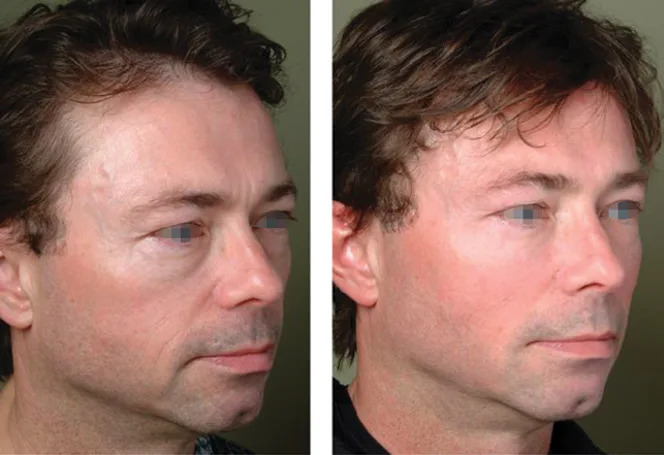 Fig 10. The oval cheek defined by a phi-point construction on the oblique view: the golden-ratio mask is used to place the point of maximal projection and to check the Ogee curve, seen here as the malar–submalar–mandibular S. The construction disciplines where support goes; it does not certify a beautiful outcome. — (Jones, 2019, p. 68) [6]
> Sources: Jones 2019 [6]
Fig 10. The oval cheek defined by a phi-point construction on the oblique view: the golden-ratio mask is used to place the point of maximal projection and to check the Ogee curve, seen here as the malar–submalar–mandibular S. The construction disciplines where support goes; it does not certify a beautiful outcome. — (Jones, 2019, p. 68) [6]
> Sources: Jones 2019 [6] [MEDLIB]
Sex-specific attractors — the metric that flips the plan
The reference values differ by sex, and reading them wrong is the commonest gender error.
| Feature | Feminine | Masculine |
|---|---|---|
| Brow | higher, peak in the lateral third, arched | straight, lower, at the orbital rim |
| Cheek | apex antero-lateral and high, inverted-triangle | apex more lateral and inferior, flatter |
| Jaw | defined but narrow, soft angle, oval chin | wide, marked gonial angle (~90°), square projected chin |
| Forehead | convex, smooth | supraorbital bossing, slight posterior slope |
| Lips | greater volume and eversion, defined Cupid's bow | longer, less everted upper lip |
| Nasolabial angle | 95–110° ⚠ | 90–100° ⚠ |
The dimorphic markers of Fig 11 are the ones to read. The commonest gender error in men is treating them on the female pattern by automatism: lifting the brow tail, projecting a high anterior cheek, tapering the jaw. The result is feminising and the patient perceives it even when he cannot name it. In the male: jaw and chin first; lateral, low cheek; brow still. These patterns are statistical and cultural, not prescriptive; in trans and non-binary patients the goal is defined by the patient, and this same grid becomes the map of feminising or masculinising manoeuvres (L3 — Special Populations & Advanced Topics) [7].
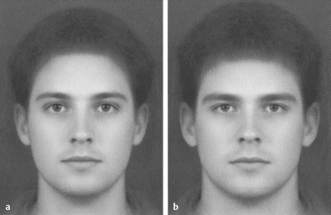 Fig 11. Two computer-manipulated faces differing only in the sexually dimorphic markers — brow height and shape, cheek apex, jaw width and chin — illustrating how small shifts in these attractors read as more feminine or more masculine. The grid is used to plan feminising/masculinising work, never to impose a sex-typical target. — (Connell, 2016, p. 108) [8]
> Sources: Connell 2016 [8]
Fig 11. Two computer-manipulated faces differing only in the sexually dimorphic markers — brow height and shape, cheek apex, jaw width and chin — illustrating how small shifts in these attractors read as more feminine or more masculine. The grid is used to plan feminising/masculinising work, never to impose a sex-typical target. — (Connell, 2016, p. 108) [8]
> Sources: Connell 2016 [8] [MEDLIB]
Symmetry, the negative vector and the skeletal metrics
Symmetry is graded, not assumed. Almost no face is symmetric, and the fifths view (Fig 7) makes the deviation measurable rather than impressionistic [4]. The clinical rule is that minor asymmetry is universal and is documented, not corrected; a marked, progressive or new asymmetry is a finding to investigate before it is a target to treat. The single most protective use of the metric is medico-legal: a pre-existing asymmetry recorded on the baseline photograph is the defence against the commonest complaint (B1.5).
The negative vector is a profile-plus-oblique reading that changes a specific decision: when the anterior globe projects beyond the malar eminence and the infra-orbital rim, the lid–cheek support is deficient, and tear-trough or malar filler in that face is prone to persistent oedema and a heavy look. Reading the vector at assessment is what stops a "simple" under-eye correction from becoming a months-long problem.
Skeletal reference points anchor the soft-tissue metrics [24]: the orbital rim and its recession, pyriform aperture width and angle, malar and zygomatic projection, the mandibular angle and the position of pogonion relative to the facial vertical. Microgenia (a small chin) and retrognathia (a retruded mandible) look similar on the front view and diverge on the profile — the distinction decides whether chin projection alone suffices or whether the whole lower jaw is deficient. The height canons that Dürer catalogued give the same message from the historical side: a wide range of "normal" proportions, not a single ideal [2][15].
The Ogee, in more detail. The oblique-view double curve is graded from full (youthful) to flat (aged); its flattening is the earliest legible sign of mid-facial deflation and descent, and its restoration — not the filling of the nasolabial fold it produces — is the correct target [3]. Reading it in the 45° view is why that photograph is one of the mandatory five (B1.4).
Ethnic variation of the metrics. The Ricketts E-line, the nasolabial angle and the fifths were derived on Caucasian populations and shift with ancestry: fuller lips sit closer to or ahead of the E-line in many African and some Asian phenotypes without being "protrusive", the nasal base commonly exceeds the intercanthal width, and dorsal height is lower [8]. Applied as a universal norm these descriptors mis-classify normal variants as defects — the mechanism of westernisation (B1.7) — so the reference values are read as one input against ancestry, sex and, above all, the patient's stated preservation constraint [34].
Brow metrics, expanded. The feminine brow peaks in the lateral third and sits above the orbital rim; the masculine brow is flatter, lower and at the rim [3][17]. The commonest ageing change read here is lateral brow descent, which mimics excess upper-lid skin — treating the "hooding" with a blepharoplasty-style plan when the problem is a descended tail is a diagnostic error the static exam prevents (B1.3).
From metric to decision — the disciplined use of the numbers
The reference values earn their place only when they change a decision, and each carries a specific one. The vertical thirds locate where on the face the imbalance sits and, because the lower third is the mobile segment, direct attention to the lip–chin relationship over the lifespan [2]. The fifths and the intercanthal/alar/oral references convert a vague "asymmetry" into a documented, photographable measurement that protects against the commonest complaint (B1.5) [4]. The profile angles decide profile-plane moves — a retruded chin read at 96° nasolabial and 90° mentocervical is weighed against sex and ancestry before a projection is planned [2][24]. The E-line and phi are restraints, not targets: they flag when a lip or a cheek is drifting away from the population relationship, and they are read with the patient's preservation constraint, never against it [34]. The single operating rule that ties the grid together is that a number is a descriptor to weigh, not a defect to erase — the metric enters the plan only where it names a real, ordered problem the patient shares, and the same number in a face the patient is happy with is left alone.
Which view carries which metric
The five views are not redundant; each carries metrics the others cannot, which is why the set is fixed (B1.4).
| View | Metrics it carries | Why it is mandatory |
|---|---|---|
| Frontal | thirds, fifths, intercanthal/alar/oral width, symmetry, brow shape | the descriptive map and the asymmetry record [2][4] |
| Oblique 45° (×2) | the Ogee curve, malar projection, the negative vector | mid-facial ageing is legible here and nowhere else [3] |
| Profile 90° (×2) | nasofrontal/nasolabial/mentocervical angles, E-line, chin projection | the entire angular canon lives on the profile [2] |
| Dynamic | frontalis/glabella/orbicularis/DAO/mentalis/platysma action | movement-only findings for any toxin plan [3] |
A worked reading. A 52-year-old woman reports looking "tired and a bit sad". Frontal: thirds roughly equal, mild left-over-right asymmetry (recorded, not treated); fifths normal. Oblique: the Ogee is flattening, submalar hollow deepening — mid-facial deflation plus early descent. Supine test partially corrects the nasolabial fold (descent component) and the digital lift corrects the tear-trough groove (support deficit, not skin). Profile: nasolabial angle 96°, within the female range; mentocervical 90°; E-line unremarkable. Dynamic: depressor anguli oris pulls the commissures down on animation — the "sad" reading is partly muscular. The ordered problem list that falls out: deep malar/mid-face support first (foundation), depressor toxin for the "sad" commissure (message), skin-quality groundwork in parallel — and not a nasolabial-fold filler, which would chase the descent. That is a diagnosis produced by the metrics and the manoeuvres together, not by any single number [1][12].
Common pitfalls: measuring the number and treating the number. A patient whose nasolabial angle reads 88° is not a defect to correct to 100°; the angle is a descriptor to weigh against the whole face, the sex, the ethnicity and — above all — what the patient asked for and asked to keep [34].
B1.7 · The Spanish particularity
Three things make the assessment different in Spain: who may do it and where is being tightened right now; whose face you are assessing is more diverse than the canons assume; and satisfaction medicine carries a heavier information duty than curative medicine.
The regulatory particularity — a moving target in 2026.
| Layer | Rule | Chair-side effect |
|---|---|---|
| State — centre | non-surgical aesthetics = unit U.48; surgical = U.47 (RD 1277/2003, Annex II) [41] | the act must be in an authorised centre |
| State — 2026 reform | RD modifying RD 1277/2003, approved 24 March 2026, in force 1 July 2026; restricts procedures to appropriately trained specialists [44] | centres have 6 months to update files; CCAA have 1 year to adapt |
| Cataluña | Decreto 151/2017 technical-sanitary requirements; Orden 18 April 2008 — all medical-aesthetic treatment directed by a physician with specific training; laser and botulinum toxin only by physicians [45][46] | in Barcelona, non-physician toxin/laser is expressly prohibited |
| Open question | may a nurse perform a hyaluronic-acid infiltration under medical indication? Court rulings diverge [47] | verify your CCAA rule before delegating any injectable |
The state baseline is RD 1277/2003, which since 2003 has defined U.48 as the unit where a physician performs non-surgical treatments with facial or bodily aesthetic purpose [41]. The pressure to tighten it has built for years: the Congreso approved a motion in September 2022 urging reform against intrusism; a September 2024 ministerial order restricting aesthetic surgery to trained specialists was suspended by the Audiencia Nacional, which required the measure to travel by real decreto rather than by order; and the resulting RD was approved on 24 March 2026 and enters force on 1 July 2026, extending the "adequate qualification and competence" duty across all sanitary care [44]. The motivation was stated as grave cases linked to unqualified operators; the sector data behind it — the Sociedad Española de Medicina Estética reported that ~65% of medical-aesthetic treatments are performed by unqualified operators and ~20% in unregulated settings such as hairdressers or homes — is the reason the assessment note now routinely records that the act is physician-performed in an authorised U.48 centre.
Autonomous communities layer their own rules, and they are not uniform: Andalucía's Orden of 3 April 2024 issues a functional and technical guide for U.48 authorisation with obligatory separate consultation/treatment/waiting rooms, sterilisation protocols and visible registration number; Cataluña — the operative jurisdiction for a Barcelona clinic — runs Decreto 151/2017 and the Orden of 18 April 2008, under which medical-aesthetic treatment must be physician-directed and laser and botulinum toxin may be applied only by physicians [45][46]; Castilla-La Mancha updated its requirements by Orden 32/2026; Murcia admits only techniques backed by at least two indexed-journal studies. The single most litigated gap is the hyaluronic-acid infiltration by nurses, on which rulings diverge — so the defensible position at assessment is to confirm the current rule of your own autonomous community and your centre's authorisation before the plan delegates any injectable or energy act [47].
The population particularity — the canons fit fewer Spanish faces every year.
Spain's population is increasingly diverse — Latin American, North African, sub-Saharan and East-Asian communities among others — and the neoclassical canons describe a narrow reference population, so applying them as a universal norm turns normal variants into "defects" and is the technical mechanism by which a treatment westernises a face without anyone deciding to [8][34]. The structural differences that change the plan, not just the conversation:
| Trait | Implication for the plan |
|---|---|
| Wider, more anterior malar and zygomatic arch (E. Asia) | less need for lateral malar volume; often the goal is to narrow, not project |
| Wider nasal base, lower dorsum | rhinomodelling seeks dorsal and tip projection, not reduction |
| Less chin projection (E. Asia, some phenotypes) | chin projection is the highest-yield manoeuvre |
| Fuller baseline lips (Afro) | the goal is usually definition and shape, not volume |
| Thicker, more sebaceous skin | fewer fine lines, more descent; Tyndall less of a problem, palpable nodule more |
| Fitzpatrick IV–VI | risk of post-inflammatory hyperpigmentation and keloid governs laser, peel and microneedling (B6 — Ethnic, Racial & Cultural Considerations in Face & Body Reshaping) [6] |
The most protective single question, asked literally before proposing anything: "what would you keep about your face, no matter what?" — it is the safeguard against both westernisation and over-treatment.
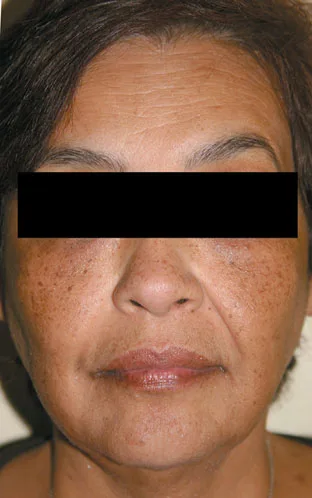 Fig 12. A patient with skin of color (higher Fitzpatrick phototype) showing facial dyschromia and hyperpigmentation. In phototypes IV–VI the risk of post-inflammatory hyperpigmentation and of keloid is the assessment finding that governs laser, peel and microneedling selection — it is read at assessment, before any device is chosen. — (Alam, 2009, p. 76) [8]
> Sources: Alam 2009 [8]
Fig 12. A patient with skin of color (higher Fitzpatrick phototype) showing facial dyschromia and hyperpigmentation. In phototypes IV–VI the risk of post-inflammatory hyperpigmentation and of keloid is the assessment finding that governs laser, peel and microneedling selection — it is read at assessment, before any device is chosen. — (Alam, 2009, p. 76) [8]
> Sources: Alam 2009 [8] [MEDLIB]
The liability particularity — satisfaction medicine, heavier duty.
Spanish jurisprudence treats aesthetic medicine as satisfaction rather than curative medicine, and consistently raises the information and documentation duty here above the curative standard [35]. The practical consequences at assessment: the consent must be exhaustive and act-specific, the elective act warrants an offered and documented reflection period, and the standardised baseline photograph with a marked pre-existing asymmetry is the evidence that the information duty was discharged. The more discretionary the procedure, the more the burden of proof of adequate information sits on the physician — which is why, in Spain more than elsewhere, the assessment paperwork is the treatment's legal foundation, not its afterthought.
The structural differences are not cosmetic footnotes; they invert specific manoeuvres [8][21]. In East-Asian faces the wider, more anterior malar and zygomatic arch often make the goal to narrow rather than to project, and the lower dorsum and less-projected chin make dorsal/tip and chin projection the high-yield moves — the opposite of the lateral-cheek-volume reflex that the Caucasian canon invites. In faces of African ancestry the fuller baseline lip is a feature to define and shape, not to inflate, and the thicker, more sebaceous skin shifts the ageing pattern toward descent over fine rhytids, which changes the tool from resurfacing toward support and energy. The global-face anatomy atlases used for planning make the same point structurally: the target is the individual's own architecture, restored, not a template imposed [18]. The single question — what would you keep, no matter what? — is the operational safeguard, because it converts an unspoken preservation constraint into a written one.
The pigmentary risk deserves its own line in the note. In Fitzpatrick IV–VI the foreseeable harms of energy and chemical injury are post-inflammatory hyperpigmentation and keloid, and these are read at assessment — phototype, prior HPI, keloid history, current pigment load — before a device or a peel depth is chosen [8]. A high-phototype face is not a contraindication to treatment; it is an instruction to sequence photoprotection and topical preparation first, to test-spot, and to choose gentler energy settings (B1.9).
The reflection period, made concrete. Spanish satisfaction-medicine practice offers and documents a reflection interval on elective acts: the phased written plan (B1.4) is handed over at the assessment visit, the treatment is booked for a later date, and the split-visit structure (B1.8) builds the interval into the workflow rather than relying on the patient's restraint [35]. This is both a consent-quality measure — a decision made under commercial pressure at the mirror is a weaker consent — and a clinical filter that lets the impulsive request cool and the complex plan be reconsidered.
Autonomic variation, at a glance. The rule that binds a given chair is the community's, and they diverge on the two questions that matter — where the act may be done and who may do it.
| Community | The distinctive rule | Effect at the chair |
|---|---|---|
| Cataluña | Decreto 151/2017 + Orden 18 Apr 2008: physician-directed; laser and botulinum toxin only by physicians [45][46] | in a Barcelona clinic, non-physician toxin/laser is prohibited outright |
| Andalucía | Orden 3 Apr 2024: U.48 functional/technical guide — separate consult/treatment/waiting rooms, sterilisation protocols, visible registration number | centre-structure requirements are explicit and inspectable |
| Castilla-La Mancha | Orden 32/2026 on technical-sanitary requirements | updated centre requirements |
| Murcia | admits only techniques backed by ≥2 indexed-journal studies; own decree announced 2025 | an evidence threshold for the technique itself |
| Most communities | no specific norm — apply RD 1277/2003 [41] | the state baseline governs until the 2026 reform lands |
The sector data that drove the reform is worth recording in the note's context: the Sociedad Española de Medicina Estética reported in 2025 that roughly 65% of medical-aesthetic treatments are performed by unqualified operators and about 20% in unregulated settings such as hairdressers or homes [44]. The assessment's routine line — physician-performed, authorised U.48, community rule verified — is the practical answer to exactly that problem, and it is the line that a 2026 inspection or a complaint will look for first.
Common pitfalls: assuming a national rule where the binding one is autonomic. "A nurse can inject in Spain" or "toxin needs no physician" are false in Cataluña and unsafe to assume anywhere — the rule that governs your chair is your community's, and in 2026 it is actively changing [44][45][47].
B1.8 · Organizational alternatives
There is more than one valid way to organise the assessment, and the right one depends on the clinic, not on fashion. Two axes matter: how the encounter is structured (single vs split visit, who does the intake, device-equipped or not) and which school of reading the physician anchors to. The schematic gives the options and their trade-off; the prose says when each fits.
How to structure the encounter.
| Model | Fits | Cost / risk |
|---|---|---|
| Single visit (assess + treat same day) | simple, low-risk acts; returning patient; clear single request | pressure to treat; weak reflection period; higher complaint risk on first-time or complex faces |
| Split visit (assessment-only, then treatment) | first visit, complex plan, elective/high-value work | slower, one more appointment; but it builds in the reflection period Spanish satisfaction medicine rewards (B1.7) [35] |
| Physician-led intake | any injectable or energy act | physician time; but consent and the three-process diagnosis are non-delegable [47] |
| Assistant-assisted intake | photography, questionnaire, history capture | efficient; the diagnosis and the shown findings stay with the physician |
| Device-equipped (VISIA/ANTERA/Vectra) | monitoring change, adherence, documentation | capital cost; risk of treating the report (B1.9) [16] |
| Clinical-exam-only | every setting; the default | none — the exam is sufficient; devices are adjuncts, never the diagnosis |
| Teleconsultation triage | pre-screening, follow-up review | cannot palpate, cannot standardise the photo, cannot consent an act; a filter, not an assessment |
The default, workable in any room with a camera and a mirror, is clinical-exam-only with a split visit for first-timers and complex plans. A skin-analysis device is an adjunct that earns its place for monitoring and adherence, not for diagnosis (B1.9); a clinic without one is not assessing worse. Teleconsultation can triage and review but cannot palpate, cannot produce a standardised baseline, and cannot lawfully consent an injectable act — so it is a filter in front of the assessment, never the assessment itself.
Which school of reading — anchor to one, borrow from the rest.
Every school reads the same face; each has a characteristic bias, and knowing the bias is how you correct for it.
| School | Thesis | Characteristic bias |
|---|---|---|
| Canonical / proportional (Swift, phi) | the goal is proportion; less is more | can under-treat; excellent against excess [34] |
| Coded (MD Codes) | reproducible points + emotional attribute [1] | very teachable; risk of applying the template without a diagnosis |
| Structural / foundation (Liew) | deep support and the profile first, surface later | needs solid anatomy; excellent on descent [11] |
| Morphometric / digital | measure before and after; decide by data | risk of treating the number, not the face [16] |
| Ethno-specific | the canon is local; the goal is the patient's identity | the correction every other school needs [8] |
The atlas position is that these are not rivals to pick between but a set to combine: anchor to structural reading for the diagnosis (it fits the three-process model of B1.3), use coded reading to communicate and phase, hold canonical/proportional as the brake against excess, add morphometric only to document, and apply ethno-specific correction to all of them. The competing-schools chapter develops each in depth (B5 — Schools of Facial Beautification — Competing Philosophies); here the organising point is that the school is a lens, and the physician who mistakes their lens for the territory produces the bias in the right-hand column.
Organising the record and the room.
Whatever the model, three organisational constants hold: the five-view photograph before touching, the act-specific plus data consent, and the phased written plan. The room is organised around the standardised photograph — fixed camera position, neutral background, controlled light — because reproducibility, not equipment cost, is what makes the assessment defensible and comparable over time (B1.4) [32]. A solo physician in a single authorised U.48 room, with a camera on a fixed mark and a mirror, discharges the assessment as completely as a device-equipped group practice; the difference is throughput and documentation richness, not diagnostic validity.
Choosing the model — a decision guide. Use a split visit whenever the patient is new, the plan is complex, the work is high-value or elective, or the request is emotionally charged; the reflection interval it builds in (B1.7) is worth the extra appointment [35]. A single visit is defensible for a returning patient with a clear, low-risk, single request and a baseline already on file. Delegate photography, history capture and the perception questionnaire to a trained assistant, but keep the three-process diagnosis, the shown findings and the consent with the physician — those are non-delegable and, for injectables in Cataluña, legally physician-only (B1.7) [47]. Reach for a device when the practice needs objective serial monitoring or a documented adherence tool, not to acquire a diagnosis [16].
The assistant-assisted workflow, concretely. A workable division of labour: the assistant greets, captures the five standardised views plus dynamics, records the structured history and hands the patient the perception questionnaire; the physician then reads the questionnaire, performs the static and dynamic exam and the palpation, runs the supine and digital-lift tests, differentiates the three processes, shows two to three findings on the standardised images, and writes the phased plan and the consent [3][26]. The efficiency comes from moving capture off the physician's time without moving the diagnosis, which stays where the law and the clinical risk require it.
Teleconsultation — where it fits and where it stops. A remote pre-visit can triage suitability, review history and set expectations, and a remote follow-up can review standardised images the patient's own clinic captured; it cannot palpate, cannot control the lighting or geometry of a self-taken photograph, cannot run the supine or digital-lift test, and cannot lawfully consent an injectable or energy act. Treated as a filter in front of the in-person assessment it saves time; treated as the assessment it produces an undocumented, unpalpated, unstandardised record — the opposite of what this chapter's whole method protects.
Records and interoperability. Whether the record is paper or a clinic system, three constants hold and must be findable at the next visit: the dated five-view baseline with pre-existing asymmetry marked, the act-specific plus data consent, and the phased plan with its review points [32][35]. A device-equipped practice adds the instrument's serial captures to that spine; a solo room adds nothing but discharges the same duty. Organising for reproducibility — same camera mark, same light, same note structure — is what makes any of these models comparable over the 12–24-month horizon the plan runs on [3].
From assessment output to the treatment lane. The assessment does not prescribe, but its ordered problem list points each finding at a lane, and organising the plan around that mapping is what keeps the tool matched to the diagnosis rather than to the operator's habit (B1.5).
| Assessment finding | Lane it points to | The error it prevents |
|---|---|---|
| Structural deflation (supine test negative, groove persists) | deep support: filler to compartments/bone, biostimulation [11][24] | filling the surface of an unsupported face |
| Positional descent (supine test corrects) | threads, energy, surgery — not volume [12] | adding weight to a descended face |
| Cutaneous change (skin pinch, device pigment/texture read) | skinbooster, peel, laser, topicals [19][20][25] | treating skin quality with volume |
| Dynamic muscular finding (DAO, glabella, mentalis) | botulinum toxin | injecting filler into a muscle problem |
| Skeletal deficiency beyond filler | implants / surgical referral [24] | over-filling to compensate for missing bone |
| Regenerative/skin-quality deficit | PRP, polynucleotides, biostimulators [22][23] | expecting filler to restore skin quality |
The lanes are developed in their own chapters; the organising point here is that a correct assessment hands the treatment plan a matched list — finding to lane — so the plan is built on the diagnosis and not on whichever product the clinic sells best. A practice organised this way treats fewer things, treats them in the right order, and produces the impact-ordered result the whole chapter is built to protect [3][26].
Common pitfalls: buying the diagnosis. Acquiring a skin-analysis or 3D imaging unit and then letting its report set the plan inverts the order — the device documents and monitors what the clinical assessment has already decided, and a clinic that treats the printout is organised around the wrong instrument [16].
B1.9 · Instrumentation and imaging devices
Skin-analysis and 3D-imaging systems are adjuncts to the assessment, not the assessment. They are excellent at two jobs — objectifying a change and winning adherence by showing the patient what the eye cannot — and poor at a third, diagnosing. The table gives what each measures and its real use; the prose gives the discipline that keeps the device an instrument rather than a sales engine, and the regulatory status that decides whether it is a medical device at all (B1.2).
| System | What it measures | Real use |
|---|---|---|
| VISIA (Canfield) | spots, pores, wrinkles, texture, porphyrins, UV spots, red areas; a percentile against age/phototype | the most widespread; strong for communication and adherence [16] |
| OBSERV (Sylton) | cross- and parallel-polarised light, UV, Wood's light | qualitative, fast, very visual |
| ANTERA 3D (Miravex) | quantitative 3D: volume, wrinkle depth, haemoglobin and melanin maps | the best for measuring a change |
| Vectra H1/H2/XT (Canfield) | 3D photogrammetry, volumetry, simulation | volumetric documentation and predictive simulation |
The reference-value trap specific to these devices. A VISIA "spot percentile" is a comparative number against a normative base for age and phototype — not a dermatological diagnosis [16]. The instrument reports where this face sits relative to a reference population; it does not report disease. Read as a diagnosis, the percentile invents pathology; read as a monitoring baseline, it is genuinely useful. The distinction is the same one that runs through the whole chapter — a metric is a descriptor, not a target — applied to machine output.
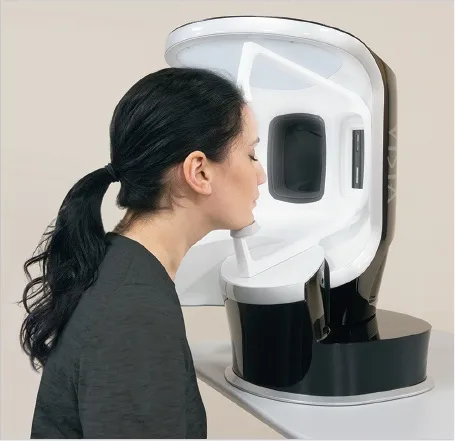 Fig 13. A patient positioning into a standardised facial-imaging booth (VISIA, Canfield): fixed head position, controlled multi-spectral light and repeatable geometry are what make the device's serial comparison valid — the same standardisation the manual photograph pursues (B1.4). — (Few, 2018, p. 435) [9]
> Sources: Few 2018 [9]
Fig 13. A patient positioning into a standardised facial-imaging booth (VISIA, Canfield): fixed head position, controlled multi-spectral light and repeatable geometry are what make the device's serial comparison valid — the same standardisation the manual photograph pursues (B1.4). — (Few, 2018, p. 435) [9]
> Sources: Few 2018 [9] [MEDLIB]
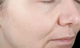 Fig 14. A skin-surface capture from a complexion-analysis system: fine texture, pores and subsurface pigment made visible under standardised light. Fig 14 is the device output the patient should be shown one parameter at a time — showing the full multi-parameter report is the commercial-iatrogenesis error of B1.5. — (Cosmetic Medicine & Surgery, 2016, p. 74) [16]
> Sources: Cosmetic Medicine & Surgery 2016 [16]
Fig 14. A skin-surface capture from a complexion-analysis system: fine texture, pores and subsurface pigment made visible under standardised light. Fig 14 is the device output the patient should be shown one parameter at a time — showing the full multi-parameter report is the commercial-iatrogenesis error of B1.5. — (Cosmetic Medicine & Surgery, 2016, p. 74) [16]
> Sources: Cosmetic Medicine & Surgery 2016 [16] [MEDLIB]
Digital morphometry and 3D. Mesh overlay, 3D analysis (Vectra and equivalents) and predictive simulation are useful to document and to communicate, and dangerous to promise (L1 — AI, Simulation & Digital Aesthetics). The rule for simulations is fixed: a simulation shown to a patient becomes, in their memory, a commitment; if you show it, annex it to the plan with the written line — this image is guidance, not a guaranteed result — and store it in the record (B1.4) [3].
The regulatory status of the box (B1.2). Whether one of these systems is a medical device under the MDR turns on its intended purpose, not its hardware: a unit sold as a cosmetic complexion analyser may sit outside the medical-device regime, whereas the same camera sold with a diagnostic or treatment-planning claim is a CE-marked medical device classified through Annex VIII, with AEMPS as the Spanish competent authority (RD 192/2023) [38][39]. The marketing claim decides the legal class, and the legal class decides your obligations.
How to use them without being used. Show the single parameter the agreed plan will treat, not the full report [16]. Use ANTERA or Vectra to measure a change objectively at follow-up, and VISIA UV to show the patient the sun damage they cannot see — that is where adherence is genuinely earned. Never let the device rank the problem list; the clinical assessment (B1.3) ranks it, and the device documents the ranking.
The parameters each device reports, and how to read them. VISIA characterises the surface across several channels — spots, pores, wrinkles, texture, UV spots, brown spots, red areas and porphyrins — and returns each as a percentile against an age- and phototype-matched base; the sub-surface brown/red separation is what shows a patient the pigment and vascular load their mirror hides [16]. OBSERV uses cross- and parallel-polarised light plus UV and Wood's-light modes to reveal pigment, vascularity and surface texture qualitatively and fast, which makes it a strong communication instrument rather than a measuring one. ANTERA 3D reconstructs the surface in three dimensions and returns quantitative maps — wrinkle depth and volume, indentation, roughness, and separated haemoglobin and melanin — which is why it, not VISIA, is the instrument for demonstrating a measured change at follow-up. Vectra photogrammetry captures a 3D surface for volumetry and for the mirror-style simulation whose risks B1.4 sets out.
Standardisation is the precondition for serial comparison. A device number is only comparable to its own prior value if capture geometry, lighting and patient positioning are held constant — the same discipline the manual photograph demands (B1.4) [32]. A change read off two differently-lit captures is an artefact, exactly as with the hand camera; the booth's fixed geometry (Fig 13) exists to remove that variable, and the resulting surface capture (Fig 14) is read one parameter at a time.
The assessment-to-plan tie-in. The pigment, vascular and texture reads feed the skin-quality lane of the plan, not a separate agenda: a high melanin/UV load flags the Fitzpatrick-driven HPI risk that governs peel and laser choice (B1.7) and points to topical and photoprotection groundwork before any energy device [19][20][25]; a wrinkle/texture and volume deficit points to the biostimulation and skinbooster groundwork of the regenerative lane [22][23]. The device does not add a diagnosis; it quantifies the cutaneous process the clinical assessment already named.
AI, morphometry and the social-media effect. Automated landmarking and AI-generated morphs make the metrics faster to compute and the simulation more photorealistic, which sharpens both the documentation benefit and the promise risk (L1 — AI, Simulation & Digital Aesthetics). Two cautions belong in the assessment. First, an AI proportion score is only as valid as its reference population, and the same population-bound problem that afflicts the hand-measured canons afflicts the automated one (B1.6) — an algorithm trained on one ancestry will "diagnose" another's normal variants as deviations [34]. Second, the patient increasingly arrives with a filtered self-image as the target, and the honest assessment measures the real face against the patient's own structure, not against a filter — a distinction that the "what would you keep?" question and the standardised, unfiltered baseline photograph both protect [32]. The device and the algorithm document and monitor; the clinician diagnoses, and the patient's own preserved architecture, not a generated ideal, is the target.
Common pitfalls: the seven-red-parameter printout handed to the patient. It manufactures seven reasons to consume and an anxious patient, and it substitutes a comparative percentile for a diagnosis — the device using the physician, not the physician using the device [16].
Coverage vs UPO
The master course (UPO) teaches facial assessment across T4 (facial proportions/analysis), G12 (planning methods), G7 (photography) and Módulo 1.2/1.3 (clinical history, photography/video). The grid maps what UPO teaches, its state here, and what the atlas adds. Extra rows list what UPO does not cover.
| UPO topic | State in this chapter | What the atlas adds |
|---|---|---|
Facial proportions: thirds, fifths, neoclassical canons (T4) [30] |
Covered — B1.6 | the explicit ⚠ that the values are population-bound; the sex-specific angle split; the evidence that phi does not predict attractiveness [34] |
| Historia clínica y diagnóstico (Módulo 1.2) [28] | Covered — B1.3, B1.4 | the verbatim/perception/preservation intake; the Spanish consent + data-protection obligations tied to article numbers [35][36] |
Fotografía y vídeo (Módulo 1.3, G7) [29] |
Covered — B1.4 | the ≥85 mm / Frankfort / fixed-light protocol as both diagnosis and lawful advertising basis [32][40] |
| Valoración facial teórica (Anido) [30] | Covered — B1.1, B1.3 | the three-process differentiation with the supine/digital-lift tests [12] |
Planning methods — MD Codes, AART/HIT (G12) [1] |
Covered — B1.3 | the explicit [D] non-evidence tagging and the 12–24-month phased plan template [3] |
| Competencias digitales (Módulo digital) [31] | Covered — B1.9 | skin-analysis/3D device reference-value trap; the MDR intended-purpose test [38] |
| Not in UPO — the regulatory map (ES + EU) | Added — B1.2 | Ley 41/2002, RGPD/LOPDGDD, MDR/RD 192/2023, RD 1907/1996, RD 1277/2003 with article numbers [35][36][38][40][41] |
| Not in UPO — the 2026 intrusism reform | Added — B1.7 | RD modifying RD 1277/2003, in force 1 July 2026; Cataluña Decreto 151/2017 and toxin/laser restriction [44][45] |
| Not in UPO — skin-of-color / ethnic assessment | Added — B1.6, B1.7 | HPI and keloid risk in Fitzpatrick IV–VI as the finding that governs energy/peel choice [8] |
| Not in UPO — errors-and-cost accounting | Added — B1.5 | the legal/financial/reputational cost per assessment error [32][35] |
UPO's two T4/G7 slides are the fastest-ageing lane and are never_sufficient_alone: a value resting only on a UPO slide (e.g. the neoclassical angles) is corroborated here against the monograph literature [2] and, where the corpus was silent (the whole regulatory map), against the primary legal instruments [35][41].
Self-assessment
- In what order do you run the first three steps of the valuation consult, and why is listening first?
Answer
Listen without examining, then ask by perception (tired/sad/angry/saggy), then ask what the patient would keep. Listening first produces the patient's own priority instead of a physician-led defect list; the preservation question guards against westernisation and over-treatment [1].- Give the three vertical thirds and the lower-third subdivision ratio.
Answer
Trichion–glabella, glabella–subnasale, subnasale–menton (ideally equal). Lower third splits subnasale→stomion 1/3, stomion→menton 2/3 [2].- State the nasofrontal, nasolabial (both sexes) and mentocervical reference angles.
Answer
Nasofrontal 115–130°; nasolabial 90–100° ♂ and 95–110° ♀; mentocervical 80–95° — all population-bound ⚠ [2].- What are the three independent ageing processes, and which manoeuvres separate them?
Answer
Structural/deflation, positional/descent, cutaneous. Supine test (descent corrects lying down), digital lift (groove corrects → descent), skin pinch (cutaneous component) [12].- Why is filling a descended, lax face the most expensive diagnostic error?
Answer
Filler adds weight, accelerates descent, reads as more ageing and invites more filler; exit needs hyaluronidase, distortion, refund and lost trust [11][12].- List the five mandatory photographic views and the two dynamics.
Answer
Frontal, oblique 45° R, oblique 45° L, profile 90° R, profile 90° L; plus maximal contraction and smile when toxin is planned [32].- Which two photographic variables most commonly fabricate a false result?
Answer
Lighting (top-lit before, front-lit after) and focal length/distance (a 30 cm phone vs an ≥85 mm clinic shot) [32].- Under Spanish law, what makes a clinical facial photograph require a separate second consent?
Answer
It is a special-category health datum (Art. 9 RGPD); teaching/marketing use needs its own, independently revocable consent beyond the care consent [36][35].- What are units U.47 and U.48, and what changes on 1 July 2026?
Answer
U.48 = non-surgical aesthetic medicine, U.47 = cosmetic surgery (RD 1277/2003). From 1 July 2026 the reforming RD restricts procedures to appropriately trained specialists [41][44].- Why is a VISIA "spot percentile" not a diagnosis, and how should the report be shown?
Answer
It is a comparative percentile against an age/phototype reference population, not disease; show one parameter at a time — the one the agreed plan will treat — not the full report [16].What's new and trends (2023-2026)
| Year | Change | Maturity class | Effect on assessment |
|---|---|---|---|
| 2021→ | MDR (UE) 2017/745 in full application; RD 192/2023 (22 Mar 2023) Spanish adaptation, AEMPS competent [38][39] | clinically actionable now | a device's intended purpose decides its class and your duties |
| 2018→ | LOPDGDD + RGPD enforcement matured for health/biometric data [36][37] | clinically actionable now | the two-consent (care vs image) structure is the documented standard |
| 2026 | RD modifying RD 1277/2003, approved 24 Mar 2026, in force 1 Jul 2026; SEME: ~65% unqualified, ~20% unregulated settings [44] | clinically actionable now | the note records physician-performed, authorised U.48; delegation tightens |
| 2024–26 | autonomic tightening: Andalucía Orden 3 Apr 2024 (U.48 guide), Castilla-La Mancha Orden 32/2026 [41] | clinically actionable now | centre-authorisation and room/sterilisation requirements formalised |
| 2024 | Singer & Papadopoulos: no universal standard of beauty (Level V review) [34] | promising but not validated | evidence backing for canons-as-description and ethno-specific correction |
| 2023→ | ANTERA/Vectra quantitative 3D monitoring and automated landmarking [16] | promising but not validated | objective serial change measurement, still population-bound |
| 2024→ | AI photorealistic simulation and ancestry-specific AI proportion scoring [34] | preclinical/speculative | documentation gain; promise-risk and algorithmic bias unquantified |
| — | the phi-mask "golden ratio guarantees beauty" and the device "diagnostic percentile" pitch [16][34] | unsupported commercial claim | rejected here — phi does not predict attractiveness; a percentile is not a diagnosis |
What did not change, and why the old references still stand. The geometric descriptors — thirds, fifths, the profile angles, the Ricketts E-line, phi — are unchanged since Leonardo and Dürer and remain descriptive vocabulary, not targets [2]. The three-process model of facial ageing and the supine/digital-lift tests that separate them are stable clinical method, and the compartmental-ageing anatomy that grounds them (Wan 2013; Update on Facial Aging 2010) has not been overturned [12][13]. Standardised five-view photography with a fixed Frankfort plane and ≥85 mm optics is the same protocol the 2016 photography literature describes [32]. The genuinely new material is regulatory (the 2026 reform, MDR/RD 192/2023, LOPDGDD maturation) and interpretive (the evidence-level rejection of a universal beauty standard) — the clinical assessment method itself is durable, and where a claim rested only on a UPO slide it is corroborated here against the monographs and the primary law.
Unexplored directions (AI speculation)
> The items below are model-generated hypotheses, not evidence or recommendations. Each is tagged [IA-ESPEC], states the anchor fact already in this chapter, the proposal, and what would settle it. None is a dose, a product or a protocol a reader may act on.
-
Ancestry-specific proportion norms.
[IA-ESPEC]Anchor: the canons are population-bound and misfire on non-Caucasian faces (B1.6, B1.7) [2][8][34]. Proposal: a reference set of thirds/fifths/angles stratified by ancestry, built from large consented photographic cohorts, replacing the single Caucasian canon as the descriptive baseline. Expected effect: fewer normal variants mis-classified as "defects" than under the single canon. Confounder: ancestry self-report and admixture blur the strata, and operator expectation may bias which variants are called defects. What would settle it: a blinded reader study comparing mis-classification rates, ancestry-stratified norms versus the single canon. -
Validating the supine and digital-lift tests against imaging.
[IA-ESPEC]Anchor: these bedside tests separate descent from deflation (B1.3) [12]. Proposal: correlate the bedside call with upright-versus-supine 3D morphometry or dynamic ultrasound of the retaining ligaments. Expected effect: agreement above chance between the manoeuvre and the imaging-measured displacement. Confounder: skin laxity and oedema affect both the test and the image; single-operator reads limit generalisability. What would settle it: a prospective blinded correlation with a pre-registered agreement threshold. -
The perception attribute as an outcome measure.
[IA-ESPEC]Anchor: the tired/sad/angry/saggy attribute gives a patient-verifiable success criterion (B1.3) [1]. Proposal: a validated patient-reported attribute score used as the primary outcome of an assessment-led plan, rather than millimetric change. Expected effect: higher correlation with satisfaction and lower regret than millimetric outcomes. Confounder: mood and social-media exposure move the attribute independently of the face; expectation effects. What would settle it: psychometric validation plus a cohort correlating the score with satisfaction and regret instruments. -
AI simulation and body-image risk.
[IA-ESPEC]Anchor: a shown simulation becomes a remembered commitment (B1.4, B1.9) [3]. Proposal: compare AI-photorealistic simulation against a schematic morph for unmet-expectation and dysmorphia-flag rates. Expected effect: photorealistic morphs raise unmet-expectation and dysmorphia flags relative to schematic morphs. Confounder: baseline body-image and platform use differ between arms; consent framing colours the response. What would settle it: a randomised comparison against validated body-image and satisfaction instruments. -
Device percentile versus clinician ranking.
[IA-ESPEC]Anchor: a skin-analysis percentile is comparative, not diagnostic, and treating the report distorts the plan (B1.5, B1.9) [16]. Proposal: compare device-led against clinician-led problem ranking on treatment count, cost and satisfaction. Expected effect: device-led ranking raises treatment count and cost without improving satisfaction. Confounder: clinic incentive structure and operator adherence to the printout. What would settle it: a two-arm trial with pre-registered treatment-count, satisfaction and regret outcomes.
Safety
Assessment is where the safety of every downstream act is decided, and the following are the safety-critical calls that must be made before, not after, a needle is loaded.
- Vascular and blindness risk is flagged at assessment. Periocular and nasal work carry occlusion and blindness risk; the consent must name it specifically, and the assessment must record the topographic anatomy and any prior filler in the danger zones [33].
- Ask actively about permanent fillers. Patients do not volunteer them; a permanent or minimally-degradable filler changes the entire plan and the risk of layering, so the direct question is mandatory and its answer recorded [11][33].
- Fitzpatrick IV–VI: pigmentary and keloid risk governs energy and peel choice. The phototype read at assessment is the finding that decides laser, peel and microneedling selection — post-inflammatory hyperpigmentation and keloid are the foreseeable harms [8].
- Never treat descent with volume. The supine and digital-lift tests are a safety step: filling a descended, lax face is foreseeable over-treatment with a costly reversal [12].
- The photograph is a safety and legal instrument. A standardised baseline with pre-existing asymmetry marked is the defence against the most common complaint; its absence is a foreseeable exposure on satisfaction medicine [32][35].
- Only a qualified physician, in an authorised centre, after real consent and a reflection period. In Spain this is unit U.48, the 2026 reform tightens it, and Cataluña restricts toxin and laser to physicians — verify your community's current rule before delegating [41][44][45][47].
- Show two or three findings, not ten. Over-disclosure is its own harm: it manufactures anxiety and over-consumption, and the full skin-analysis report is the machine version of the same error [5][16].
References
- de Maio M. MD Codes: A Methodological Approach to Facial Aesthetic Treatment with Injectable Hyaluronic Acid Fillers. 2020.
[B][MEDLIB]· DOI 10.1007/s00266-020-01762-7 - Naini FB. Facial Aesthetics: Concepts and Clinical Diagnosis. 2011.
[C][MEDLIB] - Azizzadeh B, et al. Master Techniques in Facial Rejuvenation. 2018.
[C][MEDLIB] - Farhadieh RD, et al. Plastic and Reconstructive Surgery. 2015.
[C][MEDLIB] - Liu. Atlas of Lip and Nose Plastic and Cosmetic Surgery. 2021.
[C][MEDLIB] - Jones DH. Injectable Fillers. 2019.
[C][MEDLIB] - Nouri K. Lasers in Dermatology and Medicine. 2018.
[C][MEDLIB] - Alam M, Bhatia AC, Kundu RV, Yoo SS, Chan HHL. Cosmetic Dermatology for Skin of Color. 2009.
[C][MEDLIB] - Few J, et al. Art of Combining Surgical and Nonsurgical Techniques in Aesthetic Medicine. 2018.
[C][MEDLIB] - Hong SO, et al. The Art and Science of Filler Injection. 2020.
[C][MEDLIB] - Carruthers J, Carruthers A. Soft Tissue Augmentation. 2018.
[C][MEDLIB] - Wan D, et al. Clinical Importance of the Fat Compartments in Midfacial Aging. 2013.
[B][MEDLIB]· DOI 10.1097/GOX.0000000000000035 - Update on Facial Aging. 2010.
[A][MEDLIB]· DOI 10.1177/1090820X10378696 - van Gijn DR, et al. Oxford Handbook of Head and Neck Anatomy. 2022.
[C][MEDLIB] - Erian A, Shiffman MA. Advanced Surgical Facial Rejuvenation. 2012.
[C][MEDLIB] - Cosmetic Medicine & Surgery. 2016.
[C][MEDLIB] - Cheney ML, Hadlock TA. Facial Plastic and Reconstructive Surgery. 2015.
[C][MEDLIB] - Vieira Braz A, et al. Atlas de Anatomia e Preenchimento Global da Face. 2017.
[C][MEDLIB] - Baran R, Maibach HI. Textbook of Cosmetic Dermatology. 2017.
[C][MEDLIB] - Draelos ZD. Cosmetic Dermatology: Products and Procedures. 2009.
[C][MEDLIB] - Carniol PJ, Monheit GD. Aesthetic Rejuvenation Challenges and Solutions. 2010.
[C][MEDLIB] - Hausauer AK, Jones DH. PRP and Microneedling in Aesthetic Medicine. 2019.
[C][MEDLIB] - Piccolo D, et al. A.R.T. Autologous Regenerative Therapy in Aesthetic Medicine. 2025.
[C][MEDLIB] - Yaremchuk MJ. Atlas of Facial Implants. 2020.
[C][MEDLIB] - Obagi S, et al. Chemical Peels. 2021.
[C][MEDLIB] - Parker E. Fundamentals for Cosmetic Practice. 2022.
[C][MEDLIB] - de Maio M, Wu WTL. MD Codes: Unlocking the Code. 2017.
[D][MEDLIB] - Módulo 1.2 — Historia Clínica y Diagnóstico. UPO Sorted.
[D][MEDLIB] - Módulo 1.3 — Fotografía y Vídeo. UPO Sorted.
[D][MEDLIB] - Teoría de Valoración Facial (Anido). UPO Sorted.
[D][MEDLIB] - Competencias Digitales en Medicina Estética. UPO Sorted.
[D][MEDLIB] - Nair AG, Santhanam A. Clinical Photography for Periorbital and Facial Aesthetic Practice. 2016.
[B]· PMID 27398013 · DOI 10.4103/0974-2077.184047 - Heydenrych I, De Boulle K, Kapoor KM, Bertossi D. The 10-Point Plan 2021: Updated Concepts for Improved Procedural Safety During Facial Filler Treatments. 2021.
[B]· PMID 34276222 · DOI 10.2147/CCID.S315711 - Singer R, Papadopoulos T. There Is no Universal Standard of Beauty. 2024.
[B]· PMID 39103640 · DOI 10.1007/s00266-024-04266-w - Ley 41/2002, de 14 de noviembre, básica reguladora de la autonomía del paciente y de derechos y obligaciones en materia de información y documentación clínica (BOE-A-2002-22188).
[A] - Reglamento (UE) 2016/679 (RGPD), de 27 de abril de 2016, general de protección de datos — Art. 9 categorías especiales.
[A] - Ley Orgánica 3/2018, de 5 de diciembre, de Protección de Datos Personales y garantía de los derechos digitales (LOPDGDD) (BOE-A-2018-16673).
[A] - Reglamento (UE) 2017/745 (MDR), de 5 de abril de 2017, sobre los productos sanitarios.
[A] - Real Decreto 192/2023, de 21 de marzo, por el que se regulan los productos sanitarios (BOE-A-2023-7416).
[A] - Real Decreto 1907/1996, de 2 de agosto, sobre publicidad y promoción comercial de productos, actividades o servicios con pretendida finalidad sanitaria (BOE-A-1996-18085).
[A] - Real Decreto 1277/2003, de 10 de octubre, por el que se establecen las bases generales sobre autorización de centros, servicios y establecimientos sanitarios — Anexo II, unidades U.47/U.48 (BOE-A-2003-19572).
[A] - Ley 34/1988, de 11 de noviembre, General de Publicidad.
[A] - Ley 14/1986, de 25 de abril, General de Sanidad — arts. 27, 29.1, 102.
[A] - Real Decreto por el que se modifica el Real Decreto 1277/2003 (aprobado 24 de marzo de 2026; entrada en vigor 1 de julio de 2026), restringiendo procedimientos a especialistas con formación acreditada.
[A] - Decreto 151/2017 (Cataluña), requisitos técnico-sanitarios de los centros y servicios sanitarios.
[A] - Orden de 18 de abril de 2008 (Cataluña) — tratamientos médico-estéticos dirigidos por médico; láser y toxina botulínica solo por facultativos.
[A] - Ley 44/2003, de 21 de noviembre, de ordenación de las profesiones sanitarias (LOPS).
[A]
Verification: EN canonical B1, PRACTICA template extended to 9 subchapters (B1.9 added for instrumentation/imaging devices, because the curriculum carries a ninth subchapter — skin-analysis devices — that the 8-block template would otherwise orphan). Corpus lane [MEDLIB]: retrieved over 9 per-subchapter runs (evaluation/runs/B1.1–B1.9.jsonl), graded on global_top_score; strong on assessment/photography/proportions (0.71–0.84), chronically thin on regulation_spain_eu (0.41–0.66) and contraindications_interactions (0.33–0.71), so the whole regulatory map (B1.2, B1.7) is external-lane and cited to the primary legal instruments [35]–[47], verified against the BOE and official sources; the two 2013/2010 corpus papers with DOIs were confirmed to resolve. Proprietary methods MD Codes™ [1][27] and AART/HIT [3] are printed as [D] organising methods with no evidence level. Numbers marked ⚠ (sex-specific nasolabial angles, Ricketts E-line, phi as an attractiveness predictor) are population-bound; the canons are labelled explicitly as derived from young Caucasian reference populations. 14 figures opened and captioned from what they show, not from the corpus caption (Fig 12's DB caption mismatched its image and was re-captioned to the visible finding). Salvage: mined docs/salvage/B1.prev.md (whole file), all language-neutral numbers preserved.