D11 · Mentón (chin / mentum)
> Currency and provenance — 47 references · median 2019, range 2006-2025, 23 % from 2022 on · provenance: verified external 13 % (6) · MEDLIB corpus 87 % (41, of which 4 from the UPO master's) · 4 flagged [D] never_sufficient_alone.
> Tags: [A] datasheet, guideline or consensus with year · [B] primary literature with PMID/DOI · [C] monograph · [D] slide or opinion, never sufficient alone · [MEDLIB] own corpus · (P) model reasoning (never a dose) · ⚠ disputed or stale figure.
Chapter blocks: - [x] D11.1 In 30 seconds - [x] D11.2 Layered anatomy (skin to bone) - [x] D11.3 Vessels, nerves and danger zone - [x] D11.4 Ageing of the region - [x] D11.5 Assessment - [x] D11.6 Goal and patient selection - [x] D11.7 Technique - full grid - [x] D11.8 Toxin of the region - [x] D11.9 Combination and sequence - [x] D11.10 Region-specific complications
D11.1 · In 30 seconds
The chin is the one midline bony platform of the face: a low-vascular-risk, high-reward projection target where the needle goes to periosteum and the danger is a nerve, not blindness. Diagnose first (bite test), build the skeleton with filler, relax the mentalis with toxin, and never widen a feminine chin by reflex.
| Axis | Do this | Red line |
|---|---|---|
| Diagnosis | Ask the patient to bite. Normal occlusion + small chin = microgenia → filler. Class II malocclusion = retrognathia → refer [7][16] | ⚠ Filler on a skeletal Class II camouflages a profile it cannot fix. Occlusion is the 30-second exam almost no filler course teaches [16] |
| Plane / device | Deep supraperiosteal bolus to bone, midline, needle vertical, aspirate, then optional superficial fat above mentalis for surface [12][14][16] | ⚠ Superficial product in the mentalis belly (it inserts into dermis) becomes visible on movement [16] |
| Product | High-G′/high-cohesivity HA to "mimic bone" (Volux VYC-25L 25 mg/mL) or the chin-indicated Voluma VYC-20L 20 mg/mL; CaHA (Radiesse) for projection + biostimulation [12][21][22][24] | ⚠ CaHA/PMMA are not hyaluronidase-reversible. Conservative, aspirate |
| Volume | Asymmetry 0.2-0.3 mL · mild recession 0.6-0.8 mL · moderate-severe 1.3-1.5 mL · MD Codes per-point 0.3 mL (needle) / 0.5 mL (cannula) [15][13] | ⚠ Over-projection blunts the labiomental angle and reads "witch/cocktail" chin |
| Needle vs cannula | Projection = needle to bone, midline. Contour, labiomental crease, jawline blend = cannula, subcutaneous [13][17] | Not averaged: choose by target plane (§D11.7) |
| Toxin | Mentalis 2-8 U, 1-2 low-midline deep points for cobblestone/peau d'orange. DAO 2-5 U/side, deep at mandibular border, lateral to modiolus [16][30][31] | ⚠ Mentalis too high hits orbicularis oris; DAO too medial/high hits DLI → asymmetric smile 8-12 wk |
| Vessel to respect | Stay midline for needle-to-bone; the mental artery exits laterally (below 2nd premolar). Submental / inferior-labial / facial branches are caudal-lateral [7][16][24] | ⚠ C3-point too lateral → mental artery. Intravascular HA → chin/lower-lip necrosis (rare, no blindness route) [38] |
One-line plan: bite test → profile analysis (zero-meridian, labiomental angle) → build projection with needle-to-bone HA or CaHA (0.3 mL/point, midline, aspirate) → refine crease/jawline with cannula → relax mentalis/DAO with toxin same or next session → refer the skeletal Class II and the frank skin excess. Full-face sequence in §D11.9 and L2 - Combination Protocols & Layered Rejuvenation.
Refer, do not inject, when: the bite is Class II (retrognathia); the deficiency is large or the patient wants permanence (genioplasty/implant); there is frank cervical skin excess, a true jowl or a ptotic submandibular gland; the hyoid is low; or BDD traits are present [7][16]. "Not a filler problem" is a complete answer.
One-line modality map: chin = projection (needle-to-bone HA/CaHA); jawline = continuity (cannula); submental fat = reassess after projecting the chin; cobblestone/marionette = toxin; skin laxity = energy/threads; skeletal, large or permanent = surgery.
Dose quick-card (this region): - HA per point on bone: 0.3 mL (needle); cannula lateral: 0.5 mL [13]. - Total chin: 0.2-0.3 mL (asymmetry) / 0.6-0.8 mL (mild) / 1.3-1.5 mL (moderate-severe) [15]. - Mentalis toxin: 2-8 U (12 U abobotulinum), 1-2 points, deep low midline [16][46]. - DAO toxin: 2-5 U/side (4-5 U abobotulinum), mandibular border, lateral to modiolus [30][46]. - Occlusion (HA): 500 IU hyaluronidase per area, hourly, complete within 72 h [38].
Classic trap: treating a "double chin" (papada) with lipolysis when the real deficit is an under-projected chin - projecting the mentum opens the cervicomental angle and improves the submental silhouette, so examine the chin before selling submental fat treatment [7].
D11.2 · Layered anatomy - skin to bone
Fixed 6-layer order for the chin (superficial → deep). The chin breaks the "5-compartment glide plane" rule of the midface: the mentalis binds skin to bone, so there is no free-sliding plane here.
CHIN - layered anatomy (midline, over the symphysis)
┌───────────────────────────────────────────────────────────────┐
│ 1 SKIN          thick, sebaceous, dermal-muscular tether [1]    │
│ 2 DERMIS        mentalis fibres insert directly INTO dermis [1] │  ← no glide plane
│ 3 SUPERFICIAL   pre-mental fat pad (corpo adiposo pré-mentual); │
│   FAT           thin midline, target for surface refinement [5] │
│ 4 (S)MAS/MUSCLE MENTALIS (paired), DLI, DAO - all marginal      │
│                 mandibular n.; mentalis elevates the chin pad   │
│ 5 DEEP FAT      submentalis / pre-periosteal fat over symphysis │
│ 6 PERIOSTEUM    mandibular SYMPHYSIS + parasymphysis + pogonion │  ← needle target
└───────────────────────────────────────────────────────────────┘
| Layer | Chin-specific content | Injection relevance |
|---|---|---|
| Skin / dermis | Thick, tethered; mentalis inserts into dermis [1][34] | Superficial product shows on animation; keep out of the muscle belly [16] |
| Superficial fat | Pre-mental (pré-mentual) compartment; thin at midline [3][5] | Subcutaneous cannula plane for surface/crease refinement [17] |
| Mentalis | Paired conical muscle, origin incisive fossa, inserts into chin dermis; elevates chin pad, deepens labiomental crease, everts lower lip [1][7][31] | The toxin target (§D11.8); the reason a filled chin still "cobblestones" if left untreated |
| Deep fat / pre-periosteal | Submentalis fat over the symphysis; HA injected to bone diffuses into and stays in this compartment (cadaver-proven) [20] | Where the supraperiosteal bolus actually sits |
| Bone | Symphysis, parasymphysis, body; mental protuberance (triangular) with two mental tubercles and a central depression [6] | The projection platform; needle-to-bone target |
Consensus: the chin is skin - superficial fat - mentalis - deep/pre-periosteal fat - bone, with the mentalis binding dermis to mandible so there is no free glide plane; the deep supraperiosteal compartment is the structural target and the superficial fat the finishing layer [1][5][16][20].
The mentalis is the organising muscle of the region. It is a paired conical fasciculus lying beside the frenulum of the lower lip, arising deep from the incisive fossa and inserting into the skin of the chin; contraction pulls the chin skin up, deepens the labiomental crease, raises and everts the lower lip, and in pouting produces the cobblestone or "peau d'orange" surface [1][31][34]. Because its fibres reach the dermis, any product placed superficially in the belly telegraphs on movement [16]. The depressor labii inferioris and depressor anguli oris arise from the oblique line of the mandible and insert into the lip, depressing the lower lip and the mouth corners respectively; all three (mentalis, DLI, DAO) share the marginal mandibular branch of the facial nerve [7]. The bony platform is the mandibular symphysis (the midline fusion of the two hemimandibles), flanked by the parasymphysis and continuing into the body; the mental protuberance is triangular with paired tubercles [6][42].
The bony platform, in detail. The mandible is a horseshoe-shaped bone with a horizontal anterior body that turns posterosuperiorly at the angle into the ramus [42]. The two hemimandibles fuse in the midline at the symphysis menti; a vague median ridge on the adult external surface marks the fused site and bifurcates below to enclose the triangular mental protuberance, which carries two mental tubercles and a small central depression [6]. The relevant subunits for augmentation and mentoplasty are the symphysis, parasymphysis and body [7]. Inside the bone, the mandibular canal runs from the mandibular foramen (on the medial ramus) forward and down into the body, coming to lie close to the external (labial) cortex anteriorly; near its anterior end it turns to exit at the mental foramen, and a short incisive continuation carries on to the midline [6]. This is why a deep midline supraperiosteal deposit is off the canal and its contents, while a lateral one is not.
The mental canal, in practice. The neurovascular bundle travels inside the mandibular canal and exits anteriorly at the mental foramen; a needle placed on the midline symphysis is medial to the canal and its exit, while a lateral point at the level of the premolars sits over it [6][7]. That single geometric fact is why the projection deposit is a midline gesture and any lateral (paragonion or jawline) work is a separate, more cautious one (§D11.3, §D11.7).
The mentalis and its neighbours, layer by layer. Standring describes the mentalis as a conical fasciculus lying at the side of the frenulum of the lower lip; its fibres arise from the incisive fossa, incline medially and downward, and some superficial fascicles reach the dermis while others blend toward the lower lip [2]. Because the muscle is the deepest of the lower-lip elevators and its fibres cross the midline, the cutaneous depression of a "cleft" or dimpled chin marks exactly where opposing fibres insert into skin [7][34]. Around it sit the depressor labii inferioris (medial, more superficial) and the depressor anguli oris (lateral), both from the oblique line of the mandible, both depressors, both marginal-mandibular-innervated [7][41]. The practical layer map for a needle:
| Depth (approx.) | Structure met | Needle intent |
|---|---|---|
| 0-1 mm | Epidermis / papillary dermis | Never the deposit plane (visible, nodular) |
| 1-3 mm | Reticular dermis + mentalis dermal insertions | Skin boosters/microbotox only |
| 3-6 mm | Superficial (pre-mental) fat | Subcutaneous cannula for surface/crease [17] |
| 6-10 mm | Mentalis belly | Toxin target, never filler [16] |
| 10 mm-bone | Deep / pre-periosteal fat | Structural filler diffuses and stays here [20] |
| Bone | Symphyseal periosteum | Needle-to-bone bolus, aspirate [13] |
Surface landmarks for the injector. The working landmarks are the soft-tissue pogonion (most anterior chin point), the menton (lowest midline point), the labiomental crease/sulcus (lip-to-chin junction), the mental tubercles laterally, and the melomental (marionette) folds running from the commissure toward the chin [1]. The "mandibular line" is the segment from the menton to the gonial angle, and continuity along it is what reads as a defined jaw [1]. These are the points the MD Codes map onto (§D11.7) and the references the profile analysis measures (§D11.5).
Consensus (depth): filler for projection goes to the deepest plane (periosteum / pre-periosteal fat), filler for surface goes to the most superficial fat, toxin goes to the muscle, and nothing structural goes into the mentalis belly or the dermis [16][17][20]. The layers are the same list every source gives, only the tethering differs from the midface.
The chin is a uniquely human platform, and that shapes the technique. The projecting bony chin appeared with modern humans and replaced the receding primate muzzle, giving the region a solid midline prominence rather than the thin, mobile scaffold of the midface [11]. On the lingual side of the same bone the genial (mental) tubercles anchor genioglossus and geniohyoid, so the symphysis is a dense midline block that accepts a firm to-bone bolus without the give of maxillary bone [2][6]. This is the anatomical permission for a structural supraperiosteal deposit at the chin that would over-project in the midface.
Retaining structures and the labiomental fold. The surface labiomental crease marks where the lower-lip unit meets the chin pad; its depth is set jointly by the mentalis, the bone and the mentolabial soft-tissue thickness, which is why an identical bony pogonion can read as deficient, ideal or over-creased (§D11.5) [11]. The mandibular retaining ligament tethers skin to the anterior mandibular body and is the reason the prejowl sulcus opens in front of it while tissue descends behind it [10][19]. Medial to the labiomental and nasolabial sulci the SMAS behaves as a type-2 investing fascia strongly adherent to skin rather than a free gliding sheet, so the whole lower-medial face is more tethered and less forgiving of a superficial deposit than the lateral cheek [19].
| Retaining structure | Role at the chin | Injection consequence |
|---|---|---|
| Mentalis dermal insertions | Bind muscle to skin | No structural filler in the belly; toxin only [16] |
| Mandibular retaining ligament | Anchors skin to anterior body | Treat the prejowl in front of it, not the jowl behind [19] |
| Type-2 SMAS (medial lower face) | Adherent investing fascia | Superficial product reads and migrates; go deep or cannula-subcutaneous [19] |
| Labiomental soft-tissue thickness | Sets crease depth | Read the fold, not just the mm of projection [11] |
Classic trap: injecting the chin as if it had the midface's deep-fat glide plane and depositing "deep subcutaneous" in the mentalis belly - the muscle is dermally tethered, so the correct deep target is periosteum, and the correct superficial target is the pre-mental fat above the muscle, never the muscle itself [16].
D11.3 · Vessels, nerves and danger zone
The chin is a comparatively low-risk zone because the midline platform is off the named vessels and there is no route to the ophthalmic artery - but it has one nerve you can injure and one lateral point where the artery is close. Link: J2 - Vascular Occlusion - Prevention, Recognition & Management.
| Structure | Course / depth | Variant / frequency | What fails if you hit it |
|---|---|---|---|
| Mental nerve (terminal inferior alveolar, V3) | Exits mental foramen, below/at the 2nd premolar, ~2.5 cm lateral to midline, ~1 cm above the inferior border, midway between alveolar ridge and lower border [7][17] | Foramen on the sagittal line through mid-pupil/supraorbital/infraorbital foramen; some describe it between 1st-2nd premolar [1][8] | Hypoaesthesia / dysaesthesia of the chin and lower lip - the practical chin complication, from a foramen-directed deposit [16] |
| Mental artery | Terminal branch of the inferior alveolar artery via the mandibular canal; exits the foramen with the nerve [1] | Accompanies the nerve laterally | Bleeding, bruising; theoretical intravascular event if a lateral bolus enters it [13] |
| Inferior labial artery | Branch of the facial artery; originates ~2.5 cm from the labial commissure, runs to the lower lip [1] | Crosses midline, anastomoses | Lower-lip ischaemia; supplies the mentalis with the mental artery [9] |
| Submental artery | Branch of the facial artery, runs along the inferior mandibular border toward the midline | Superficial near the border; the cannula-avoidance target for jawline blending [40] | Submental/chin skin ischaemia; the reason cannula + border work stays subcutaneous and aspirates |
| Facial artery | Crosses the mandibular border at the antegonial notch (just anterior to the masseter); palpable [20] | Palpable pulse in most patients | Bruising/occlusion at the jawline junction - palpate the pulse before any border deposit |
Consensus: keep the projection deposit midline and on bone, where the named vessels are not; the real chin hazard is mental-nerve dysaesthesia from a foramen-directed injection, not catastrophic occlusion, and there is no ophthalmic (blindness) route from the chin [7][16][40]. When you must go lateral (paragonion, jawline), switch to cannula, aspirate, and respect the mental foramen and the palpable facial artery.
Why the chin reads "safe" yet can still necrose. The lower-lip and chin skin are supplied by the inferior-labial and submental branches of the facial artery plus the mental branch, all anastomosing across the midline [1][9]; an intravascular filler bolus can still produce chin or lower-lip skin ischaemia and necrosis, uncommon relative to the midface and nose but real, and managed as an HA vascular event with high-dose pulsed hyaluronidase (§D11.10) [38]. The absence of a retrograde ophthalmic pathway is what removes the blindness risk that dominates nose and glabella work [39] - a genuine categorical difference, not a reason to skip aspiration.
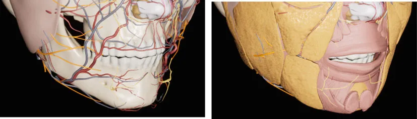 Fig 1. Left: skull with the arterial (red), venous (blue) and nerve (yellow) network draped over the mandible and mental region; right: the same field with mentalis and the lip depressors overlying the pre-periosteal fat. The midline symphysis (needle target) sits medial to the named vessels - (Hong, 2020, p. 180).
> Sources: [MEDLIB] Hong 2020 [14]. Corpus figure opened before captioning; the Fig 1 panels document why the midline-to-bone deposit is off the vascular corridor while lateral points are not.
Fig 1. Left: skull with the arterial (red), venous (blue) and nerve (yellow) network draped over the mandible and mental region; right: the same field with mentalis and the lip depressors overlying the pre-periosteal fat. The midline symphysis (needle target) sits medial to the named vessels - (Hong, 2020, p. 180).
> Sources: [MEDLIB] Hong 2020 [14]. Corpus figure opened before captioning; the Fig 1 panels document why the midline-to-bone deposit is off the vascular corridor while lateral points are not.
Marking and palpation before the needle. Mark the facial midline and both mental foramina, palpate the facial-artery pulse at the antegonial notch, and for any lateral or jawline point either stay off the marked foramen or map the vessel on Doppler [16][20][40]. The mental foramen sits roughly two finger-breadths lateral to the midline and one above the lower border, but its variability is precisely why you palpate and mark rather than trust a fixed measurement [7][8]. A five-second pulse check at the antegonial notch is the cheapest vascular safety step in the region [20].
The arterial tree, traced. The chin and lower lip are fed by two systems that anastomose freely across the midline. From the facial artery come the inferior labial artery (to the lower lip, arising roughly 2.5 cm from the commissure) and the submental artery (running medially along the inferior mandibular border); from the inferior alveolar artery (a branch of the maxillary) comes the mental artery, which travels in the mandibular canal and exits the mental foramen with the nerve [1][9]. The mentalis itself is supplied by the inferior labial and mental arteries [1]. Before the inferior alveolar artery enters the mandibular canal it gives the mylohyoid artery, and its proximity to the inferior alveolar nerve at the mandibular foramen is why an intraoral inferior-alveolar block has a high positive-aspiration rate [37]. For the injector the message is simple: the named vessels are lateral and caudal; the midline symphysis is not on them.
| Structure | System | Depth at the chin | Aspiration/technique note |
|---|---|---|---|
| Inferior labial a. | Facial a. | Submucosal/subcutaneous, near the wet-dry lip | Low-pressure, small aliquots near the labiomental crease [9] |
| Submental a. | Facial a. | Subcutaneous along the inferior border | Cannula + aspirate for jawline blend [40] |
| Mental a. + nerve | Inferior alveolar (maxillary) | Exit foramen, below 2nd premolar | Keep lateral points off the foramen [7][16] |
| Facial a. (antegonial) | External carotid | Crosses the border, palpable | Palpate the pulse before a border deposit [20] |
| Retromandibular v. / facial v. | Venous | Angle / along the border | Bruising risk; not an occlusion route |
Mental-foramen landmarks and variants. The foramen sits on the sagittal line that also passes through the supraorbital and infraorbital foramina and the mid-pupil, at the midpoint of the mandibular body vertically; most describe it below the second premolar, though some place it between the first and second premolars, and it lies roughly 2.5 cm lateral to the midline and 1 cm above the inferior border [7][8][17]. That variability is the argument for keeping the projection point strictly midline and for palpating rather than trusting a fixed millimetre rule. The mental nerve, a terminal branch of the inferior alveolar (V3), is sensory to the chin, lower lip and jowl, so a foramen-directed deposit produces a dermatomal hypoaesthesia the patient notices immediately [1][8].
Regional nerve block. A mental-nerve block anaesthetises the ipsilateral chin and lower lip; it is placed intraorally at the labial vestibule opposite the second premolar or transcutaneously toward the foramen, and bilateral blocks cover the whole chin [7]. It is useful for a painful or extensive treatment but numbs the dynamic field, so complete the animation exam first (§D11.5). Because the block targets the foramen, it also marks the exact point a filler deposit must avoid.
Why "safe" still means aspirate and inject low-pressure. Filler travels retrograde into a vessel far more easily at high flow and high pressure, and scarred tissue from prior treatments is a documented conduit; the countermeasure is small aliquots, low pressure, aspiration at bone, and avoidance of a single large bolus [13][38]. The chin's genuine safety margin is anatomical (no ophthalmic route, vessels off the midline), not a licence to abandon the vascular precautions that apply to every filler.
The pressure argument, quantified. A representative facial artery segment holds a very small volume (the supratrochlear artery averages ~0.085 mL), so even a fraction of a millilitre delivered under pressure can fill a length of vessel and travel; injecting at low pressure with the needle moving, and aspirating before a bolus at bone, are the practical defences [38]. High flow and a fixed needle tip in one spot are the two conditions that turn a deposit into an embolus.
Venous and lymphatic drainage. The chin and lower lip drain to the facial vein and, at the angle, the retromandibular vein; venous outflow at the chin is redundant, so venous obstruction is a non-issue here unlike in axial-pattern territory, and the bruising risk is cosmetic rather than ischaemic [38]. Lymphatic drainage of the central lower lip and chin runs to the submental nodes, which is why persistent chin/submental swelling after filler warrants examination rather than reflexive massage.
Doppler ultrasound of the region. On colour Doppler the normal facial/submental artery shows a clean pulsatile trace; an obstructed vessel shows chaotic back-and-forth flow with both colours, and intravascular HA appears as a hypoechoic (dark) deposit downstream of the chaotic segment [44]. A trained operator can therefore both map the vessel before injecting laterally and confirm reperfusion after dissolving an embolus, using far less hyaluronidase than the blind protocol (§D11.5, §D11.10) [40][44].
Classic trap: aiming a "chin" bolus at soft-tissue pogonion but drifting laterally toward the mental foramen - the deposit that should have been midline-on-bone ends up over the foramen, producing lower-lip numbness; the fix is to mark the midline and keep the C3/pogonion point medial [13][16].
D11.4 · Ageing of the region - what is lost, and in what order
Bone and ligament change first and drive the soft-tissue stigmata; the skin thins last. Restore in the same order you lost: skeleton before surface.
ORDER OF LOSS (chin / lower third)
1 BONE        symphyseal resorption → less projection + less vertical height   [10]
2 DEEP FAT /  pre-periosteal fat deflation + ligament laxity → deep labiomental
  LIGAMENT    crease, opening prejowl sulcus                                    [10][19]
3 SUPERFICIAL pre-mental (pré-mentual) fat descent/deflation → surface laxity   [4][5]
4 SKIN        dermal thinning, chin dimpling with hyperkinetic mentalis (last)  [10][34]
| Tissue | Change with age | Aesthetic result |
|---|---|---|
| Bone (symphysis) | Resorption, reduced projection and vertical height [10] | Recession; the platform itself shrinks - filler on top cannot recreate lost bone geometry, only mask it |
| Deep fat + ligament | Pre-periosteal deflation, mandibular-ligament laxity [10][19] | Deep labiomental crease, prejowl sulcus opens, jawline becomes a wave |
| Superficial fat | Pre-mental compartment descent/deflation [4][5] | Surface irregularity, "chin pad" ptosis |
| Skin + muscle | Dermal thinning + collagen loss + hyperkinetic mentalis [34] | Uncontrollable cobblestone dimpling, mentolabial crease against the gums |
Consensus: the chin "deflates" from the bone outward - superficial and deep fat compartments lose volume while the symphysis resorbs - so the ageing chin is a combined skeletal + soft-tissue loss, and bone/ligament change precedes and drives the crease-and-sulcus stigmata [10][19]. This is why a structural (high-G′) product to bone outperforms a soft gel to the surface in the ageing chin, and why a hyperkinetic mentalis must be relaxed or the surface keeps crimping over any new volume [34].
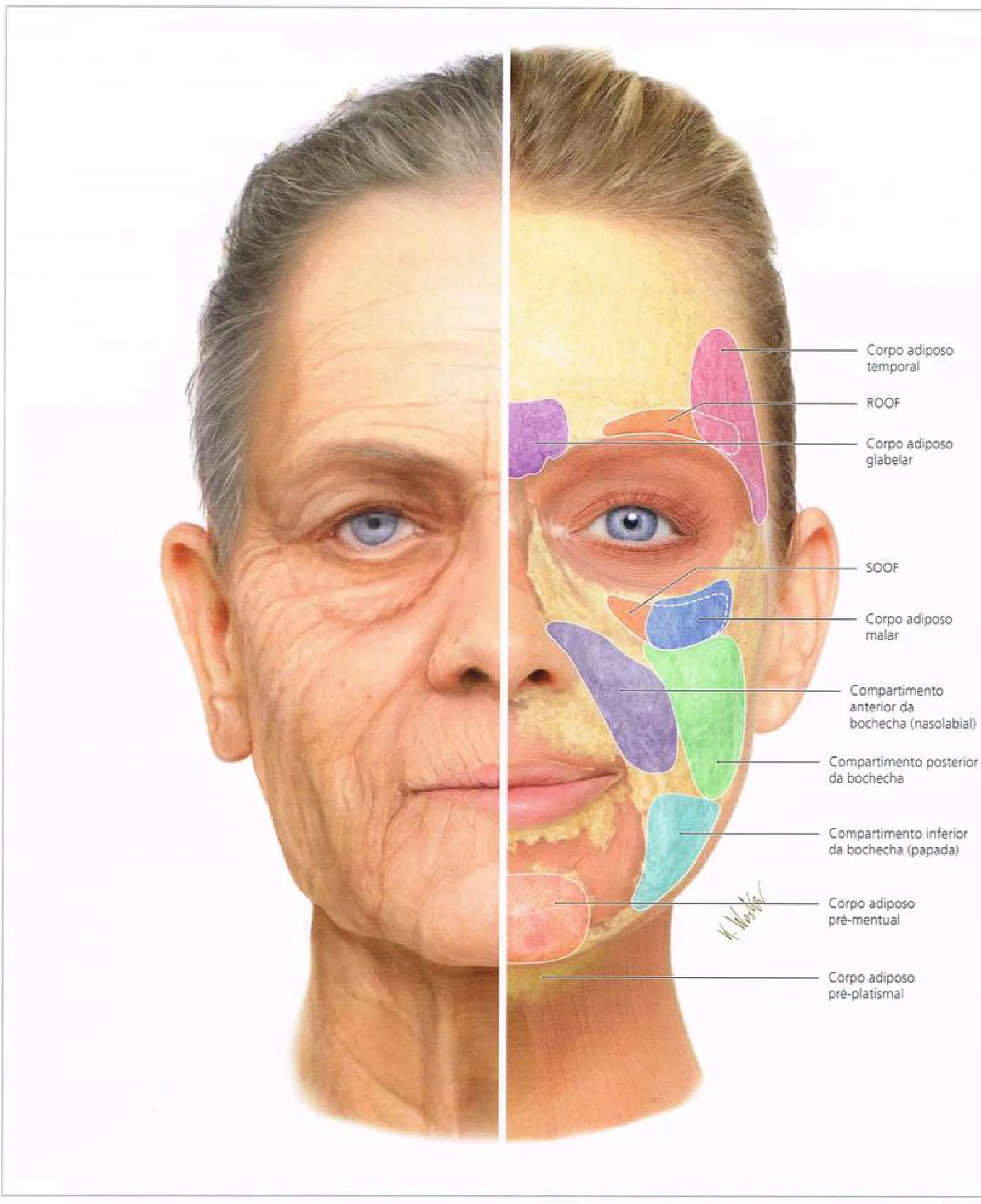 Fig 2. Split-face map of the superficial fat compartments; note the labelled corpo adiposo pré-mentual (pre-mental fat) and pré-platismal (pre-platysmal) pads of the lower third that deflate and descend with age, alongside the malar and buccal compartments - (Radlansky, Atlas Ilustrado de Anatomia Clínica da Face, p. 58).
> Sources: [MEDLIB] Radlansky [5]; corroborated Cotofana 2016 [10], Schenck 2018 [4]. The Fig 2 panel is referenced to locate the pre-mental pad named in the order-of-loss diagram.
Fig 2. Split-face map of the superficial fat compartments; note the labelled corpo adiposo pré-mentual (pre-mental fat) and pré-platismal (pre-platysmal) pads of the lower third that deflate and descend with age, alongside the malar and buccal compartments - (Radlansky, Atlas Ilustrado de Anatomia Clínica da Face, p. 58).
> Sources: [MEDLIB] Radlansky [5]; corroborated Cotofana 2016 [10], Schenck 2018 [4]. The Fig 2 panel is referenced to locate the pre-mental pad named in the order-of-loss diagram.
Why restoration mimics the loss, not the youthful shape. Because the deep structural loss (bone, deep fat) leads, restoring it with a supraperiosteal structural deposit reverses the dominant driver, whereas chasing the surface stigmata with soft gel treats the symptom and leaves the cause - the mechanistic basis for "skeleton first" [10][19]. In the younger, congenitally small chin there is no loss to restore, only a feature to add, so the identical deposit is augmentation and the durability, expectation and screening differ (§D11.6) [7]. In both cases the mentalis contribution means the surface may still need toxin rather than more volume [34].
Skeletal ageing, specifically. The mandible loses height and projection with age, the gonial angle opens (becomes more obtuse), and the symphysis resorbs, so the bony platform on which the chin sits literally shrinks and rotates [10]. Filler restores a soft-tissue silhouette over that changed skeleton but cannot rebuild lost bone geometry; a large skeletal loss is a genioplasty or implant problem, not a filler one (§D11.6). The prejowl sulcus opens because the mandibular retaining ligament tethers the skin at the anterior body while the tissue behind it descends and the bone in front of it resorbs, turning a straight jawline into a wave - which is why the correct target is the sulcus and the lateral chin, not the jowl itself [10][19].
Soft-tissue ageing, specifically. Both the superficial (pre-mental) and deep fat compartments of the chin deflate, so the chin "deflates like other facial areas" while the symphysis reabsorbs beneath it [19]. The skin thins, collagen falls, and a mentalis that becomes relatively hyperkinetic against a shrinking platform produces the uncontrollable cobblestone and the mental crease pressed against the gums [10][34]. Gray's atlas links these changes across the layers: bone, ligament, fat and skin age together but on different clocks, and the earliest and most correctable driver is the deep structural loss [2][10].
Contrast with the midface. In the midface, fat-compartment deflation and descent dominate and bone is a slower player; in the chin, bone and ligament lead, so the restorative order inverts the instinct to "add volume where it looks empty." Restore projection to bone first, relax the muscle, and treat the surface last (§D11.9) [10][19][34].
Ageing vs augmentation are different consents. A young patient with a congenitally small chin wants augmentation (adding a feature that was never there), while an older patient wants restitution (rebuilding what resorbed); the anatomy of the deposit is similar but the expectation, the screening and the durability planning differ, and a young augmentation warrants a BDD screen before any structural change [7]. Naming which one you are treating prevents the mismatch of restoring "youth" to a chin that was always deficient.
The knock-on effects of chin ageing. As the symphysis resorbs and the chin pad descends, three neighbouring problems appear together: the prejowl sulcus opens, the marionette line deepens from the commissure toward the chin, and the cervicomental angle becomes obtuse as projection is lost [7][10][19]. Treating the chin therefore improves the jawline transition and the submental silhouette at once, which is why the chin is assessed before the jowl and before any submental fat plan (§D11.6, §D11.9). The mentalis, meanwhile, becomes relatively hyperkinetic against the shrinking platform and presses the mental crease against the gums, so the surface stigma is muscular as much as volumetric [34].
The clinical stigmata, read as a set. The ageing chin and lower third present together as a recognisable pattern: a deepening labiomental crease, an opening prejowl sulcus, a jawline that waves rather than lines, marionette shadows from the commissure, a mentalis-crimped surface, and an obtuse cervicomental angle from lost projection [7][10][19]. Reading them as one process (skeleton and ligament leading, fat and skin following) is what lets a single structural correction improve several of them at once, and what warns against treating each stigma as a separate filling target [19].
Classic trap: reading the ageing chin as "just needs volume" and layering soft filler on a resorbing symphysis with a hyperkinetic mentalis - the projection washes out and the surface still crimps; the sequence is structural product to bone plus mentalis toxin, not soft gel alone [16][34].
D11.5 · Assessment - measure, photograph, test, scan
The chin is diagnosed on the profile and treated from the front, which is exactly why it is diagnosed badly. Four lanes: the bite, the profile lines, the dynamic exam, and the scan.
| Lane | What you do | Decision it drives |
|---|---|---|
| Occlusion | Ask the patient to bite; inspect the dental relationship | Microgenia (normal occlusion) vs retrognathia (Class II) → filler vs referral [7][16] |
| Profile / cephalometric | Zero-meridian, labiomental angle, E-line, Riedel plane, facial angle | Degree and vector of deficiency; realistic ceiling [11] |
| Dynamic | Lip closure (peau d'orange), speech, lip competence, marionette pull | Mentalis strain, DAO overactivity, need for toxin [16][34] |
| Palpation + scan | Mental foramen, facial-artery pulse, hyoid, submental masses; ultrasound | Danger points, poor-prognosis anatomy, vessel/plane map [40] |
Profile analysis - the reference lines, used as description, never as a numeric target.
| Line / metric | Definition | Norm (orientation only) |
|---|---|---|
| Zero-degree meridian (González-Ulloa) | Vertical from soft-tissue nasion, perpendicular to Frankfort horizontal; soft-tissue pogonion (Pog′) should meet it [11] | Retrusion <10 mm = 1st degree, 10-20 mm = 2nd, >20 mm = 3rd (⚠ the same monograph's text elsewhere cites >30 mm for 3rd degree - internal discrepancy, preserved) [11] |
| Subnasale-perpendicular (Wolford) | Horizontal distance Pog′ to a line perpendicular to FH through subnasale | Mean −3 ± 3 mm (3 mm behind the line) [11] |
| True-vertical through nasion | Natural head position, vertical through soft-tissue nasion | Pog′ 0 ± 2 mm [11] |
| Lower-vermilion drop (practical) | Line from lower vermilion perpendicular to FH; pogonion should approach it | Simplest chairside profile check [7] |
| Labiomental angle / fold | Angle/depth of the mentolabial crease; the lip-to-chin transition | Descriptive norm ~110-130°; ⚠ population-variable, orientation only, never a numeric goal [11] |
Consensus: define the aetiology first (skeletal base vs soft-tissue chin pad vs occlusion), classify sagittal deficiency by a profile line (zero-meridian is the most cited), read the labiomental transition, and photograph a standardised frontal + oblique + profile in natural head position; de Maio adds assessment on animation, oblique, profile and head tilted down because a chin that looks balanced at rest can pucker or recede on movement [11][12][13].
Discrepancy that changes the gesture: the mentolabial fold morphology is arguably more important than the raw sagittal projection - three chins with an identical pogonion position can read as deficient, ideal or over-creased depending on fold depth alone [11]. So the plan is not "how many mm behind the line" but "what does the lip-to-chin transition need," which is why over-projecting to hit a number blunts the labiomental angle and worsens the profile.
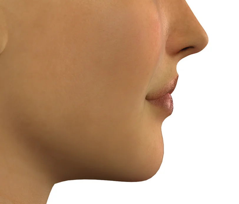 Fig 3. The lateral profile is the working view for chin analysis: nasal tip, upper and lower lip, labiomental fold and soft-tissue pogonion are read against the zero-meridian and lower-vermilion drop; the chin is then treated from the front - (de Maio, MD Codes, p. 41).
> Sources: [MEDLIB] de Maio 2017 [12]. Fig 3 anchors the profile lanes below.
Fig 3. The lateral profile is the working view for chin analysis: nasal tip, upper and lower lip, labiomental fold and soft-tissue pogonion are read against the zero-meridian and lower-vermilion drop; the chin is then treated from the front - (de Maio, MD Codes, p. 41).
> Sources: [MEDLIB] de Maio 2017 [12]. Fig 3 anchors the profile lanes below.
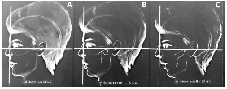 Fig 4. Three profiles gridded on the vertical (zero-meridian) and horizontal reference: (A) first degree, retrusion <10 mm; (B) second degree, 10-20 mm; (C) third degree, >20 mm - the classification that separates a filler-appropriate deficit from a skeletal one - (Naini, Facial Aesthetics, p. 344).
> Sources: [MEDLIB] Naini 2011 [11]. Fig 4 documents the degree scale cited in the profile table.
Fig 4. Three profiles gridded on the vertical (zero-meridian) and horizontal reference: (A) first degree, retrusion <10 mm; (B) second degree, 10-20 mm; (C) third degree, >20 mm - the classification that separates a filler-appropriate deficit from a skeletal one - (Naini, Facial Aesthetics, p. 344).
> Sources: [MEDLIB] Naini 2011 [11]. Fig 4 documents the degree scale cited in the profile table.
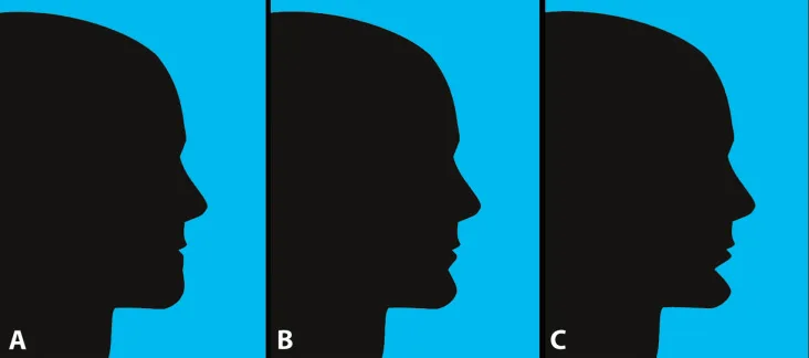 Fig 5. Three silhouettes with the same pogonion position but different mentolabial folds: (A) reduced/obtuse, (B) ideal, (C) deep/acute - proof that the lip-to-chin transition, not the raw projection number, decides the plan - (Naini, Facial Aesthetics, p. 351).
> Sources: [MEDLIB] Naini 2011 [11]. Fig 5 is the visual argument for the "discrepancy" note above.
Fig 5. Three silhouettes with the same pogonion position but different mentolabial folds: (A) reduced/obtuse, (B) ideal, (C) deep/acute - proof that the lip-to-chin transition, not the raw projection number, decides the plan - (Naini, Facial Aesthetics, p. 351).
> Sources: [MEDLIB] Naini 2011 [11]. Fig 5 is the visual argument for the "discrepancy" note above.
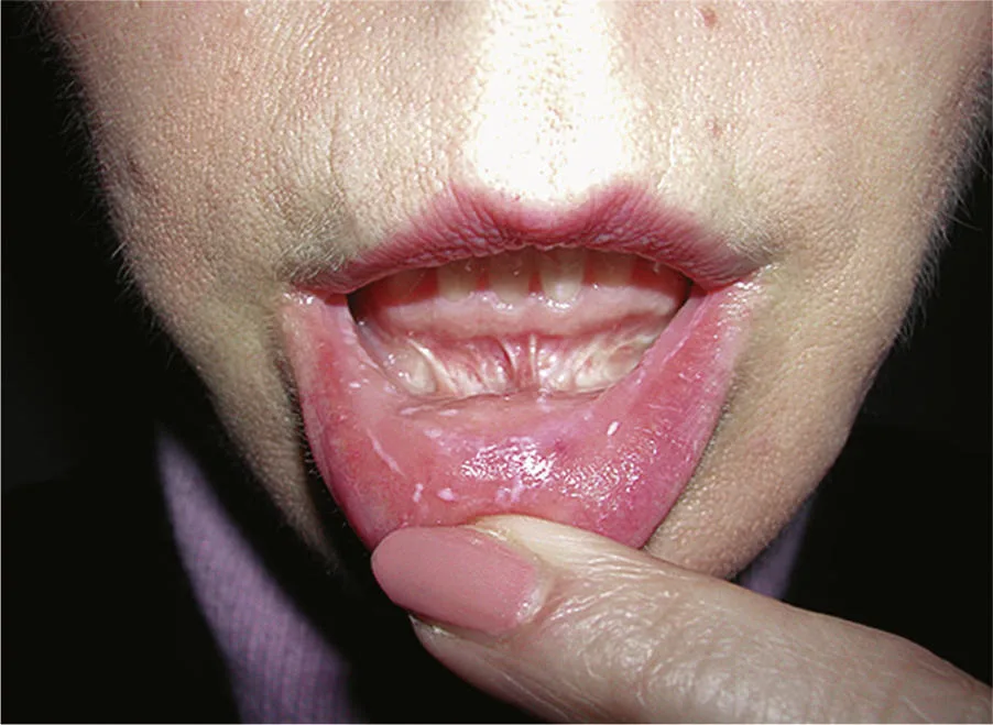 Fig 6. Lower-lip retraction exposing the labial mucosa: part of the dynamic chin exam, assessing lip competence, mentalis tone and the lower-lip depressor complex before deciding filler-versus-toxin balance - (Yaremchuk, Atlas of Facial Implants, p. 141).
> Sources: [MEDLIB] Yaremchuk 2020 [36]. Fig 6 illustrates the dynamic/lip-competence lane of the assessment table.
Fig 6. Lower-lip retraction exposing the labial mucosa: part of the dynamic chin exam, assessing lip competence, mentalis tone and the lower-lip depressor complex before deciding filler-versus-toxin balance - (Yaremchuk, Atlas of Facial Implants, p. 141).
> Sources: [MEDLIB] Yaremchuk 2020 [36]. Fig 6 illustrates the dynamic/lip-competence lane of the assessment table.
Anthropometry and the numbers you record. Beyond the profile lines, document the vertical chin height and the labiomental fold depth on a simple ordinal grade so the plan and the outcome are measured against the same scale [16]. Record baseline asymmetry (a deviated chin point is often skeletal and will persist), the hyoid position and the cervicomental angle, and note whether the deficit is sagittal, vertical or both, because the vector sets the product distribution (§D11.7). These measurements double as the consent record: the patient sees on their own profile photograph what the plan will and will not change [11][12].
Measurements to record (chin visit): occlusal class (bite); sagittal projection against the zero-meridian and the lower-vermilion drop; vertical chin height grade; labiomental fold depth; width and any deviation; hyoid position and cervicomental angle; mentalis strain on lip closure; DAO/marionette on the "sad face"; the mental-foramen and facial-artery landmarks; and standardised frontal/oblique/lateral plus animation photographs [7][11][16]. The list is simultaneously the treatment plan and the consent record.
Ultrasound - corpus-grounded, currency from the external lane. High-frequency B-mode with colour Doppler maps the facial and submental arteries, the deep fat plane and any prior filler, and guides hyaluronidase if occlusion occurs; the corpus carries the 2024 facial-aesthetics ultrasound protocol [9], and the dedicated lower-third scan-while-injecting best-practice protocol is 2025 external material (§What's new) [40].
Ultrasound findings that change the plan. On scan you can measure the true depth of the pre-periosteal plane, see whether prior product sits deep or superficial, detect an unsuspected asymmetry of the bony platform, and map the submental artery before a lateral or jawline deposit; the same probe confirms reperfusion after treating an occlusion and guides intravascular hyaluronidase with minimal drug [40][44]. It moves from optional to advisable for the revision chin, the previously-treated field, and any point placed off the midline (§D11.3, §D11.10). Ultrasound is optional for a routine midline bolus but changes the risk profile for lateral/jawline work and for revision of a lumpy or previously treated chin.
The wider analysis toolbox (choose by facial type, not dogma). Beyond the zero-meridian, the chin position can be read against several reference systems, none of which agree exactly and all of which are population-derived, so they orient rather than dictate [11]:
| Analysis | Reference | Reads |
|---|---|---|
| Ricketts E-line | Nasal tip to soft-tissue pogonion | Lower-lip and chin position relative to the aesthetic line |
| Steiner S-line | Nasal midpoint to pogonion | Lip protrusion vs chin |
| Holdaway H-line / facial angle | Upper-lip to pogonion; soft-tissue facial angle | Lip-chin harmony |
| Riedel plane | Through the most anterior upper and lower lip | Whether pogonion falls on the lip plane |
| Legan-Burstone | Angle of facial convexity | Skeletal convexity/concavity |
| Merrifield Z-angle | Profile line to Frankfort horizontal | Chin-lip-nose balance |
Classification you assign before treating.
| Axis | Categories |
|---|---|
| Sagittal | Microgenia/retrogenia (under-projected) · normal · macrogenia (over-projected) |
| Occlusal | Class I (normal) · Class II (retrognathic) · Class III (prognathic) |
| Vertical height | Short · ideal · long chin |
| Width | Narrow (feminine) · square/wide (masculine) |
| Symmetry | Symmetric · deviated (functional or skeletal) |
| Soft tissue | Chin-pad thickness reduced / normal / increased; mentolabial fold shallow/ideal/deep |
Wong grades the two operative dimensions on a simple ordinal scale (e.g. vertical chin height 1 = mildly deficient up to an ideal 2, and a lip-fullness scale) so the plan and the result are measured against the same yardstick before and after [16].
Photography and dynamic exam, standardised. Take frontal, oblique (45°) and true lateral views in natural head position, plus a smile/animation frame and a head-tilted-down view, because a chin that balances at rest can recede or pucker on movement [12][13]. Dynamically, test: lip closure (peau d'orange = mentalis strain), speech and pursing (mentalis and DAO overactivity), lip competence (can the lips seal at rest without mentalis effort), and the marionette pull (DAO). Palpate the mental foramen, the facial-artery pulse at the antegonial notch, the hyoid position and the submental region for the submandibular gland or any mass, because a firm submental fullness that does not pinch is not fat and must not be treated as such [7][16].
Ultrasound, in practice. High-frequency B-mode plus colour/Doppler ultrasound maps the facial and submental arteries, distinguishes the deep fat plane from muscle, detects prior filler as a hypoechoic deposit, and lets a trained operator perform Doppler-ultrasound-guided hyaluronidase with far less drug than the blind protocol; slow-flow Doppler shows the chaotic back-and-forth signal of an obstructed vessel, unlike the clean pulsatile arterial trace [40]. The 2024 corpus protocol grounds the anatomy; the 2025 lower-third best-practice protocol adds the scan-while-injecting workflow (§What's new) [40].
Standardised photography protocol. Consistent light and framing are what make the before/after profile comparison meaningful, and the chin is the region where an inconsistent angle most easily fakes or hides a result [12]: 1. Neutral background, even lighting, no chin makeup. 2. Natural head position (self-balanced gaze), which is preferred over a forced Frankfort plane for the true-vertical analysis [11]. 3. Views: frontal at rest, frontal smiling, both obliques (45°), both true laterals, and a lateral with the head tilted down. 4. Animation frames: lip closure (mentalis), "sad face" (DAO), full smile. 5. Fixed camera height and distance each visit; the profile is the decisive view.
Read the chin relative to the lip and nose, not in isolation. Because the E-line, S-line and H-line all judge the chin against the lips and the nasal tip, an "under-projected" chin can be an illusion created by a protrusive lip or a prominent nose; correcting that perception (lip, or rhinoplasty referral) may solve the profile without a single millilitre in the chin [11]. The analysis is of the lower-face unit.
What the assessment must actively exclude. A firm submental fullness that does not pinch is the submandibular gland or a mass, not fat; a constitutionally low hyoid with an obtuse angle predicts a poor outcome for everything and must be declared before charging; and unexplained lymphadenopathy or a thyroid mass is investigated, not treated [7][16]. These are the findings that turn a filler consult into a referral, and missing them is the assessment error with the highest cost.
Classic trap: analysing the chin only at rest and only from the front - the under-projection hides on the frontal photo and the mentalis strain only appears on lip closure, so the treatment is planned for the wrong problem; profile plus animation is mandatory [11][13].
D11.6 · Goal and patient selection - who benefits, who is referred
One complaint, opposite treatments by phenotype and sex. "I want a stronger chin" means add for the deficient soft-tissue chin, and means refer for the skeletal Class II.
| Candidate | Do | Refer / avoid |
|---|---|---|
| Mild-moderate microgenia (normal occlusion) | Supraperiosteal HA/CaHA to bone [7][16][22] | - |
| Deep labiomental crease, soft chin pad ptosis | Subcutaneous crease support + structural base [11][17] | - |
| Feminising contour (narrow, pointed, forward) | Midline projection + apex; keep width down [13] | ⚠ Widening (C5/lateral) masculinises |
| Masculinising contour (wide, square, projected) | Add lateral tubercles (C5) + projection [13] | - |
| Skeletal Class II / significant malocclusion | - | Orthognathic / orthodontic referral [7][16] |
| Large skeletal deficiency / permanent goal | - | Genioplasty or alloplastic implant [7] |
| Frank skin excess, true jowl, submental gland ptosis | - | Surgery; not a filler problem |
| Low hyoid / short hyoid-mentum, BDD traits | Counsel; conservative or decline | ⚠ Poor prognosis; screen BDD before touching |
The three vectors, decided explicitly:
| Vector | Changes | When / caution |
|---|---|---|
| Anterior projection | The profile; opens the cervicomental angle [7] | Most requested, highest yield |
| Vertical height | Lower-third proportion (short chin) | ⚠ Over-lengthening ages the face |
| Width | The frontal shape | ⚠ Widening a feminine chin masculinises; irreversible if done with a biostimulator |
Consensus: the ideal candidate is mild-to-moderate microgenia with normal occlusion and a soft-tissue (not skeletal) deficit; treat projection deep-on-periosteum and the crease subcutaneously, decide the width vector on purpose against the patient's sex, and refer the Class II, the large skeletal deficiency and the frank skin excess [7][16]. Chin augmentation reliably increases the hyoid-to-mentum distance and sharpens an obtuse cervicomental angle, which is why the chin is the forgotten treatment for a complaint of "double chin" [7].
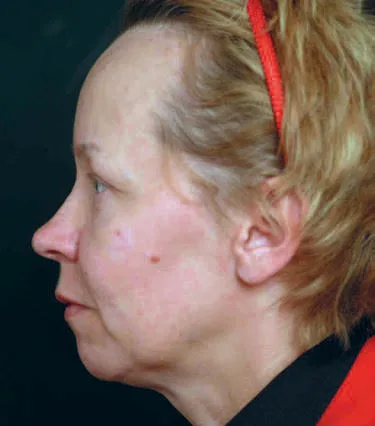 Fig 7. Profile of microgenia: the under-projected chin shortens the hyoid-to-mentum distance and deepens the prejowl sulcus, the exact deficit that filler (or, when skeletal, a chin implant) corrects by pushing pogonion toward the reference line - (Azizzadeh, Master Techniques in Facial Rejuvenation, p. 325).
> Sources: [MEDLIB] Azizzadeh 2018 [7]. Fig 7 shows the selection target the table above describes.
Fig 7. Profile of microgenia: the under-projected chin shortens the hyoid-to-mentum distance and deepens the prejowl sulcus, the exact deficit that filler (or, when skeletal, a chin implant) corrects by pushing pogonion toward the reference line - (Azizzadeh, Master Techniques in Facial Rejuvenation, p. 325).
> Sources: [MEDLIB] Azizzadeh 2018 [7]. Fig 7 shows the selection target the table above describes.
Filler does not treat the skeletal base. In a true Class II retrognathia the whole mandible is retruded and the occlusion is abnormal; filler can partially camouflage the profile but leaves the functional problem, so the correct move is orthognathic/orthodontic referral, and the bite test is the 30-second exam that separates the two [7][16]. In genuine microgenia with normal occlusion, filler (or a chin implant for larger or permanent needs) restores projection well.
Sex dimorphism, target by target. The male and female chin are different shapes, and the same complaint ("stronger chin") means opposite gestures, so the vector is decided against the patient's sex on purpose [13]:
| Feature | Feminine target | Masculine target |
|---|---|---|
| Frontal shape | Oval, narrow, pointed | Square, wide |
| Projection | Moderate, forward but soft | Greater, more anterior |
| Vertical height | Lower | Greater |
| Width / lateral tubercles (C5) | Kept minimal | Deliberately added |
| Transition to mandibular border | Smooth, tapered | Marked, angular |
| Product distribution | Central apex/pogonion | Central + lateral support |
Who benefits most, ranked. Highest yield: mild-to-moderate microgenia with normal occlusion and good skin, wanting a profile improvement. Next: the patient with a soft labiomental crease and a hyperkinetic mentalis (filler and toxin). Then: the "double chin" patient whose real deficit is projection [7][16]. Lowest yield, and the referrals: the skeletal Class II, the large or permanent-goal deficit, the frank skin excess, and the low hyoid - injectables there produce the cost without the result [7][19]. Rank the candidate before quoting, because the ceiling of the non-surgical result is fixed by which of these the patient is.
Expectation-setting that prevents the common disappointment. The two mismatches to pre-empt are the patient who expects a filler to fix a bite or rebuild a jaw (it cannot; that is surgery) and the patient who expects a dramatic single-visit change on a resorbed platform (structure is built in layers and settles over weeks) [7][16]. State the off-label status, the reversibility of HA versus the permanence of biostimulators and PMMA, and show the profile photograph, because the chin is the feature a patient least often sees of themselves and most often misjudges [11][12].
The chin as the forgotten answer to "double chin". A meaningful share of patients requesting submental fat treatment actually have an under-projected chin; projecting the mentum increases the hyoid-to-mentum distance and opens the obtuse cervicomental angle, improving the submental silhouette without touching the fat [7]. Examine projection and the hyoid position before selling deoxycholic acid or liposuction. A genuinely low hyoid gives a constitutionally short neck and an obtuse angle that responds poorly to everything; say so before charging (§D6).
Contraindications and cautions (region-relevant).
| Type | Examples |
|---|---|
| Absolute (HA) | Active infection/inflammation at the site; known hypersensitivity to HA/lidocaine/gram-positive bacterial proteins; injection into vessels [12] |
| Product-specific | PMMA/bovine-collagen carrier → skin allergy test first [27]; CaHA/PMMA in a revision-prone patient (irreversible) |
| Relative | Anticoagulation/antiplatelet + herbal supplements (bruising); autoimmune disease; keloid/hypertrophic-scar tendency; pregnancy/breastfeeding; recent laser/peel/dermabrasion [12] |
| Anatomical | Prior permanent filler in the field (unknown behaviour, layered vascular risk); heavy scarring (retrograde-flow conduit) [38] |
| Psychological | Body dysmorphic traits → screen before any structural change; young augmentation is augmentation, not restitution, and warrants different consent |
Consent essentials for the chin: most chin filler and all chin toxin is off-label; biostimulators and PMMA are not reversible; over-projection and mentalis over-treatment have specific, visible downsides; and a skeletal Class II will be only partly camouflaged. Ethnic and cultural norms for chin shape differ and are a descriptive input, never a fixed target [13].
The bite test, as a protocol. Ask the patient to bite together and look at the dental occlusion: normal bite + small chin = microgenia (treat), Class II malocclusion = retrognathia (refer) [7][16]. It is the single most cost-effective exam in the chapter and is absent from most filler courses.
Ethnic and cultural variation. Ideal chin shape and projection differ across populations and the profile reference lines derive from specific normative samples, so they orient rather than dictate; a chin that is "retruded" against a Caucasian norm may be balanced within another facial framework [11]. The width, projection and pointedness a patient wants are a cultural and individual preference to be documented, and imposing one population's numeric target is both clinically and ethically wrong [13].
The revision or previously-treated chin. A chin with prior filler (especially unknown or permanent product), palpable nodules, or asymmetry is a higher-risk field: prior product is a retrograde-flow conduit, scarring distorts planes, and a permanent filler cannot be dissolved [38]. Map it with ultrasound before re-injecting, favour a reversible HA, and set the expectation that correcting an over-projected or migrated result may mean dissolving before rebuilding [40][44].
Realistic-expectation counselling. State plainly that filler restores a soft-tissue silhouette over the existing skeleton and does not rebuild bone or fix a bite; that most chin filler and all chin toxin is off-label; that biostimulators and PMMA are not reversible; and that the result is graded on the profile the patient rarely sees of themselves, so before/after profile photography is part of consent, not just documentation [7][11][12].
Classic trap: applying the male jawline/chin map to a woman and widening the chin or gonial region "for definition" - it masculinises the lower third, and if a biostimulator was used it cannot be dissolved; width is a deliberate, sex-specific decision, not a default add [13].
D11.7 · Technique - the full grid
Point-by-point injection map (MD Codes + freehand). Every point states plane, device, entry, volume and the alert. Projection lives at C2/C3 on bone; the crease and jawline are subcutaneous.
| Code / point | Area | Plane | Device | Vol/point | Effect · alert |
|---|---|---|---|---|---|
| Ck1 (anchor) | Deep chin start point | Supraperiosteal, to bone | Needle | 0.1 mL | Anchoring deposit; ⚠ a single 0.3 mL bolus in one spot swells and looks unnatural [13] |
| C1 | Mental crease / labiomental angle | Subcutaneous + muscle | Needle | ~0.1-0.3 | Reduces lower-lip protrusion, supports commissure, elongates chin [13] |
| C2 | Chin apex | Supraperiosteal (bone) | Needle | 0.3 | Improves vertical dimension / chin height [13] |
| C3 | Anterior chin / soft-tissue pogonion | Supraperiosteal | Needle | 0.3 (midline only) | Anterior projection; ⚠ not too lateral → mental artery [13] |
| C4 | Anterior chin / soft-tissue pogonion | Subcutaneous | Needle | 0.3 (midline only) | Enhances C2/C3, rotates chin up [13] |
| C5 | Lateral lower chin | Supraperiosteal | Needle | 0.3 | Widens/squares chin, lateral support; ⚠ masculinising [13] |
| C6 | Prejowl sulcus / lateral chin | Subcutaneous | Cannula | 0.5 | Softens prejowl; jawline blend [13] |
| Jw4 / Jw5 | Prejowl / anterior-inferior chin (jawline) | Subcutaneous | Cannula | 0.5 | Continuity to the mandibular line → §D6 jawline [13] |
Consensus: anchor and project at the midline on bone (Ck1/C2/C3), aspirate, then refine the crease (C1/C4 subcutaneous) and the jawline transition (C6/Jw cannula); aspiration is explicitly recommended when a needle is at bone level, and one large single bolus is discouraged in favour of layered smaller deposits [13][16]. Volume per point on bone is ~0.3 mL; total chin correction is typically 0.6-1.5 mL, scaled to the degree of deficiency.
Volume ladder (chin, HA):
| Indication | Volume | Source |
|---|---|---|
| Asymmetry / touch-up | 0.2-0.3 mL | Garg 2021 [15] |
| Mild recession / flattening | 0.6-0.8 mL | Garg 2021 [15] |
| Moderate-severe recession | 1.3-1.5 mL | Garg 2021 [15] |
| Feature augmentation (cheeks + chin + jawline, one young patient) | 2.5 mL total | Carruthers 2018 [19] |
| Structural chin (single HA) | ~2 mL (e.g. Definisse Core) | Wong 2022 [16] |
| Lateral jawline (per side, min) | 0.5 cc, up to 1-2 mL as jowl grows | Illustrated Manual 2011 [29] |
| Bilateral prejowl + angle definition | 3-4 mL | Illustrated Manual 2011 [29] |
Product grid - enumerate the whole shelf, choose by rheology and reversibility.
| Class / product | Key spec | Chin use | Reversible? |
|---|---|---|---|
| HA, high-G′/cohesivity - Volux (VYC-25L) | 25 mg/mL, highest G′/cohesivity of the Juvederm line, "bone-mimicking"; FDA jawline, chin off-label but evidenced [21][24][26] | Projection to bone; structural | Yes (hyaluronidase) |
| HA, structural - Voluma (VYC-20L) | 20 mg/mL HA + lidocaine 3 mg/mL; FDA chin profile; deep dermis / subQ / upper-periosteal only [12][22][23] | Projection + vertical height | Yes |
| HA, other high-G′ | Restylane Lyft/Defyne; Belotero Volume/Intense (26 mg/mL, polydensified malleable); Teosyal Ultra Deep; RHA-4 [21][19] | Projection / contour by school | Yes |
| HA, soft (contrast, NOT for structure) | Volbella G′ 271 Pa, cohesivity 19 gmf - softest Vycross [19] | Only surface/labiomental micro-refinement; ⚠ will not project | Yes |
| CaHA - Radiesse | 30 % CaHA microspheres 25-45 µm in 70 % CMC gel carrier; 1.5 / 3 mL syringes; FDA 2006; duration ~1-2 yr (some report 2-5); higher elasticity than HA [1][27][28] | Supraperiosteal projection + biostimulation; hyperdilute/subdermal for skin/jawline blend | No - conservative, aspirate |
| PLLA - Sculptra | Poly-L-lactic acid, biostimulatory neocollagenesis | Gradual projection over sessions; ⚠ aspirate/avoid vessel | No |
| PCL - Ellansé | Polycaprolactone, biostimulatory + longer-lasting | Structural + collagen | No |
| PMMA - Bellafill/Artefill | Permanent PMMA microspheres in bovine-collagen carrier | Permanent projection; allergy test required | No |
| Autologous fat | Lipofilling: volume + biostimulation | Volume in the right hands; operator-dependent take | No |
| Polynucleotides / PDRN | Plinest, Jalupro - fibroblast renewal | Skin quality of the chin/lower face (adjunct) | n/a |
| Skin boosters / mesotherapy | Non-crosslinked HA, cocktails | Surface hydration/quality; microbotox for pores (§D11.8) | n/a |
Skin-quality adjuncts for the chin. Beyond volume, the chin skin is treated with polynucleotides/PDRN (fibroblast renewal), non-crosslinked HA skin boosters and mesotherapy cocktails, and intradermal microbotox for pores and oiliness; these form a finishing surface layer, not a structural technique, and are staged after the projection is built [16][47]. The corpus grounds these generically for the face; the chin/lower-face-specific application is an adjunct choice rather than a region-validated protocol, so it is offered as quality refinement, not projection.
Instrument grid.
| Instrument | Spec | Where |
|---|---|---|
| Sharp needle | 27G 1.25-inch (Garg); 25G/30G alternatives | Midline supraperiosteal bolus to bone; C1-C5 [15][13] |
| Blunt cannula | 25G 40 mm (Vieira Braz); 22G/27G | Subcutaneous crease/jawline; C6/Jw; submental-artery avoidance [20][13] |
| Entry needle (pertuis) | 21G bevel, very superficial | Cannula entry port at angle or line end [20] |
| Ultrasound | HF B-mode + colour Doppler | Optional for midline; recommended for lateral/revision [40] |
Plane and movement grid.
| Plane | Movement | Purpose |
|---|---|---|
| Supraperiosteal | Bolus / microbolus to bone, aspirate | Projection, vertical height (C2/C3) |
| Deep / pre-periosteal fat | Bolus | Structural fill where product diffuses and stays [20] |
| Superficial fat (above mentalis) | Retrograde linear / microbolus | Surface definition when deep alone is insufficient [14] |
| Subcutaneous / supra-SMAS | Retrograde linear threading, fanning, cross-hatching (cannula) | Crease, jawline blend, parallel technique [17][18] |
| Subdermal | Serial puncture / fanning | Skin-quality, hyperdilute CaHA |
| Intradermal | Microdroplet / blanching | Skin boosters, microbotox |
Bevel rule: with the needle to bone, cephalic bevel to project, caudal bevel to elongate; if the deep deposit alone under-corrects, add a medium-viscoelastic gel into the superficial fat over the muscle [14].
Treating the labiomental crease specifically. A deep mental crease has two components: a dynamic one from mentalis pull, softened by toxin, and a static one from a fixed fold, filled with a small subcutaneous retrograde thread of a medium HA placed superficial to the muscle, never into the belly [16][17]. Over-filling the crease flattens the natural lip-to-chin transition and reads as an operated chin, so the crease is refined, not erased, and only after the projection is set (§D11.9).
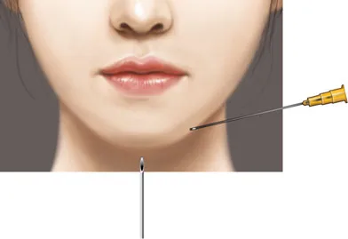 Fig 8. Injection illustration for chin augmentation: a needle enters the midline vertically toward the symphyseal periosteum while a second, lateral point addresses contour; the deep supraperiosteal deposit is the projection layer, the superficial fat an optional surface layer - (Hong, 2020, p. 181).
> Sources: [MEDLIB] Hong 2020 [14]. Fig 8 shows the needle entry/plane described in the point map.
Fig 8. Injection illustration for chin augmentation: a needle enters the midline vertically toward the symphyseal periosteum while a second, lateral point addresses contour; the deep supraperiosteal deposit is the projection layer, the superficial fat an optional surface layer - (Hong, 2020, p. 181).
> Sources: [MEDLIB] Hong 2020 [14]. Fig 8 shows the needle entry/plane described in the point map.
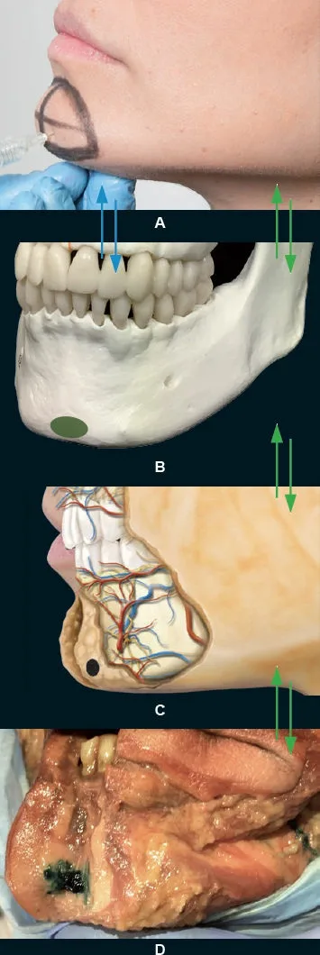 Fig 9. Four panels: (A) clinician placing a supraperiosteal chin deposit on profile; (B) skull model with the needle to the symphysis and the bony target marked; (C) illustration of the mandibular vasculature relative to the point; (D) fresh-cadaver hemiface with green-stained HA injected to periosteum - the dye shows the product entering and remaining in the deep pre-periosteal chin compartment even when delivered onto bone - (Vieira Braz, Atlas de Anatomia e Preenchimento Global da Face, p. 624).
> Sources: [MEDLIB] Vieira Braz 2017 [20]. Fig 9 is the anatomical proof that a needle-to-bone bolus stays deep, panel by panel.
Fig 9. Four panels: (A) clinician placing a supraperiosteal chin deposit on profile; (B) skull model with the needle to the symphysis and the bony target marked; (C) illustration of the mandibular vasculature relative to the point; (D) fresh-cadaver hemiface with green-stained HA injected to periosteum - the dye shows the product entering and remaining in the deep pre-periosteal chin compartment even when delivered onto bone - (Vieira Braz, Atlas de Anatomia e Preenchimento Global da Face, p. 624).
> Sources: [MEDLIB] Vieira Braz 2017 [20]. Fig 9 is the anatomical proof that a needle-to-bone bolus stays deep, panel by panel.
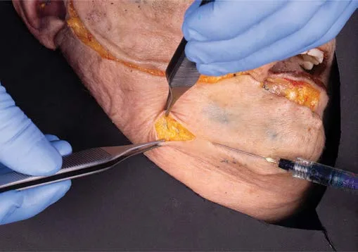 Fig 10. Cadaver dissection of the chin with the skin-fat flap lifted and a syringe delivering into the subcutaneous plane: the parallel/cannula technique used for contour, the labiomental crease and jawline blending, where submental-artery avoidance and a soft even spread matter more than bony projection - (Pirayesh, Aesthetic Facial Anatomy Essentials, p. 237).
> Sources: [MEDLIB] Pirayesh 2020 [1]. Fig 10 documents the subcutaneous plane of the needle-versus-cannula controversy below.
Fig 10. Cadaver dissection of the chin with the skin-fat flap lifted and a syringe delivering into the subcutaneous plane: the parallel/cannula technique used for contour, the labiomental crease and jawline blending, where submental-artery avoidance and a soft even spread matter more than bony projection - (Pirayesh, Aesthetic Facial Anatomy Essentials, p. 237).
> Sources: [MEDLIB] Pirayesh 2020 [1]. Fig 10 documents the subcutaneous plane of the needle-versus-cannula controversy below.
Controversy 1 - needle-to-bone vs cannula. Not averaged: chosen by target plane.
Consensus: everyone anchors projection deep and everyone aspirates before a bolus [13][16][17]. Discrepancy: - A - Needle-to-bone (Hong, de Maio, Wong): vertical needle to the symphyseal periosteum, midline, deep supraperiosteal bolus; argument = the bony midline is off the named vessels, so risk is low and projection is precise [12][14][16]. - B - Blunt cannula (Jones; Vieira Braz; 2025 lower-third ultrasound best-practice): subcutaneous/soft-tissue placement with submental-artery avoidance, favoured for the jawline, prejowl and labiomental crease and under ultrasound; argument = a cannula reduces intravascular risk in the lateral soft tissue [17][20][40]. - Decide by: the target. Projection = needle to bone (B places product too superficially to project). Contour, crease, jawline, revision = cannula (needle-to-bone there risks the mental artery and the submental artery). Never average the two into a mid-depth deposit that neither projects nor stays safe.
Controversy 2 - which high-G′ HA: Volux (VYC-25L) vs Voluma (VYC-20L). Not averaged: choose by G′/cohesivity and regulatory indication.
Consensus: the chin needs a high-G′, high-cohesivity, structural HA to hold projection; a soft gel diffuses and wastes [19][21]. Discrepancy: - A - Volux VYC-25L (25 mg/mL): the highest G′/cohesivity Juvederm, "bone-mimicking," FDA-cleared for jawline definition and used off-label for chin projection; the 2024-2025 evidence extends it to chin retrusion [21][24][26]. - B - Voluma VYC-20L (20 mg/mL + lidocaine 3 mg/mL): the FDA chin-indicated structural HA, lower G′ than Volux, well-studied chin data [12][22][23]. - Decide by: how much projection versus how much regulatory cover you want. Both are now chin-evidenced (VYC-20L pivotal chin data [22][23]; VYC-20L + VYC-25L reinforcement [26]; VYC-25L chin-retrusion cohort [24]). Do not pick the number "in between."
The chin HA evidence base, in one place. VYC-20L (Voluma) has the pivotal chin-augmentation data behind its chin indication [22] and a companion analysis showing higher responder rates on live than photographic assessment [23]; VYC-25L (Volux) has jawline-restoration RCT data [24] and a chin-retrusion cohort [25]; and the two are combined for chin reinforcement in the newest work [26]. The practical reading: both are validated structural HAs for the mentum, VYC-25L brings more G′ for a strong projection, VYC-20L brings the on-label chin position, and the choice is projection-need versus regulatory-cover, not a compromise gel. Other lines follow the same logic on their own rheology - Restylane's firm gels (Lyft/Defyne) and Belotero Volume/Intense (polydensified, malleable) project by a different feel - so the "school" is a preference within the same high-G′ requirement [19][21].
Controversy 3 - HA vs CaHA for projection.
Consensus: both project supraperiosteally; CaHA adds biostimulation [1][27]. Discrepancy: HA is immediate and reversible (hyaluronidase rescue if occluded or overfilled); CaHA (Radiesse 30 %/70 %) is structural + collagen-stimulating but not reversible and has no rescue, so it demands more conservative technique, mandatory aspiration and a lower threshold to refer. Choose HA when reversibility matters (first chin, revision-prone patient), CaHA when durability and skin quality are the goal in a confident hand.
Non-injectable alternatives - when the correct answer is NOT a filler.
| Option | Mechanism | Choose when |
|---|---|---|
| Sliding / osseous genioplasty | Skeletal repositioning of the symphysis | Large skeletal deficiency, malocclusion, permanent structural goal [7] |
| Alloplastic chin implant (silicone / Medpor / ePTFE) | Adds fixed projection; submental approach allows periosteal suture fixation (less migration than intraoral, less infection) | Moderate-large microgenia wanting permanence [7] |
| Autologous fat transfer | Volume + biostimulation, often with implant across the labiomental sulcus | Volume deficit + skin-quality goal, surgical setting [7] |
| Threads | Suspension | Jawline lifting - not chin projection; do not sell as chin augmentation |
| Energy / EBD, RF, HIFU | Skin tightening | Skin laxity dominates, not volume; before adding weight to a lax lower third |
Consensus: filler treats soft-tissue projection and camouflages a modest skeletal deficit; when the deficiency is skeletal, large, or the patient wants permanence, the correct answer is genioplasty or an implant, and when the dominant problem is skin laxity, energy precedes volume [7]. Refer rather than stack injectable on a problem injectables do not solve.
Schools of chin augmentation (positions, not averages).
| School | Thesis | Where it fails |
|---|---|---|
| Structural-sequence | Project midline-to-bone first, then crease, then jawline; least product, best line | Slower, less dramatic in a single visit |
| MD Codes point-map (de Maio) | Coded points C1-C6 with fixed plane/device/volume [13] | Rigid if applied without the profile analysis |
| Cannula + ultrasound (Jones; 2025 lower-third best-practice) | Subcutaneous, submental-artery avoidance, scan-guided [17][40] | Under-projects where bone contact is needed |
| High-load volumetric | The chin needs a lot of product | Costly, heavy, over-projects, blunts the labiomental angle |
| Biostimulator-led (CaHA/PLLA) | Projection + collagen, durable [27] | Not reversible; gradual; operator-dependent |
| Surgical-first | Skeletal/large deficits are genioplasty/implant [7] | Excludes the many who need only soft-tissue projection |
Needle-vs-cannula, resolved per target.
| Target | Device | Plane |
|---|---|---|
| Midline projection (C2/C3, pogonion) | Needle | Supraperiosteal, aspirate |
| Vertical height (chin apex, C2) | Needle | Supraperiosteal |
| Labiomental crease (C1/C4) | Needle or cannula | Subcutaneous |
| Lateral chin / prejowl (C5/C6) | Cannula | Subcutaneous |
| Jawline continuity (Jw) | Cannula | Subcutaneous |
| Skin quality | Needle (serial puncture) | Subdermal / intradermal |
Genioplasty vs implant vs filler - the surgical menu. When the answer is surgical, the options graduate. An alloplastic implant (silicone, porous polyethylene/Medpor, or ePTFE) adds fixed projection and, placed by a submental approach, is sutured to the periosteum to prevent shifting and vertical migration, whereas an intraoral approach risks oral-flora infection [7]. A sliding (osseous) genioplasty repositions the symphysis itself and can correct vertical height and asymmetry an implant cannot. Autologous fat, often laid across the labiomental sulcus, adds volume plus biostimulation in the surgical setting [7]. Filler is the reversible, in-office alternative for soft-tissue projection; it is not a substitute for skeletal repositioning, and the honest consult names the surgical option when the deficit is skeletal or large.
Rheology, and why it decides the product. Projection is a mechanical problem: the gel must resist the compression of the soft tissue and the mimetic muscle without spreading. The properties that matter are G′ (elastic modulus, resistance to deformation), cohesivity (resistance to fragmentation/spread), and viscosity; a high-G′, high-cohesivity gel "mimics bone" and holds a projected apex, while a soft, low-G′, low-cohesivity gel (Volbella, G′ 271 Pa, cohesivity 19 gmf) spreads and is only appropriate for surface or micro-crease work [19]. Elasticity and viscosity together determine a filler's capacity for volume and lift, and CaHA sits above the HAs on elasticity, which is why it projects and biostimulates but cannot be reversed [1][27]. Choose the product by the plane and the job, not by brand loyalty.
| Product | Class / spec | Chin role by rheology |
|---|---|---|
| Volux (VYC-25L) | HA 25 mg/mL, highest G′/cohesivity Vycross [21][24] | First-line bony projection; jawline |
| Voluma (VYC-20L) | HA 20 mg/mL + lidocaine, high G′ [12][22] | Projection + vertical height; FDA chin |
| Restylane Lyft / Defyne | NASHA / XpresHAn, firm | Projection / contour by school [17] |
| Belotero Volume / Intense | 26 mg/mL, polydensified malleable [21] | Structural yet malleable; deep support |
| Teosyal Ultra Deep | High-G′ | Deep bolus projection |
| RHA-4 | Resilient HA | Dynamic structural areas |
| MLT 25 mg/mL voluminiser | 25 mg/mL, BDDE 10%, very-high cohesivity, high G′, 25G×1″, 9-20 mo | Supraperiosteal chin/cheek/jawline structural support [43] |
| Radiesse (CaHA) | 30% CaHA 25-45 µm / 70% CMC, 1.5-3 mL [27][1] | Projection + biostimulation; not reversible |
| Sculptra (PLLA) | Reconstituted, biostimulatory | Gradual projection over sessions |
| Ellansé (PCL) | Polycaprolactone, biostimulatory | Durable structure + collagen |
| Bellafill/Artefill (PMMA) | 32-40 µm PMMA in bovine collagen + 0.3% lidocaine, FDA 2006 [27] | Permanent; allergy test first |
Step-by-step procedure (needle-to-bone, midline projection). 1. History, occlusion (bite test), profile analysis, standardised photographs; mark the midline and the mental foramina; palpate the facial-artery pulse [7][16]. 2. Antisepsis; topical or infiltrative anaesthesia (the FDA products carry lidocaine; a mental-nerve block is optional for comfort but numbs the field you assess). 3. Recline the patient to ~45° [15]. 4. Attach a 27G 1.25-inch needle firmly; extrude a bead to confirm flow [15]. 5. Enter vertically at the midline chin apex/pogonion, advance to periosteal bone contact, aspirate, deposit a small anchor (~0.1 mL, Ck1), then the projection aliquot (~0.3 mL) under low pressure [13][15]. 6. Bevel: cephalic to project, caudal to lengthen; if the deep deposit under-corrects, add medium-viscoelastic gel into the superficial fat over the muscle [14]. 7. Withdraw with a retrograde touch if needed; mould the product against the bone with finger pressure; reassess in profile mid-procedure, not only at the end [16][20]. 8. Refine the labiomental crease (C1/C4 subcutaneous) and jawline (C6/Jw cannula) as separate gestures. 9. If blanching or disproportionate pain occurs at any point, stop and treat as occlusion (§D11.10) [13].
Cannula technique (contour, crease, jawline). Make a 21G entry port at the angle or the end of the marked line, introduce a 25G 40 mm microcannula in the subcutaneous plane, and deliver by retrograde linear injection, changing direction to cover the demarcated area, then mould against the mandibular bone with the index finger; keep off the pre-jowl ptosis, which is not filled [20]. The multilayer technique (MLT) reaches three planes from one entry point in sequence: structural support (supraperiosteal), dynamic volumisation, and surface redensification, using products matched to each layer [43].
Jawline continuation (the chin is one end of the line). The MD Codes jawline points extend the chin plan: Jw1 mandibular angle (supraperiosteal needle 0.5 / subcutaneous cannula 0.5), Jw2 pre-auricular (subcutaneous cannula 1.0, beware the parotid), Jw3 mandibular body (subcutaneous cannula, beware the superficial temporal artery and parotid), Jw4 prejowl (subcutaneous cannula 0.5), Jw5 anterior-inferior chin (subcutaneous cannula 0.5) [13]. Treat only the angle and you widen without defining; the line needs continuity from angle to chin (§D6).
Biostimulator technique, specifically. CaHA is placed undiluted (or lightly diluted) supraperiosteally for projection and biostimulation, and hyperdiluted (mixed 1:1 or more with saline/lidocaine) subdermally for skin quality and jawline blending; mixing CaHA with lidocaine improves comfort and moulding but is an off-label reconstitution in the EU [27]. Course material notes the three lower/central regions where CaHA is placed supraperiosteally rather than subdermally - the prejowl, the chin and the nasal ala - because those are structural bony targets [27]. PLLA is reconstituted and injected supraperiosteally in a series of sessions with aspiration, building projection through neocollagenesis rather than immediate volume; PMMA (Artefill) is permanent and preceded by a bovine-collagen allergy test [27]. None of the biostimulators has a hyaluronidase rescue, so the technique is conservative and the product choice is a reversibility decision (§D11.7 controversy 3).
Movement vocabulary at the chin.
| Movement | What it is | Chin use |
|---|---|---|
| Bolus / microbolus | A single deposit to bone (or small deposits) | Projection at C2/C3, aspirate first |
| Tower / column | Stacked deposits from periosteum upward | Maximal midline projection |
| Retrograde linear | Depositing while withdrawing | Labiomental crease, jawline (cannula) |
| Fanning | Multiple retrograde passes from one port | Broad subcutaneous contour |
| Cross-hatching | Perpendicular fanning grids | Even subcutaneous support |
| Serial puncture / microdroplet | Small superficial deposits | Skin-quality, hyperdilute CaHA, boosters |
Anaesthesia. The FDA-indicated HAs carry lidocaine, which is usually enough; topical anaesthetic covers a cannula entry port. A mental-nerve block gives excellent anaesthesia of the chin and lower lip but numbs the field you use for dynamic assessment, so if used, complete the animation exam first [7]. Vasoconstriction from epinephrine is generally avoided in the field where you are watching skin colour for occlusion.
Product-by-plane matrix.
| Plane | Best product class | Rationale |
|---|---|---|
| Supraperiosteal (projection) | High-G′ HA (Volux/Voluma) or CaHA | Resists compression, holds an apex [19][22][24] |
| Deep/pre-periosteal fat | High-G′ HA | Diffuses and stays deep [20] |
| Superficial fat (surface) | Medium HA | Smooths without projecting [14] |
| Subcutaneous (crease/jawline) | Medium HA by cannula | Contour, submental-artery avoidance [17] |
| Subdermal (skin quality) | Hyperdilute CaHA / PLLA | Biostimulation, not volume [27] |
| Intradermal | Skin booster / microbotox | Surface hydration, pores [47] |
Classic trap: using a soft or mid-viscosity gel (or averaging needle and cannula into a mid-plane deposit) to "project" the chin - it spreads, under-projects, and wastes product; projection needs a high-G′ structural product placed by needle onto bone, and contour needs a cannula subcutaneously, as two separate gestures [16][19].
D11.8 · Toxin of the region
The lower third is where toxin causes the most functional complications because the muscles are small, adjacent and control oral competence, speech and smile. All chin toxin is off-label. Start at the minimum of the range and review at 2 weeks.
| Target | Dose (ona) | Points | Plane / depth | Antagonist NOT touched |
|---|---|---|---|---|
| Mentalis (cobblestone / peau d'orange / deep mental crease) | 2-8 U total (2-5 U Wong; 4-8 U total corpus map) [16] | 1 low-midline, or 2 low points | Deep, near bone, low chin (~1 cm above the border, below the mental sulcus) | ⚠ Orbicularis oris - a point too high weakens it and drops the lower lip |
| DAO (downturned corners / marionette) | 2-5 U/side [31]; or 2 U × 3 locations [30] | 1 (or lower 2) per side | Deep, 3-5 mm above the mandibular border, lateral/posterior to the modiolus | ⚠ DLI - a point too medial/anterior/high weakens it → asymmetric smile 8-12 wk [30] |
| Chin dimple ("golf-ball" chin) | 2-4 U | 1 midline | Deep, as mentalis | ⚠ Same as mentalis |
| (Boundary) Lip flip (orbicularis oris) | 2-4 U total upper lip | 2-4 points | Very superficial | Adjacent unit, not chin → §D5; ⚠ speech/straw/wind instruments |
Consensus: for chin cobblestone/crimping, place a deep, low, midline deposit into the mentalis (one or two points), start at the low end (2-5 U), and add filler for a deep static crease; the effect lasts 4-6 months [16][30][31][34]. For the DAO, inject deep at the mandibular border, lateral and posterior to the modiolus, on the vertical dropped from the commissure or just behind it [30][35]. Locate the DAO by asking the patient to show the "sad" gesture or tense the neck and palpating the contracting band; the DAO is lateral to the modiolus, the DLI medial and above [16][35].
Safety distances and the muscle you must miss. The single rule that prevents the classic lower-face complication: keep the mentalis point low and midline (too high spreads to orbicularis oris → lower-lip incompetence) and keep the DAO point deep at the border and lateral (too medial/high spreads to the DLI → the lower lip cannot be depressed symmetrically, and the patient shows a crooked smile for 8-12 weeks) [30][35]. Too much DAO toxin also over-weakens the depressor system and can cause upper-lip ptosis, smile asymmetry and excessive lengthening of the upper lip; these injections are relatively contraindicated in professional singers, actors and wind instrumentalists [35].
Skin-quality toxin (adjunct): intradermal microbotox micro-droplets over the chin reduce pore appearance and surface oiliness; a skin-quality use, not a muscle-relaxing dose, kept separate from the mentalis target [16].
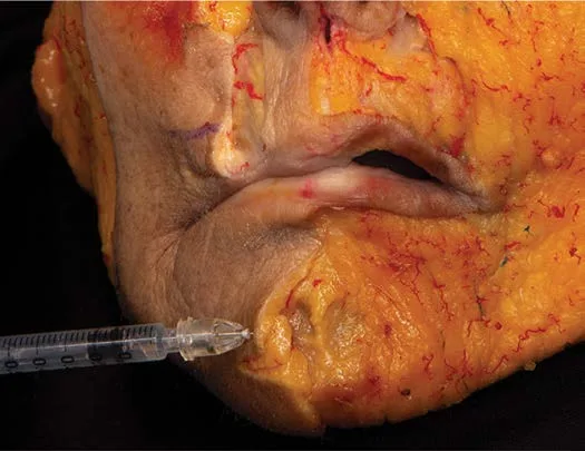 Fig 11. Dissection of the perioral/chin region with a syringe delivering into the mentalis: the toxin target is reached by a deep, perpendicular pass low in the chin, near bone, away from orbicularis oris above and the lip depressors laterally - (Pirayesh, Aesthetic Facial Anatomy Essentials, p. 235).
> Sources: [MEDLIB] Pirayesh 2020 [1]. Fig 11 documents the deep perpendicular mentalis approach in the dose table.
Fig 11. Dissection of the perioral/chin region with a syringe delivering into the mentalis: the toxin target is reached by a deep, perpendicular pass low in the chin, near bone, away from orbicularis oris above and the lip depressors laterally - (Pirayesh, Aesthetic Facial Anatomy Essentials, p. 235).
> Sources: [MEDLIB] Pirayesh 2020 [1]. Fig 11 documents the deep perpendicular mentalis approach in the dose table.
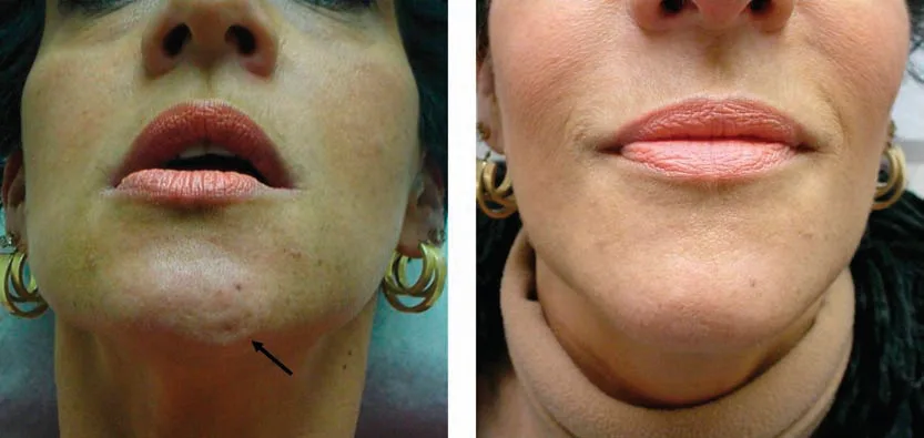 Fig 12. Frontal views: (left) a hyperkinetic mentalis producing a focal chin dimple/pucker on animation, arrowed; (right) the effaced, smooth chin three weeks after a mentalis deposit - the surface result that filler alone cannot achieve when the cause is muscular - (Benedetto, Botulinum Toxins in Clinical Aesthetic Practice, p. 181).
> Sources: [MEDLIB] Benedetto 2011 [30]. Fig 12 shows the mentalis-toxin result described above.
Fig 12. Frontal views: (left) a hyperkinetic mentalis producing a focal chin dimple/pucker on animation, arrowed; (right) the effaced, smooth chin three weeks after a mentalis deposit - the surface result that filler alone cannot achieve when the cause is muscular - (Benedetto, Botulinum Toxins in Clinical Aesthetic Practice, p. 181).
> Sources: [MEDLIB] Benedetto 2011 [30]. Fig 12 shows the mentalis-toxin result described above.
The golden rule of the lower third: start at the minimum of the range and review at 2 weeks. The cost of under-dosing is one extra visit; the cost of over-dosing is a patient who cannot speak or smile normally for three months [16][30].
Brand-specific dosing (units are not interchangeable across toxins). Course and consensus material gives the mentalis and DAO in the two common unit systems, which do not convert 1:1 [16][30][31]:
| Muscle | onabotulinum / abobotulinum-"BX/BC" family | Abobotulinum (Dysport/Azzalure, "AZ") | Points / depth |
|---|---|---|---|
| Mentalis | 4-8 U total (Villanueva course) [46]; 2-5 U (Wong) [16] | 12 U [46] | Central, ~1 cm above the bone line, deep; ± 2 lateral points [16][46] |
| DAO | 2-4 U per side (VB/BC); 2 U × 3 (Benedetto) | 4-5 U per side | Mandibular border, lateral to modiolus, deep [30][31] |
| Chin dimple | 2-4 U | - | Midline, deep |
Mentalis technique in detail. Infiltrate the central chin roughly 1 cm above the mandibular border, deep; a strong or broad muscle sometimes needs two lateral points in addition to the midline; for a residual "orange-peel" surface, reinforce with small intradermal papules [33][46]. The mentalis can be genuinely strong in Class I occlusion, where it helps close the mouth, so a conservative dose preserves function while softening the crimp [46].
DAO localisation and technique. Ask the patient to make the "sad" face; the DAO is the triangular muscle from the mandibular bar to the commissure, its fibres interlacing with the platysma and lying just lateral to the depressor labii inferioris. Inject either the muscle belly (needle a third of the way in) or the mandibular border on the imaginary line through the nasal ala, the commissure and the mandible; stay off the DLI medially [16][30][32]. The characteristic complication is a visible, poorly tolerated mouth asymmetry from DLI spread [16].
Skin-quality toxin (mesobotox / microbotox), with a real dilution. For dilated pores and surface oiliness of the chin (and nose), an intradermal micro-papule technique uses 20 U of toxin diluted in 2 mL of a polyvitamin carrier delivered into the mid-dermis; the same 20 U in 5 mL is the wider "meso-lift" dilution. Cadence is biweekly initially, then monthly [47]. This is a dermal, low-unit application distinct from the deep neuromodulating mentalis dose and must not be confused with it.
Diffusion, dilution and the safety distances. A larger reconstitution volume spreads the toxin further, so a high dilution invites unwanted spread to orbicularis oris or the DLI; a diluent volume above ~1 mL per field is associated with adverse spread in the lower face [30]. Keep the mentalis point ≥1 cm above the mandibular border and on the midline, and the DAO point at the border, lateral to the modiolus, so even the expected halo of diffusion stays inside the target muscle [16][30][35]. The lower third is the region where diffusion, not just placement, decides the complication rate, which is another reason to start low and review.
Reconstitution and product equivalence. The unit is meaningless without the product: reconstitute per the manufacturer, keep the diluent volume modest in the lower face to limit spread, and never assume a stored cross-brand conversion, because ona-, inco- and abobotulinumtoxinA differ in potency scale and diffusion [16][30]. When switching brands, re-derive the dose from the product's own guidance rather than converting the number you remember.
Onset, duration, review. The effect appears over about two weeks and lasts 4-6 months for the mentalis; review at 2 weeks and top up conservatively rather than front-loading the dose [16][30]. Reconstitution and the exact unit depend on the product; never carry a remembered cross-brand conversion into the lower third, where a small excess is a functional complication.
The antagonist map - the muscle you deliberately spare. Lower-lip position is a balance of elevators and depressors; the point of chin toxin is to weaken the right muscle and leave its antagonist intact, so every dose is chosen as much for what it must not reach [16][35]:
| Injected (target) | Must be spared | Failure if it spreads |
|---|---|---|
| Mentalis (deep, low, midline) | Orbicularis oris (above) | Lower-lip incompetence, lip drop, "witch's chin" |
| Mentalis | DLI (lateral) | Asymmetric lower lip |
| DAO (border, lateral to modiolus) | DLI (medial) + lip levators | Crooked smile, upper-lip ptosis, over-lengthened upper lip |
Units do not convert across toxin brands. The mentalis takes ~4-8 U on the ona/inco "BX/BC" scale but ~12 U on the abobotulinum "AZ" scale, and the DAO ~2-4 U versus ~4-5 U per side; these are not interchangeable, and carrying a remembered conversion into the small, closely-packed lower-face muscles is precisely how a minor excess becomes a functional complication [16][46]. Dilution and unit are read off the product each time.
Dynamic dimple vs fixed cleft. An animated cobblestone from an overactive mentalis responds to toxin; a congenital bony/soft-tissue cleft chin is structural and does not, so separate the dynamic dimple (toxin) from the fixed cleft (not toxin) before dosing, and co-treat a deep static mental crease with filler while a dynamic one resolves with toxin alone (§D11.9) [16][34].
Spacing and top-ups. Increase the dose between sessions, not within one; a 2-week review lets you add a conservative top-up rather than front-load a dose you cannot take back [16][30]. The lower third rewards patience and punishes the extra unit.
Classic trap: chasing the surface dimple with a high or lateral mentalis point - a high deposit reaches orbicularis oris and drops the lip, a lateral one spreads to the DLI; the safe deposit is deep, low and midline, and a deep static crease is co-treated with filler, not more toxin [16][30].
D11.9 · Combination and sequence
Build the skeleton first, relax the muscle second, finish the surface last. The chin is one node of the lower-face unit; treat it in that order. Full sequence: L2 - Combination Protocols & Layered Rejuvenation.
| Order | Step | Why here | Interval |
|---|---|---|---|
| 1 | Filler projection (chin C2/C3 ± jawline, prejowl) | Structure-bearing regions first; sets the platform the rest is judged against [13][19] | Immediate effect |
| 2 | Toxin mentalis / DAO (same or next session) | Relax the muscle that would crimp over new volume; unmask the true crease [16][34] | Peaks ~2 weeks |
| 3 | Perioral / marionette / lips | Refine the mid-lower face once the chin sets the proportion [19] | Same or staged |
| 4 | Skin quality (polynucleotides, boosters, microbotox, energy) | Finishing layer; never the foundation | Staged |
Consensus: across the whole face the order is structure-bearing regions first (cheek, chin, jaw), then perioral and periocular refinement, then skin quality and toxin [19]. Within the lower face, project the chin before deciding submental fat treatment, because projecting the mentum opens the cervicomental angle and improves the submental silhouette on its own [7]. Toxin can be placed in the same visit as filler (many do) or staged; it acts over about two weeks while filler is immediate, so review at 2 weeks lets you judge both.
Sequencing that inverts the plan: if the lower third is wide because of masseter hypertrophy, definition is achieved by subtracting (masseter toxin), not adding chin/jaw filler; adding volume enlarges what the patient wanted narrowed (§D6, C3 - Botulinum Toxin - Full Technique Map). And if the border shows real skin laxity (positive pinch test), energy or threads precede volume, because filler adds weight to a descending tissue.
Follow-up cadence: review injectables at 2-4 weeks, review toxin at 2 weeks, and re-photograph against baseline annually; record region, plane, product, lot, volume per side and cumulative volume [16].
Region combinations, by complaint.
| Presenting complaint | Chin's role | Combine with |
|---|---|---|
| Weak profile / "double chin" | Project the chin first | Jawline (Jw), then reassess submental fat (§D6) |
| Sad/downturned mouth | Support prejowl + chin | DAO toxin + marionette filler (§D5) |
| Cobblestone chin + short chin | Structural filler to bone | Mentalis toxin, staged or same visit |
| Wide lower face wanting definition | Add only if the border is lost | ⚠ Masseter toxin if hypertrophy (subtract, do not add) (§C3) |
| Ageing lower third, lax skin | Volume only after tightening | Energy/threads first, then structural filler [19] |
| Lip + chin imbalance | Rebalance proportion | Lip filler after the chin sets lower-third height [16] |
Same session or staged? Filler and toxin are commonly placed in one visit; the toxin acts over about two weeks while the filler is immediate, so a 2-week review lets you judge both and titrate. Stage when the plan is large, when you want to see the effect of projection on the crease before dosing the mentalis, or when the patient is toxin-naïve. Biostimulators (PLLA, CaHA, PCL) build gradually over sessions and are planned as a series, not a single correction.
Layering in one region. The multilayer approach places structural product supraperiosteally, dynamic volume in the mobile subcutaneous plane, and a redensifying/skin-quality layer superficially, from a single entry, so the chin is built as a stack rather than a single depth [43]. Skin-quality adjuncts (polynucleotides, boosters, microbotox) are the finishing layer, never the foundation.
Documentation and follow-up. Record region, plane, product, lot number, volume per side and cumulative volume, and for toxin the muscle, units and dilution; review injectables at 2-4 weeks, toxin at 2 weeks, and re-photograph against baseline annually [16]. A cumulative-volume record is what prevents the slow over-projection that creeps in across visits.
Why the whole-face order is structure-first. Structure-bearing regions (cheek, chin, jaw) are treated before perioral and periocular refinement because they set the frame the rest is judged against, and because midface support unloads the tear trough, nasolabial and marionette that would otherwise be over-filled; skin quality and toxin finish the plan [19]. Treating the chin out of order - surface before structure, or lips before the chin sets the lower-third height - produces corrections that must be redone once the frame changes.
Timing of the modalities.
| Modality | Onset | Peak / settle | Plan around it |
|---|---|---|---|
| HA filler | Immediate | Settles ~2 wk | Judge projection at once; review 2-4 wk |
| Toxin | 2-3 days | ~2 weeks | Review at 2 wk before any top-up |
| CaHA / PLLA biostimulation | Weeks | 2-3 months, over sessions | Plan as a series, not one correction |
| Threads / energy | Immediate lift / gradual tightening | Weeks | Before adding weight to a lax lower third |
Combination protocols by goal.
| Goal | Chin's part | Combine |
|---|---|---|
| Profile correction | Projection to bone | Jawline continuity ± lip rebalance [13][19] |
| Downturned mouth | Prejowl + chin support | DAO toxin + marionette filler (§D5) |
| Wide lower face (masseter) | Add only if border lost | Masseter toxin first (subtract), then decide (§C3) |
| Ageing, lax skin | Volume after tightening | Energy/threads → structural filler → skin quality [19] |
| "Double chin" | Project the chin first | Reassess submental fat only after (§D6) |
The chin is rarely treated alone; it is the keystone of the lower-face plan, and the sequence is what turns separate procedures into one coherent result (§L2).
Staging examples. A common two-visit plan: visit 1 builds chin projection and jawline continuity with HA and places a conservative mentalis dose; visit 2 at two to four weeks reviews the projection, tops up the crease, and adds DAO toxin if the marionette persists. A biostimulator plan runs over three sessions spaced weeks apart, with the chin re-photographed each time. The rule that governs both: do not chase a result before the previous layer has settled, because the frame keeps moving until the structure is set [16][19].
Classic trap: placing toxin in the mentalis before building projection and then over-filling to chase a crease that the relaxed muscle has already softened - sequence the structure and the muscle, review at 2 weeks, and add filler to the residual static crease only, not to a dynamic one that toxin will resolve [16][34].
D11.10 · Region-specific complications
Not the generic bruise and swelling: the complications that only happen at the chin because of its muscles, its one nerve, and its non-reversible product options. Cross-refs: J1 - Filler Complications Overview, J2 - Vascular Occlusion.
| Complication | Where / mechanism | Recognition | Management |
|---|---|---|---|
| Lip incompetence / "witch's chin" (chin ptosis) | Mentalis over-weakened, dose too inferior/high, drops the soft-tissue chin pad | Lower-lip drooping/drooling, chin pad ptosis on animation | Wait it out (4-6 mo); prevent with conservative deep midline dose [16][34] |
| Asymmetric smile / lower-lip weakness | DAO toxin too medial/high → DLI spread | Crooked smile, cannot depress lower lip symmetrically, 8-12 wk | Reassure, self-limited; balance with micro-dose contralaterally if severe [30][35] |
| Upper-lip ptosis / over-lengthening | Excess DAO dose over-weakens the depressor system | Long upper lip, altered smile | Self-limited; avoid in voice/wind professionals [35] |
| Over-projection ("cocktail/witch" chin) | Too much volume; blunts the labiomental angle | Ball-like or witch contour on profile | Dissolve (HA) or wait/refer (CaHA/PMMA) |
| Mental-nerve dysaesthesia | Deposit over/into the mental foramen (too lateral) | Numbness/tingling of chin + lower lip | Usually transient; keep the projection point midline-on-bone [7][16] |
| Filler migration along the mandibular border | Product tracks along the jawline plane | Palpable/visible fullness away from injection site | Massage, dissolve if HA; avoid over-volume in the subcutaneous border plane |
| Palpable nodules / late granuloma | Superficial or excessive deposit; product-specific | Firm lumps, early (technique) or late (inflammatory) | HA → hyaluronidase; CaHA/PMMA → intralesional steroid/excision, no dissolution |
| Vascular occlusion (chin/lower lip) | Intravascular into submental / inferior-labial / facial / mental artery | Pain out of proportion, blanching then dusky livedo, slow capillary refill | HA → HDPH below; no blindness route from the chin; CaHA/PMMA not reversible [38] |
Consensus: the chin's own complications are functional (muscle/nerve) and product-irreversibility, not the catastrophic ocular events of the upper face; prevent lip incompetence and smile asymmetry with conservative, deep, correctly-placed toxin, prevent nerve injury by keeping the projection deposit midline-on-bone, and prefer a reversible HA in any patient where you might need to undo the result [7][16][34].
Complications by onset (chin-relevant).
| Onset | Complication | First move |
|---|---|---|
| Immediate (minutes) | Vascular occlusion: blanching, pain out of proportion | Stop; HDPH for HA (below) [38] |
| Hours-days | Mentalis/DAO functional effect (toxin) | Reassure; self-limited; review 2 wk [16][35] |
| Days-2 wk | Bruising, oedema, early nodule | Massage; hyaluronidase for an HA nodule [44] |
| Weeks-months | Delayed inflammatory nodule, biofilm, migration | Antibiotics/steroid; hyaluronidase (HA); ultrasound [26][44] |
| Persistent | Over-projection, Tyndall, asymmetry | Dissolve (HA) or refer (CaHA/PMMA) [17][44] |
Vascular occlusion - chin protocol (HA). Recognise early: pain out of proportion, blanching then dusky reticulate mottling, prolonged capillary refill along the vessel [38]. Treat as an HA vascular event with high-dose pulsed hyaluronidase (DeLorenzi): ~500 IU per affected area, repeated every 60-90 minutes until skin colour and capillary refill normalise; 1000 IU for two areas, 1500 IU for three, aiming to complete within 72 hours, keeping the patient in clinic (typically three sessions over about three hours) [38][1]. Massage to spread the hyaluronidase; roughly 5-10 U dissolve 0.1 mL of 20 mg/mL HA for simple correction, but far more is used for an intravascular event [1]. Do not apply nitroglycerin paste until the offending HA is dissolved (day 2-3), since dilating adjacent pathways can propagate the embolus [38]. The chin has no retrograde ophthalmic pathway, so unlike the nose/glabella there is no blindness risk, but CaHA and PMMA are not reversible and have no hyaluronidase rescue, which is the single strongest argument for a reversible HA in a first or revision-prone chin. Hyaluronidase allergy is rare (~1/1000); a skin test is skipped in the emergency but considered for elective dissolution in bee/wasp-allergic patients [17].
Vascular occlusion - the full chin algorithm. 1. Recognise (minutes to hours): pain out of proportion, blanching, then dusky reticulate mottling/livedo along the vessel, prolonged capillary refill; livedo without pain can still be occlusion [38][44]. 2. Stop injecting immediately. 3. First response: aspirin (a 650 mg oral loading dose is a common recommendation), effective analgesia, warm compress, and massage to move the embolus distally [44]. 4. Dissolve: HDPH ~500 IU per affected area, repeated every 60-90 minutes until skin colour and capillary refill normalise; escalate to 1000 IU (two areas) / 1500 IU (three), aiming to finish within 72 hours; van Loghem's practical variant is 500 U reconstituted in 1 mL of 2% lidocaine per 5×5 cm of affected skin, hourly, and reconstitution with lidocaine (no epinephrine) adds vasodilation [38][44]. Inject widely across the whole ischaemic field, not only the injection point, because intravascular filler forms columns, not a single embolus [14]. 5. Ultrasound-guided hyaluronidase restores flow with far less drug where available [40][44]. 6. Do not apply nitroglycerin paste until the HA is dissolved (day 2-3) [38]. 7. Adjuncts (low evidence): LMWH, pentoxifylline, hyperbaric oxygen; keep an updated filler-emergency kit and protocol to hand [44][45]. 8. Follow up at 7 days and 4 weeks; start antibiotics if infection supervenes on the infarcted tissue [44].
Recognising it early is most of the cure. The earliest signs are pain out of proportion to the procedure and blanching, followed within minutes to hours by a dusky reticulate mottling (livedo) along the vessel's territory and a prolonged capillary refill; livedo can precede pain, so a mottled chin or lower lip after injection is occlusion until disproved [38][44]. The chin territory to watch is the lower lip and the chin skin along the inferior-labial and submental distribution; because there is no ophthalmic route, vision is not at risk, but the tissue window is still only about 72 hours [38].
Nodules, granulomas and delayed reactions. Early nodules are usually technical (superficial or excess deposit) and respond to massage or hyaluronidase (HA); late nodules may be inflammatory or biofilm-related and, with HA, are treated with hyaluronidase ± antibiotics/steroid, while CaHA and PMMA cannot be dissolved and may need intralesional steroid or excision [44]. Delayed inflammatory reactions are a recognised, product-specific phenomenon of the highest-G′ Vycross gels (reported distinctly for VYC-25L, sometimes in a patient who tolerated VYC-20L), which is a consideration when choosing the chin product and when counselling [26]. A superficial HA deposit can also produce a bluish Tyndall discoloration, corrected by dissolving it.
Overcorrection and migration. Over-projection blunts the labiomental angle and reads as a "ball" or "witch" chin on profile; HA is dissolved, CaHA/PMMA is waited out or referred. Migration along the mandibular border comes from over-volume in the mobile subcutaneous plane; keep the border deposits measured and mould against bone.
Infection, biofilm and delayed nodules. Early infection (redness, warmth, tenderness, sometimes fluctuance) is treated with antibiotics and drainage if abscessed; a biofilm presents later as a low-grade inflammatory nodule and is managed with antibiotics while avoiding an incision that can seed it, plus hyaluronidase for HA [44]. Delayed-onset nodules (weeks to months, sometimes triggered by illness or vaccination) are inflammatory: HA responds to hyaluronidase ± steroid/antibiotic, while CaHA and PMMA nodules cannot be dissolved and may need intralesional steroid (with a 5-fluorouracil adjunct in refractory cases) or excision [44]. The highest-crosslink Vycross gels carry a distinct delayed-inflammatory signal that feeds back into product choice and consent [26].
Tyndall and superficial irregularity. A superficial HA deposit can scatter light to a bluish Tyndall hue or sit as a visible ridge; both are corrected by dissolving the superficial product and re-placing it deeper, and both are prevented by keeping structural product deep and surface product minimal and even [17][44].
The filler emergency kit, chin version. Stock and rehearse: hyaluronidase in quantity for a high-dose pulsed protocol, aspirin, warm compresses, nitroglycerin paste (for after the HA is dissolved) and a written algorithm; ultrasound, if available, cuts the drug needed [44][45]. The chin variation of the standard posology is simply the per-area HDPH dosing above; there is no retrobulbar step, because there is no ophthalmic route from the chin [45].
Prevention checklist (region-specific): midline-on-bone for projection, cannula and aspiration for lateral/border work, small aliquots and low pressure, reversible product where reversibility might matter, conservative and correctly-placed toxin, ultrasound for revision/lateral work, and a written occlusion protocol with hyaluronidase stocked [38][40][44][45].
Management by product when it goes wrong.
| Product | Overcorrection / nodule | Occlusion |
|---|---|---|
| HA | Hyaluronidase dissolves | Hyaluronidase (HDPH) [38] |
| CaHA | No dissolution; wait, steroid, or excise | No rescue; supportive [44] |
| PMMA | Permanent; steroid/5-FU or excise | No rescue [44] |
| PLLA | Wait / steroid for nodules | No rescue [44] |
This table is the argument for reversibility: an HA problem is recoverable in the chair, a biostimulator or permanent problem is not, so the product is chosen partly for how it fails [1][27][44].
Classic trap: treating a chin vascular event as "just bruising" and waiting - the window is hours; blanching plus pain out of proportion is occlusion until proven otherwise, and the response is immediate high-dose pulsed hyaluronidase (for HA), not observation [38].
Coverage vs UPO
UPO teaches the chin as two techniques (supraperiosteal filler, mentalis/DAO toxin) with brand-specific doses; the atlas adds the injection map, the schools that disagree, the profile analysis, the regulatory evidence and the referral thresholds. UPO doses are [D] never_sufficient_alone.
| UPO topic (what the course teaches) | Status in this chapter | What the atlas adds |
|---|---|---|
| Chin projection, supraperiosteal plane (Arenas, T8.1) [27] | Covered (D11.2, D11.7) | The point-by-point MD Codes map, per-point volume, bevel rule, cadaver proof [13][20] |
| Mandibular line, subdermal microcannula (Arenas) [27] | Covered (D11.7) | Jw1-Jw5 codes with depth/device, submental-artery avoidance, needle-vs-cannula rule [13][40] |
| Marionette lines = DAO + platysma; fan/cross-hatch filler + DAO toxin (Arenas) [27] | Covered (D11.8, D11.9) | DAO localisation, DLI-spread complication, dose in two unit systems [30][35] |
| Mentalis toxin 4-8 U (BX/BC), 12 U (AZ), central ~1 cm above bone, deep, ± intradermal papules (Villanueva, T8.2) [46] | Covered (D11.8) | Cross-checked against Wong (2-5 U) and Benedetto; antagonist map; 4-6 mo duration [16][30][31] |
| DAO toxin 2-4 U (VB/BC), 4-5 U (AZ) per side (Villanueva) [46] | Covered (D11.8) | Landmark line, "sad face" localisation, upper-lip-ptosis caution [30][35] |
| Mesobotox chin pores, 20 U in 2 mL, biweekly→monthly (Ordiz, T8.3) [47] | Covered (D11.8) | Framed as a dermal skin-quality use distinct from the neuromodulating dose |
| High-G′ 25 mg/mL structural voluminiser for chin/cheek/jaw, 25G, 9-20 mo (Rosso, T9.6 MLT) [43] | Covered (D11.7) | Rheology (G′/cohesivity) explained; Volux/Voluma named; multilayer technique [19][21][43] |
| PMMA Artefill (bovine collagen, allergy test) (Arenas) [27] | Covered (D11.7) | Placed in the reversible-vs-permanent product grid with CaHA/PLLA/PCL |
| Not in UPO: microgenia vs retrognathia bite test | Added (D11.6) | The 30-second occlusion exam that flips the indication [7][16] |
| Not in UPO: cephalometric profile analysis (zero-meridian, degrees) | Added (D11.5) | González-Ulloa/Wolford/E-line, degree classification [11] |
| Not in UPO: genioplasty / chin-implant referral thresholds | Added (D11.6, D11.7) | When the correct answer is surgery, not filler [7] |
| Not in UPO: needle-to-bone vs cannula, Volux vs Voluma controversies | Added (D11.7) | Both preserved with authors, never averaged [13][17][22][24] |
| Not in UPO: ultrasound-guided injection / HDPH occlusion protocol | Added (D11.5, D11.10) | Malherbe + Vasconcelos-Berg; DeLorenzi HDPH dosing [38][40] |
| Not in UPO: VYC-20L/VYC-25L trial evidence, order-of-loss ageing | Added (D11.4, D11.7, What's new) | 2020-2025 chin RCT data; skeleton-first ageing model [10][22][24][26] |
UPO is the fastest-ageing lane in the chapter: it grounds the technique and the toxin doses, but its slides are [D] and cannot alone support a dose or a currency claim, which is why every UPO number here is corroborated by a textbook or a dated primary source.
Self-assessment
Ten active-recall questions, built only from facts stated above. Answers folded.
- What single 30-second exam separates a filler-appropriate chin from a referral, and what does each result mean?
Answer
The bite test: **normal occlusion + small chin = microgenia** (treat with filler); **Class II malocclusion = retrognathia** (refer for orthognathic/orthodontic care) [7][16].- Give the chin's six anatomical layers from skin to bone.
Answer
Skin, dermis (with mentalis insertions), superficial (pre-mental) fat, mentalis/muscle, deep/pre-periosteal fat, periosteum over the symphysis [1][5].- Why can you not deposit structural filler "deep subcutaneous" in the chin the way you might in the midface?
Answer
The mentalis inserts into the dermis, so there is no free glide plane; superficial product in the belly shows on movement. The deep target is periosteum, the superficial target is the pre-mental fat above the muscle [16].- Where is the mental foramen, and what is the practical complication of injecting over it?
Answer
Below/at the 2nd premolar, ~2.5 cm lateral to the midline, ~1 cm above the inferior border; a deposit there causes hypoaesthesia/dysaesthesia of the chin and lower lip [7][17].- State the HA chin volume ladder for asymmetry, mild recession and moderate-severe recession.
Answer
0.2-0.3 mL (asymmetry), 0.6-0.8 mL (mild recession), 1.3-1.5 mL (moderate-severe) [15].- In the needle-vs-cannula controversy, what decides the choice?
Answer
The target plane: needle-to-bone for midline projection; cannula subcutaneously for contour, the labiomental crease and the jawline (submental-artery avoidance). Never averaged into a mid-plane deposit [13][17].- Give the mentalis and DAO toxin doses and the muscle each must avoid.
Answer
Mentalis 2-8 U, deep low midline, avoid **orbicularis oris** (too high). DAO 2-5 U/side, deep at the mandibular border lateral to the modiolus, avoid the **DLI** (too medial/high → asymmetric smile 8-12 wk) [16][30][31].- What is the zero-degree meridian, and how is chin retrusion graded on it?
Answer
A vertical from soft-tissue nasion perpendicular to Frankfort horizontal; soft-tissue pogonion should meet it. Retrusion <10 mm = 1st degree, 10-20 mm = 2nd, >20 mm = 3rd (⚠ the source's text elsewhere says >30 mm) [11].- Which chin products are NOT hyaluronidase-reversible, and why does it matter?
Answer
CaHA (Radiesse) and PMMA (Bellafill/Artefill); there is no rescue for occlusion or overcorrection, so they demand conservative technique and favour a reversible HA in first/revision-prone chins [1][27].- Outline the first steps of a chin HA vascular occlusion.
Answer
Recognise (pain out of proportion, blanching, dusky livedo, slow refill); stop; aspirin + analgesia + warm compress + massage; HDPH ~500 IU per area every 60-90 min (1000/1500 for two/three areas) within 72 h; no nitroglycerin until the HA is dissolved. No blindness route from the chin [38][44].What's new and trends (2024-2026)
| Year | Development | Maturity | Effect on practice |
|---|---|---|---|
| 2020 | VYC-20L (Voluma) chin-augmentation pivotal data [22] | clinically actionable now | Underpins the FDA chin indication; structural HA validated for the mentum |
| 2023 | VYC-20L live-vs-photographic responder analysis [23] | clinically actionable now | Higher responder rates on live assessment; supports in-person outcome grading |
| 2024 | VYC-25L (Volux) jawline-restoration RCT [24] | clinically actionable now | Highest-G′ Vycross validated for the jawline; chin used off-label but evidenced |
| 2024 | Chin reinforcement with VYC-20L + VYC-25L combined [26] | promising but not validated | Layering the two structural gels for the chin unit |
| 2025 | VYC-25L for chin retrusion cohort [25] | promising but not validated | Direct chin-projection evidence for the highest-G′ gel |
| 2025 | HF-ultrasound best-practice protocol for the lower third [40] | clinically actionable now | Formalises scan-while-injecting for the chin/jawline; vessel mapping + guided hyaluronidase |
| emerging | Periosteal biostimulator to offset symphyseal bone resorption | preclinical/speculative | Hypothesis only (§Unexplored); no chin bone-longevity data yet [10] |
| ⚠ | "Permanent liquid genioplasty" / "risk-free cannula" marketing | unsupported commercial claim | Rejected: no permanent filler is risk-free and a cannula does not abolish occlusion [38][44] |
What did NOT change, and why the older references still stand. The core technique is stable: needle-to-bone supraperiosteal projection at the midline with aspiration remains the standard (de Maio 2017/2020, Hong 2020), and the anatomy (Pirayesh, Standring, Naini) does not date [1][2][11][13][14]. The toxin doses for the mentalis and DAO are unchanged across a decade of sources [16][30][31]. The occlusion backbone is still DeLorenzi's 2017 HDPH protocol [38]. The genuine 2020-2025 movement is regulatory and evidentiary (chin/jawline HA now has RCT-grade data instead of expert opinion) and procedural (ultrasound moving from optional to best-practice for lateral/revision work), not a change in where the needle goes. The corpus-anchored consensus is therefore current, with the external lane supplying the trial evidence and the ultrasound protocol the textbooks predate.
Unexplored directions (AI speculation)
> [IA-ESPEC] The following are model-generated research directions, not evidence and not clinical guidance. Each names a cited anchor already in this chapter, a proposal, and what would settle it. None contains a dose, product or protocol a reader could act on. Tag: [IA-ESPEC].
-
[IA-ESPEC]Region-specific occlusion-risk stratification for the chin. Anchor: the chin has no ophthalmic route and its vessels anastomose across the midline off the bony platform [1][38]. Proposal: a chin/jaw-specific vascular-risk score and consent, distinct from the midface/nose. Expected effect: earlier recognition and a lower necrosis rate for chin/jaw occlusion. Confounder: chin occlusions are rare, so incidence differences need large samples, and operator experience varies. What would settle it: a prospective multicentre chin/jaw filler registry recording occlusion incidence, plane and device. -
[IA-ESPEC]Simultaneous vs sequential mentalis toxin plus structural filler for cobblestone chin. Anchor: the mentalis is dermally tethered and its relaxation unmasks the true static crease [16][34]. Proposal: first-line combined micro-dose toxin + bony filler versus the sequential approach. Expected effect: better crease resolution and satisfaction at fewer visits. Confounder: filler settling and toxin onset differ in time, confounding the crease endpoint, and blinding is difficult. What would settle it: a split-face or parallel randomised trial with blinded crease-depth and patient-satisfaction endpoints. -
[IA-ESPEC]Periosteal biostimulator to counter symphyseal resorption. Anchor: bone resorption drives the ageing chin and precedes the soft-tissue stigmata [10][19]. Proposal: long-term supraperiosteal biostimulator to slow or offset symphyseal loss. Expected effect: measurable slowing of symphyseal bone loss versus untreated. Confounder: bone loss is slow and multifactorial (systemic, dental), and repeat CBCT radiation limits sampling. What would settle it: a longitudinal cone-beam-CT cohort measuring symphyseal bone over years, treated vs untreated. -
[IA-ESPEC]Doppler-first routine for chin injection. Anchor: ultrasound-guided hyaluronidase restores flow with far less drug and maps the submental artery [40][44]. Proposal: pre-injection Doppler mapping as routine, not just for revision. Expected effect: fewer intravascular events and lateral-point complications. Confounder: operator ultrasound skill varies, and the added time and cost may bias adoption and outcomes. What would settle it: a randomised comparison of ultrasound-mapped versus landmark chin/jaw injection for occlusion and accuracy. -
[IA-ESPEC]Delayed-inflammatory-reaction phenotyping for the highest-G′ gels. Anchor: delayed inflammatory reactions are reported distinctly for VYC-25L, sometimes in VYC-20L-tolerant patients [26]. Proposal: pre-treatment markers that predict which patients react to the highest-crosslink chin gels. Expected effect: patient-specific product selection that avoids delayed nodules. Confounder: delayed reactions are rare and triggered by intercurrent illness or vaccination, confounding causation. What would settle it: a case-control immunophenotyping study of reactors versus tolerant controls.
§ Safety
The chin's own red lines, each with its evidence. Generic filler/toxin safety lives in J1 - Filler Complications Overview to J8.
- Bite first. A Class II malocclusion (retrognathia) is a skeletal/functional problem; filler only camouflages it. Do the occlusion exam before treating and refer the Class II [7][16].
- Midline, on bone, aspirate. Keep the projection deposit midline and supraperiosteal, aspirate at bone, use small aliquots and low pressure; the mental artery and mental foramen are lateral [13][16].
- Mentalis point stays low and midline. Too high reaches orbicularis oris and drops the lower lip (lip incompetence / "witch's chin"); dose conservatively and review at 2 weeks [16][34].
- DAO point stays deep, at the border, lateral to the modiolus. Too medial/high weakens the DLI and gives an asymmetric smile for 8-12 weeks; avoid entirely near a critical voice/wind-instrument profession without full counselling [30][35].
- Choose reversibility deliberately. CaHA and PMMA are not hyaluronidase-reversible and have no occlusion rescue; prefer a reversible HA in first-time or revision-prone chins [1][27].
- Occlusion is minutes-to-hours, not "bruising". Blanching plus pain out of proportion is occlusion until proven otherwise; treat HA events with high-dose pulsed hyaluronidase (~500 IU/area hourly, escalating, within 72 h), no nitroglycerin until the HA is dissolved. There is no blindness route from the chin, but chin/lower-lip necrosis is real [38][44].
- Do not widen a feminine chin by reflex, and do not use a biostimulator for width you might want to reverse; masculinisation with a permanent product cannot be undone [13].
- Do not over-project. Respect the labiomental angle; a "ball/witch" chin on profile is an aesthetic complication, dissolvable only if HA [11].
- PMMA/bovine-collagen products need a skin allergy test before use [27].
- Refer, don't stack. Large skeletal deficiency, frank skin excess, true jowl or a low hyoid are surgical or non-injectable problems; injectables there spend money without the result [7][19].
References
Tags: [A] datasheet/guideline/consensus · [B] primary literature (PMID/DOI) · [C] monograph · [D] slide/opinion, never sufficient alone · [MEDLIB] own corpus.
- Pirayesh A. Aesthetic Facial Anatomy Essentials for Injections. 2020.
[C][MEDLIB] - Standring S (ed). Gray's Anatomy. 41st ed. 2016. ISBN 9780702052309.
[C][MEDLIB] - Cotofana S, et al. Anatomy of the facial fat compartments and their relevance in aesthetic surgery. 2019. DOI 10.1111/ddg.13737.
[B][MEDLIB] - Schenck TL, et al. Functional anatomy of the superficial fat compartments of the face. 2018. DOI 10.1097/PRS.0000000000004364.
[B][MEDLIB] - Radlansky RJ, Wesker KH. Atlas Ilustrado de Anatomia Clínica da Face. ISBN 9788578890773.
[C][MEDLIB] - Watanabe K, et al. Anatomy for Plastic Surgery of the Face, Head, and Neck. 2016. ISBN 9781626230910.
[C][MEDLIB] - Azizzadeh B, Murphy MR, Johnson CM, et al. Master Techniques in Facial Rejuvenation. 2nd ed. 2018. ISBN 9780323358767.
[C][MEDLIB] - Seckel BR. Facial Danger Zones: Avoiding Nerve Injury in Facial Plastic Surgery. 2nd ed.
[C][MEDLIB] - Malherbe C, et al. Ultrasound Protocol for Facial Aesthetics. 2024.
[C][MEDLIB] - Cotofana S, et al. Anatomy of the Aging Face: A Review. 2016. DOI 10.1055/s-0036-1582234.
[B][MEDLIB] - Naini FB. Facial Aesthetics: Concepts and Clinical Diagnosis. 2011. ISBN 9781405181921.
[C][MEDLIB] - de Maio M. MD Codes: Unlocking the Code. 2017.
[C][MEDLIB] - de Maio M. MD Codes: A methodological approach to facial aesthetic treatment with injectable hyaluronic acid fillers. 2020. DOI 10.1007/s00266-020-01762-7.
[B][MEDLIB] - Hong KH, et al. Art and Science of Filler Injection. 2020. ISBN 9789811306105.
[C][MEDLIB] - Garg S. Dermal Fillers for Dental Professionals. 2021. ISBN 9780867158304.
[C][MEDLIB] - Wong V. Decision Making in Aesthetic Practice. 2022. ISBN 9781032046037.
[C][MEDLIB] - Jones DH, Beleznay K, et al. Injectable Fillers: Facial Shaping and Contouring. 2nd ed. 2019. ISBN 9781119046967.
[C][MEDLIB] - Kontis TC, Lacombe VG. Cosmetic Injection Techniques: A Text and Video Guide to Neurotoxins and Fillers. 2nd ed. 2019.
[C][MEDLIB] - Carruthers J, Carruthers A. Soft Tissue Augmentation. 4th ed. 2018. ISBN 9780323476584.
[C][MEDLIB] - Vieira Braz A, et al. Atlas de Anatomia e Preenchimento Global da Face. 2017. ISBN 9788527732482.
[C][MEDLIB] - Injection Techniques for Esthetic Lip Treatment (Lips-45). ISBN 9781786981097.
[C][MEDLIB] - Beer K, Kaufman-Janette J, Bank D, et al. Safe and effective chin augmentation with the hyaluronic acid injectable filler, VYC-20L. 2020. DOI 10.1097/DSS.0000000000002795.
[B] - Dayan S, Green JB, Schlesinger T, et al. Higher responder rates observed with live participant assessment versus photographic assessment after VYC-20L hyaluronic acid treatment for chin augmentation. 2023. DOI 10.1093/asj/sjad348.
[B] - Safe and effective restoration of jawline definition with hyaluronic acid injectable gel VYC-25L: a randomized controlled study. 2024. DOI 10.1093/asj/sjae147.
[B] - VYC-25L is safe and effective for enhancing the chin and jawline by correcting chin retrusion in Chinese adults. 2025. DOI 10.1093/asj/sjaf033.
[B] - Mohammed GF, Al-Dhubaibi MS, Bahaj SS. Chin reinforcement using the hyaluronic acid injectable filler VYC-20L and VYC-25L. 2024. DOI 10.1111/jocd.16290.
[B] - Arenas D. Revisión y Técnica Avanzada de Materiales de Relleno (UPO Sorted master course, T8.1).
[D][MEDLIB]- never sufficient alone. - Avram MR, et al. Fat Removal: Invasive and Non-invasive Body Contouring. 2015.
[C][MEDLIB] - Illustrated Manual of Injectable Fillers. 2011.
[C][MEDLIB] - Benedetto AV. Botulinum Toxins in Clinical Aesthetic Practice. 3rd ed. 2018. ISBN 9781841845098.
[C][MEDLIB] - Benedetto AV. Botulinum Toxin in Clinical Dermatology. 2006. ISBN 9780203495056.
[C][MEDLIB] - Anatomy of the Lower Face and Botulinum Toxin Injections. 2015. DOI 10.1097/PRS.0000000000001787.
[B][MEDLIB] - Haney B. Aesthetic Procedures: Nurse Practitioner's Guide to Cosmetic Dermatology. 2020. ISBN 9783030199470.
[C][MEDLIB] - Draelos ZD. Cosmetic Dermatology: Products and Procedures. 2009.
[C][MEDLIB] - Blitzer A, Benson BE, Sadick N. Botulinum Neurotoxin for Head and Neck Disorders. 2020.
[C][MEDLIB] - Yaremchuk MJ. Atlas of Facial Implants. 2nd ed. 2020. ISBN 9780323624763.
[C][MEDLIB] - Fehrenbach MJ, Herring SW. Illustrated Anatomy of the Head and Neck. 5th ed. 2016.
[C][MEDLIB] - DeLorenzi C. New high dose pulsed hyaluronidase protocol for hyaluronic acid filler vascular adverse events. 2017. DOI 10.1093/asj/sjw251.
[B][MEDLIB] - Yang Q, et al. Fatal cerebral infarction and ophthalmic artery occlusion after nasal augmentation with hyaluronic acid. 2019. DOI 10.1007/s00266-019-01589-x.
[B][MEDLIB] - Vasconcelos-Berg R, et al. Best practices for the use of high-frequency ultrasound to guide esthetic filler injections - Part 3: lower third of the face. 2025. DOI 10.3390/diagnostics15070921.
[A][B] - Parker E. Fundamentals for Cosmetic Practice. 2022. ISBN 9781032057163.
[C][MEDLIB] - Netter FH. Atlas of Human Anatomy. 8th ed. 2022.
[C][MEDLIB] - Rosso P. Reestructuración MLT 3.1 - Multilayer Technique (UPO Sorted master course, T9.6).
[D][MEDLIB]- never sufficient alone. - van Loghem J, et al. Soft Tissue Filler Complications. 2023. ISBN 9781032440460.
[C][MEDLIB] - Fakih-Gomez N, et al. Updated filler emergency kit: next-generation emergency solution. 2023. DOI 10.1007/s00266-023-03722-3.
[B][MEDLIB] - Villanueva. Toxina Botulínica Avanzada - tercio inferior (UPO Sorted master course, T8.2).
[D][MEDLIB]- never sufficient alone. - Ordiz. Mesoterapia Facial y Microneedling (UPO Sorted master course, T8.3).
[D][MEDLIB]- never sufficient alone.
Verification: 2026-08-24. Region chapter D11 (Mentón / chin), lower third, pass 3, authored EN-canonical from the generated brief + Phase-0 scope contract (scouts/D11/D11.scope.jsonl, 24 admissible cells, 0 gaps). All 10 required blocks (D11.1-D11.10) present plus the gated closing sections. Corpus pass: 10 subchapter retrieval runs on disk (evaluation/runs/D11.1-D11.10.jsonl, medrag generic + aesthetic-regenerative overlay, k 8 / figure-k 6; doctor medlib VERDICT usable, 419k text chunks / 145k figures). Thin facets (contraindications/dose per the brief) carried by DOI-verified external primary sources. Figures: 12 region-specific figures, each opened with Read before captioning and copied to _images/D11/ (Hong lower-face neurovascular + injection layers; Radlansky aged/young fat compartments with pre-mental pad; Naini zero-meridian degrees + mentolabial fold morphology; Yaremchuk lip-competence exam; Azizzadeh microgenia profile; Vieira Braz cadaver supraperiosteal; Pirayesh parallel subcutaneous + mentalis toxin; Benedetto chin-dimple pre/post). figure_pick receipt on disk. References: 47 total, 13 DOI/PMID-verifiable (3,4,10,13,22-26,32,38,39,40,45) written as doi.org links for RM refverify (Crossref existence + Retraction-Watch); the VYC-20L/VYC-25L chin/jawline DOIs and the 2025 lower-third ultrasound protocol [40] discovered via Crossref/Europe PMC, not memory; corpus monographs cited by ISBN, UPO slides tagged [D] never_sufficient_alone. Salvage: prior content mined from D6 - Lower Face - Chin, Jawline & Neck.es.md (D6.1 chin) and C3 - Botulinum Toxin - Full Technique Map.es.md (C3.4 mentalis/DAO); microgenia-vs-retrognathia bite test, three vectors, gender targets, supraperiosteal high-G′ technique, mental-foramen/mentalis cautions, peau-d'orange→mentalis-toxin link, and the toxin doses (mentalis 4-8 U BX/12 U AZ, DAO 2-4 U/4-5 U AZ) all integrated. Additions/why: no extra subchapter added - the 10-block region template fit the chin exactly; the décolleté/neck/submental material stays in D6/D14 and is cross-linked, not duplicated. Controversies preserved, never averaged: needle-to-bone vs cannula; Volux VYC-25L vs Voluma VYC-20L. RM-hook (refverify/orphan_check) runs at close; deep style/antifab off by default.