K3 · Geroscience Foundations: Hallmarks, Biomarkers & Diagnostics
> Currency and provenance — 33 references · median 2022, range 2000-2025, 48 % from 2022 on · provenance: verified external 94 % (31) · MEDLIB corpus 6 % (2, of which 2 from the UPO master's).
> Clinical interface: measurement, interpretation, uncertainty, and referral. Systemic longevity drugs, hormones, nutraceuticals, fasting regimens, and exercise prescriptions are federated to Medicina Integrativa.
Evidence legend
- [A] consensus, guideline, systematic review, or validation framework.
- [B] peer-reviewed primary study or focused narrative review.
- [C] own MEDLIB corpus source, read in full for this chapter.
- [D] UPO master's material; useful for curriculum coverage, never sufficient alone for a clinical claim.
- (P) explicit model reasoning; never quantitative, prescriptive, or bibliographic.
- [MATERIAL GAP] requested facet not adequately supported by the retrieved corpus; external evidence carries the claim.
Block checklist
- [x] K3.1 Measurement boundary and red lines
- [x] K3.2 Twelve hallmarks mapped to skin
- [x] K3.3 Cellular senescence, SASP, and skin senotype
- [x] K3.4 DNA-methylation clocks
- [x] K3.5 Biomarkers beyond clocks
- [x] K3.6 Mitochondrial and oxidative readouts
- [x] K3.7 Practical longevity diagnostics and hand-off
K3.1 · In 30 seconds: the measurement boundary
| Immediate decision | Measurement rule | Diagnostic boundary |
|---|---|---|
| Name the construct | State whether the output estimates chronological pattern, phenotypic risk, mortality risk, pace, tissue state, or another target. | “Biological age” without an operational target is uninterpretable. |
| Name the specimen | Blood, saliva, buccal cells, skin, or another tissue must remain visible in every conclusion. | A systemic specimen is not facial skin. |
| Lock the method | Record platform, preprocessing, algorithm, version, comparator, and units. | Cross-platform values cannot form one trend. |
| Quantify uncertainty | Use assay-specific analytical and biological variation. | A precise integer is not proof of precision. |
| Seek corroboration | Pair novelty output with history, examination, standard diagnostics, and a relevant local endpoint. | One score cannot authorize diagnosis or treatment. |
| Keep claims matched | Distinguish observation, population association, individual diagnosis, response, and surrogacy. | Movement in a surrogate is not rejuvenation or longevity. |
| Predefine action | Order only when confirmation, no action, standardized repeat, or referral is already specified. | Curiosity does not justify an actionable label. |
| Preserve federation | K3 owns measurement, interpretation, uncertainty, and hand-off. | Systemic drugs, hormones, nutraceuticals, diet, fasting, and exercise prescriptions remain outside K3. |
Current frameworks treat aging biomarkers as qualified tools with defined use cases, not one universal diagnostic age. The original hallmarks framework supplies mechanistic coverage but not an individual score. [A][1–4]
In 30 seconds
| Chair-side question | Defensible answer | Red line |
|---|---|---|
| “What is my biological age?” | A test estimates one defined construct from one specimen, platform, algorithm, and comparator population. Report the construct and uncertainty, not a universal age. [A][1,2,3] | Never convert one proprietary score into a diagnosis, prognosis, aesthetic indication, or longevity promise. |
| “Does an older score explain facial aging?” | It may justify a broader history and routine risk review. It does not establish that the measured systemic process caused wrinkles, laxity, dyschromia, or poor healing. [A][1][B][3,4] | Never infer facial tissue state from blood or saliva without a skin-specific endpoint. |
| “Did treatment rejuvenate me?” | Only if the test is analytically repeatable, the change exceeds expected technical and biological variation, and an independent clinical endpoint changes concordantly. [A][1,2] | A lower repeat score alone is not proof of rejuvenation, slower aging, or longer life. |
| “Which treatment follows?” | None follows automatically. Define the abnormal clinical domain, confirm it with standard diagnostics, and refer when it exceeds aesthetic scope. [A][1,2] | Systemic drugs, hormones, nutraceuticals, and lifestyle prescriptions are outside K3. |
| “Can this be advertised?” | Claims must match the validated endpoint, population, specimen, and time horizon. [A][1] | “Reverses biological age,” “extends lifespan,” and “repairs all hallmarks” are unsupported unless the exact claim was prospectively validated. |
| “What can an aesthetic clinic safely do?” | Record phenotype, exposome, healing history, routine clinical risk, photographs or validated skin measures, and the assay's own metadata. Use the result as context, not authority. [B][3,4] | Do not let a novelty biomarker displace a history, examination, standard laboratory work-up, or indicated referral. |
Operating rule: every biological-age result is a tuple:
construct + specimen + preanalytics + platform + algorithm/version + comparator + uncertainty + intended use
A result missing any element is incomplete. Moqri 2023 separates biomarker categories and use cases; Justice 2018 requires reliability, feasibility, aging relevance, outcome prediction, and responsiveness before a marker is promoted into geroscience trials. [A][1,2]
What geroscience contributes, and what it does not
| Layer | What it can answer | What it cannot answer | Aesthetic use |
|---|---|---|---|
| Mechanistic framework | Which interacting processes are plausibly involved in aging biology. [A][1][B][4] | Which hallmark is the unique cause of one person's visible phenotype. | Organize hypotheses; avoid one-mechanism marketing. |
| Biomarker association | Whether a measured feature covaries with age, morbidity, function, or mortality in a studied population. [A][1,2,3] | Whether the feature is causal or actionable in an individual. | Select corroborating questions, not a procedure. |
| Risk prediction | Whether an algorithm improves prediction in its validation population. [A][1] | Whether it is calibrated for a different ancestry, tissue, disease burden, or clinic. | Ask for external validation and calibration. |
| Longitudinal monitoring | Whether repeat values change under standardized collection and a stable assay. [A][1,2] | Whether a small change represents altered aging rather than noise, cell composition, or regression to the mean. | Repeat only when the result could change a defined medical decision. |
| Skin measurement | Whether skin structure, function, or molecular state changed at the sampled site. [B][4] | Whether systemic lifespan changed. | Pair standardized imaging or validated skin endpoints with clinical examination. |
| Routine diagnostics | Whether recognized disease or risk-factor pathways need assessment. | Whether a commercial “age” output replaces standard diagnostic thresholds. | Use normal clinical pathways and refer beyond scope. |
| Intervention selection | A biomarker may enrich a trial or monitor a validated target. [A][1,2] | It does not authorize systemic longevity treatment. | Hand off to Medicina Integrativa or the relevant specialty. |
(P) The safest hierarchy is clinical problem → validated standard test → optional aging biomarker, never the reverse.
Construct first: the word “age” hides different targets
| Construct | Typical output | Appropriate reading | Common category error |
|---|---|---|---|
| Chronological-age estimator | Years | Pattern similarity to age in the training data. | Calling prediction error “biological acceleration.” |
| Phenotypic-risk estimator | Years or age acceleration | Composite relation to clinical chemistry, morbidity, or mortality target. | Treating the output as tissue age. |
| Mortality-risk surrogate | Years, acceleration, or risk score | Relative ranking under the model's assumptions. | Communicating years of life lost or gained. |
| Pace-of-aging measure | Rate-like value | Estimated speed of multisystem decline over a defined interval. | Calling pace an attained biological age. |
| Organ- or tissue-specific model | Tissue-specific score | Pattern in the sampled tissue or organ proxy. | Generalizing to the whole person. |
| Senescence burden assay | Marker panel or cell fraction | Evidence compatible with a senotype in a specified tissue. | Equating one positive marker with senescent-cell burden. |
| Telomere assay | Mean, distribution, or relative length | Telomere property of the sampled cell mixture and method. | Translating one leukocyte result into remaining lifespan. |
| Omics clock | Platform-specific score | Model output derived from selected proteins, metabolites, glycans, transcripts, or combined features. | Comparing values across platforms as the same unit. |
| Functional age composite | Standardized score | Performance relative to a reference population. | Relabelling disability or disease burden as a molecular age. |
Jylhävä 2017 found distinct predictor families rather than one privileged universal measure; the families included epigenetic, telomere, transcriptomic, proteomic, metabolomic, and composite predictors. Longitudinal confirmation remained a central requirement. [A][3] The original nine-hallmark framework supplies mechanistic coverage, not an individual diagnostic score. [A][4]
The eight-question order form
Do not order until every field has an answer.
| # | Question | Acceptable answer | Stop condition |
|---|---|---|---|
| 1 | What clinical decision could change? | A named decision: investigate, refer, or monitor a defined domain. | Curiosity alone when the result is likely to be over-interpreted. |
| 2 | What construct does the assay estimate? | Chronological pattern, phenotypic risk, mortality surrogate, pace, tissue state, or another explicit target. | “Biological age” without an operational definition. |
| 3 | Which specimen and cell mixture? | Blood, saliva, buccal cells, skin biopsy, or another named specimen, with cell-composition handling. | Specimen omitted from the report. |
| 4 | Which platform and algorithm version? | Platform, preprocessing, normalization, model name, and version. | Black-box score with no technical documentation. |
| 5 | What reference population applies? | Age range, sex distribution, ancestry, health status, and geography are reported. | No relevant external validation. |
| 6 | What are repeatability and expected variation? | Laboratory and biological variation are available for the exact assay. | No way to distinguish signal from noise. |
| 7 | Is the intended use validated? | Screening, prognosis, stratification, or response monitoring matches the study use. | Borrowing validation from a different use case. |
| 8 | What happens after an extreme result? | Standard clinical confirmation and a referral route are pre-specified. | Automatic supplement, hormone, drug, or procedure recommendation. |
Minimum report that can be signed
| Report field | Required content | Why it matters |
|---|---|---|
| Identity | Patient identifier, collection date/time, specimen, laboratory. | Prevents sample and temporal ambiguity. |
| Assay | Platform, algorithm, version, units, laboratory accreditation where applicable. | Algorithms and preprocessing can change the output. |
| Intended use | Research, risk stratification, adjunctive monitoring, or another stated use. | Association is not equivalent to diagnosis. |
| Comparator | Reference cohort and relevant demographic/clinical constraints. | A percentile has meaning only inside its comparator. |
| Point estimate | Raw output without persuasive relabelling. | Preserves what was measured. |
| Uncertainty | Analytical variation, test-retest information, confidence interval if supplied, and known biological modifiers. | A point estimate alone exaggerates precision. |
| Concordance | Standard history, examination, routine measures, and skin-specific findings. | Corroboration is the red line. |
| Interpretation | “Consistent with,” “discordant with,” or “indeterminate,” plus the tested construct. | Avoids causal or lifespan language. |
| Action | Confirm, repeat under standardized conditions, refer, or no action. | Prevents biomarker-to-treatment shortcuts. |
| Limitations | Tissue, ancestry, disease, medication, inflammatory state, and platform transport limits. | Makes non-transportability visible. |
Three levels of claim
| Level | Wording | Evidence needed | Status in an aesthetic clinic |
|---|---|---|---|
| Observation | “The assay returned X under the named model.” | Valid sample and report. | Permitted. |
| Association | “In the validation population, higher values were associated with Y.” | Published external validation with effect direction. | Permitted with population and uncertainty. |
| Individual diagnosis or response | “This patient is aging faster,” “treatment reversed aging,” or “lifespan improved.” | Calibrated individual reference, validated clinical utility, repeatable change beyond error, and concordant meaningful endpoint. | Usually unsupported; do not claim. |
Chair-side discrepancy grid
Consensus: aging biomarkers may support research, risk stratification, or longitudinal observation when the construct and assay are qualified. They do not replace standard clinical diagnosis. [A][1,2,3]
Discrepancy: translational view: qualified panels can enrich trials and may eventually support individual stratification [A][1,2] · clinical-caution view: present platforms lack universal individual reference standards, interchangeable units, and proven treatment actionability [A][1][3] · decide by intended use, calibration, repeatability, and whether an established clinical decision changes.
| Situation | Interpretation | Next step | Forbidden leap |
|---|---|---|---|
| Score “older,” routine evaluation normal | Discordant novelty biomarker. | Review specimen, preanalytics, model limits, and repeatability; no systemic intervention from K3. | “Hidden accelerated aging” as a diagnosis. |
| Score “younger,” disease risk abnormal | False reassurance risk. | Act on established disease-risk pathways and refer. | Delaying standard care because the age score is favorable. |
| Repeat score improves, photos unchanged | Molecular output changed without aesthetic endpoint concordance. | Check technical variation and collection comparability. | Advertising visible rejuvenation. |
| Photos improve, score worsens | Local aesthetic response and systemic assay are measuring different domains. | Document the local endpoint; do not force concordance. | Calling the procedure systemically pro-aging. |
| Two commercial clocks disagree | Different targets, preprocessing, training data, or reliability. | Interpret each within its own validation; do not average. | Creating a “consensus age” from incompatible outputs. |
| Skin biopsy marker positive | Marker-compatible tissue finding. | Require a senotype panel and clinicopathological context. | Whole-body senescent-cell burden. |
Communication that preserves uncertainty
| Avoid | Use instead |
|---|---|
| “Your real age is 58.” | “This model estimated a value equivalent to 58 years in its reference dataset; it is not a universal biological age.” |
| “You gained four years.” | “The repeat output changed; whether the change exceeds assay and biological variation must be established.” |
| “Your skin is biologically old.” | “The measured specimen was blood/saliva; the result does not directly measure facial skin.” |
| “This proves the treatment worked.” | “The result is concordant or discordant with the pre-specified clinical endpoint.” |
| “Your mitochondria are old.” | “The assay measured an indirect or direct mitochondrial readout in a named specimen.” |
| “Your inflammation age is high.” | “The measured inflammatory pattern is nonspecific and needs clinical context.” |
| “We can reverse the score.” | “Systemic intervention is outside K3 and requires a separate medical assessment.” |
Failure signatures
- Construct laundering: a chronological-age predictor is sold as a morbidity, treatment-response, or lifespan test. [A][1][3]
- Specimen laundering: a blood, saliva, or buccal result is represented as facial-skin biology. [B][4]
- Precision laundering: a single integer is reported without repeatability or interval.
- Outcome laundering: movement in a surrogate is represented as improved function, healthspan, or survival. [A][1,2]
- Platform laundering: two clocks with different targets are averaged or trended as one series.
- Causality laundering: association with age or mortality is interpreted as a modifiable causal mechanism.
- Aesthetic laundering: improvement in appearance is represented as reversal of systemic aging.
- Commercial laundering: a proprietary report is treated as independent validation.
(P) A useful result narrows uncertainty around a real decision; an impressive result with no decision pathway only creates a new label.
| Classic trap: | Ordering a biological-age panel before defining what would change, then converting an extreme score directly into a longevity or aesthetic treatment plan. |
|---|---|
K3.2 · Hallmarks of aging: from nine to twelve, mapped to skin
| Framework move | Defensible skin translation | Overreach blocked |
|---|---|---|
| Start with nine | Genomic instability, telomere attrition, epigenetic alteration, proteostasis loss, nutrient-sensing change, mitochondrial dysfunction, senescence, stem-cell exhaustion, and altered communication form the 2013 baseline. | The categories are not equal-weight clinical scores. |
| Expand to twelve | Disabled macroautophagy, chronic inflammation, and dysbiosis join the 2023 model. | New categories do not create twelve diagnostic tests. |
| Map many-to-many | Each visible phenotype can involve several hallmarks; each hallmark can influence several phenotypes. | No one-to-one wrinkle-to-hallmark map. |
| Respect tissue | Skin has epidermal, dermal, vascular, immune, adnexal, pigment, ECM, and microbial compartments. | Blood or stool does not become a facial-skin assay. |
| Separate intrinsic/extrinsic | Chronological biology overlaps with UV, pollution, smoking, disease, and procedure effects. | Photoaging is not pure systemic aging. |
| Measure the phenotype | Standardized imaging, barrier, color, mechanics, histology, or tissue assays answer local questions. | A pathway story cannot substitute for a measured outcome. |
| Use interaction logic | Damage, mitochondrial state, senescence, inflammation, autophagy, and communication can reinforce each other. | A single upstream cause is rarely established. |
| Keep action separate | The framework organizes coverage and research questions. | It does not select a product, device, injectable, or systemic intervention. |
The 2013 and 2023 frameworks plus skin-specific reviews support an interconnected, organ-specific map; skin macrophage biology adds local immune context. [A][4–5][C][6–7][B][8]
At-a-glance framework
| 2013 hallmark | 2023 status | Skin-facing manifestations and measurable readouts | Interpretation limit |
|---|---|---|---|
| Genomic instability | Retained | UV-associated DNA lesions; somatic mutations; DNA-damage-response foci; impaired repair in keratinocytes and fibroblasts. [A][4,5][C][6,7] | Damage markers are not specific to aging and vary by exposure and lesion timing. |
| Telomere attrition | Retained | Telomere length/distribution; telomere dysfunction-induced foci; cell-type-specific shortening. [A][4,5][C][7] | Leukocyte telomeres do not equal dermal or epidermal telomeres. |
| Epigenetic alterations | Retained | DNA-methylation drift, chromatin remodeling, altered histone marks, cell-state signatures. [A][4,5][C][7] | A blood clock is not a direct skin clock. |
| Loss of proteostasis | Retained | Reduced protein quality control, damaged proteins, altered collagen/elastin turnover, impaired heat-shock response. [A][4,5][C][6] | ECM abundance is a downstream tissue phenotype, not a one-to-one proteostasis measure. |
| Deregulated nutrient sensing | Retained | AMPK, mTOR, insulin/IGF-related signaling in experimental skin models. [A][4,5][C][7] | Systemic intervention is federated; pathway markers do not prescribe treatment. |
| Mitochondrial dysfunction | Retained | Respiration, membrane potential, mtDNA damage/deletions, redox imbalance, altered mitochondrial dynamics. [A][4,5][C][6,7] | ROS alone does not establish mitochondrial dysfunction. |
| Cellular senescence | Retained | Stable arrest plus p16/p21-related signals, reduced lamin B1, SA-β-gal, lipofuscin, DDR, SASP, morphology. [A][5][C][7] | No single universal senescence marker. |
| Stem-cell exhaustion | Retained | Reduced renewal capacity; impaired epidermal and follicular maintenance; delayed repair. [A][4,5][C][6] | Clinical thinning or slow healing is multifactorial. |
| Altered intercellular communication | Retained | SASP, fibroblast-keratinocyte-melanocyte signaling, neuroimmune and vascular communication. [A][4,5][C][6,7] | Circulating cytokines are not skin-source specific. |
| Disabled macroautophagy | Added in 2023 | Autophagic flux, lysosomal function, accumulation of damaged organelles and lipofuscin. [A][5][C][7] | Static autophagy proteins may not represent flux. |
| Chronic inflammation | Added in 2023 | Local inflammatory milieu, macrophage state, cytokines, barrier-linked immune activation. [A][5][B][8] | Systemic CRP or IL-6 cannot localize inflammation to facial skin. |
| Dysbiosis | Added in 2023 | Skin microbial ecology and host-microbe interaction; barrier and inflammatory context. [A][5] | Composition is not function; sampling site and product exposure dominate results. |
López-Otín 2013 defined nine interconnected hallmarks grouped as primary, antagonistic, and integrative processes. López-Otín 2023 retained those nine and added disabled macroautophagy, chronic inflammation, and dysbiosis, yielding twelve. [A][4,5]
The framework is causal logic, not a checklist
The 2013 proposal required candidate hallmarks to satisfy three broad premises: manifestation during normal aging, experimental aggravation that accelerates aging, and experimental amelioration with the opportunity to decelerate aging. The 2023 update retained this mechanistic ambition while emphasizing interconnection. [A][4,5]
| Misuse | Why it fails | Correct use |
|---|---|---|
| One visible sign assigned to one hallmark | Wrinkles, laxity, dyschromia, dryness, vascular change, and impaired repair share multiple upstream and downstream processes. [C][6,7] | Build a many-to-many map and measure the clinical phenotype directly. |
| Hallmark count used as severity score | The framework does not provide equal weights, a clinical scale, or a validated additive total. [A][4,5] | State which process and readout are being studied. |
| Commercial ingredient mapped to a hallmark | Pathway plausibility is not clinical efficacy, target engagement, or durable skin benefit. | Require human endpoint data for the exact formulation and intended claim. |
| Blood assay used to rank skin hallmarks | Tissue, cell composition, exposure, and local microenvironment differ. [C][6,7] | Pair systemic context with skin-specific measurement. |
| “All hallmarks addressed” claim | No accepted panel proves comprehensive modulation in a person. [A][1][5] | Treat breadth as a research program, not a patient result. |
| Hallmark used as a diagnosis | Hallmarks are biological processes, not ICD-style disease entities. | Diagnose recognized disease with established criteria. |
(P) For aesthetic medicine, the hallmarks are best used as a coverage matrix: they prevent omission of plausible biology but do not select the chair-side intervention.
Intrinsic aging versus facial photoaging
| Domain | Intrinsic-dominant pattern | Exposome/photoaging-dominant pattern | What can be measured |
|---|---|---|---|
| Epidermis | Thinning, altered renewal, dryness, reduced barrier resilience. [C][6,7] | Variable thickness, dysplasia risk, pigment irregularity, accumulated photodamage. | Standardized examination, barrier metrics, histology when clinically indicated. |
| Dermal ECM | Reduced collagen synthesis, fragmented fibers, reduced mechanical support. [C][6] | Solar elastosis, marked disorganization, MMP-linked degradation. [C][6,7] | Imaging, validated elasticity measures, histology or research molecular assays. |
| Pigment system | Declining melanocyte number in protected skin; heterogeneous regulation. [C][7] | Lentigines, mottled pigmentation, melanocyte activation, melanosome retention. [C][6,7] | Standardized photography, colorimetry, dermoscopy where indicated. |
| Vascular/immune | Reduced adaptive reserve and altered surveillance. [B][8] | UV- and pollution-related inflammatory activation, telangiectatic phenotype. [C][6] | Examination, imaging, clinically indicated inflammatory work-up. |
| Senescence | Accumulation with age and reduced clearance. [C][7] | Stress-induced senescence amplified by UV and local damage. [C][6,7] | Multi-marker tissue senotype; not a single blood marker. |
| Mitochondrial/redox | Age-associated decline and altered quality control. [A][4,5] | UVA-linked mtDNA injury and ROS signaling. [C][7] | Direct cellular function in research; oxidative products are indirect. |
| Clinical tempo | Gradual, patterned by genetics, hormones, and time. | Patchy, site-specific, and exposure-dependent. | Compare protected and exposed sites when the question is photoaging. |
Shin 2023 emphasizes overlap: middle-aged aesthetic patients usually show intrinsic and extrinsic aging simultaneously, while histological distinctions remain useful but imperfect. [C][6] Bulbiankova 2023 maps UV exposure to DNA damage, ROS, cell-cycle exit, p16 expression, lamin B1 loss, MMP expression, inflammatory cytokines, lipofuscin accumulation, collagen disorganization, and solar elastosis. [C][7]
Organ-specific translation grid
| Hallmark | Systemic readout often offered | Skin-specific corroboration | Chair-side conclusion if discordant |
|---|---|---|---|
| Genomic instability | Blood-derived mutation or damage marker | Lesion-directed dermatologic evaluation; research biopsy markers | Do not infer cutaneous genomic damage from blood alone. |
| Telomere attrition | Leukocyte telomere length | Skin-cell telomere assay or dysfunction foci in research | Treat values as different tissues, not competing truths. |
| Epigenetic alteration | Blood/saliva methylation clock | Validated skin methylation or cell-state model | Report systemic-model output and local phenotype separately. |
| Proteostasis | Circulating protein panel | Skin ECM structure/function and tissue markers | Clinical ECM change does not identify the upstream proteostasis lesion. |
| Nutrient sensing | Metabolic and signaling proxies | Skin-cell pathway assay in research | Refer systemic metabolic abnormalities; do not prescribe from K3. |
| Mitochondrial dysfunction | PBMC or platelet bioenergetics | Keratinocyte/fibroblast function or skin tissue assay | Specimen-specific result only. |
| Senescence | Circulating SASP-like panel | Multi-marker skin senotype | Circulating cytokines cannot quantify skin senescent-cell burden. |
| Stem-cell exhaustion | No routine universal test | Renewal, wound-healing, follicular or lineage-specific research measures | Slow healing requires ordinary differential diagnosis first. |
| Communication | Cytokine/proteomic pattern | Local cell-cell and ECM signaling readouts | Do not localize a systemic signal without tissue evidence. |
| Macroautophagy | Static proteins or metabolite proxies | Autophagic flux in a defined model | Static abundance may reflect induction or blocked clearance. |
| Chronic inflammation | CRP, cytokines, immune-cell composition | Local examination, histology, tissue or surface sampling | Identify infection, inflammatory dermatosis, UV injury, or systemic disease. |
| Dysbiosis | Stool or broad microbiome panel | Site-specific skin sampling under standardized conditions | Gut findings do not define facial-skin dysbiosis. |
Many-to-many phenotype map
| Visible/functional phenotype | Hallmarks plausibly involved | Required local measurement | Major confounders |
|---|---|---|---|
| Fine wrinkles | ECM/proteostasis, senescence, mitochondrial/redox, stem-cell renewal, signaling. [C][6,7] | Standardized lighting/position, validated wrinkle scale or 3D topography. | Dehydration, expression, prior filler/toxin, recent procedures. |
| Coarse photodamage | Genomic instability, chronic inflammation, senescence, mitochondrial dysfunction, altered communication. [C][6,7,8] | Photodamage scale, dermoscopy or histology if indicated. | Cumulative UV, smoking, pollution, occupational exposure. |
| Laxity | ECM/proteostasis, fibroblast state, stem-cell exhaustion, altered communication. [C][6] | Validated laxity assessment and device-specific imaging if used. | Fat and bone change, weight loss, edema, positioning. |
| Dyschromia | Genomic/epigenetic change, senescent fibroblast-melanocyte signaling, inflammation. [C][6,7] | Standardized colorimetry/photography; diagnosis of pigment disorder. | Hormones, drugs, inflammation, visible light, phototype. |
| Dryness/barrier impairment | Proteostasis, stem-cell renewal, communication, dysbiosis, inflammation. [C][7] | Transepidermal water loss or corneometry under controlled conditions. | Cleansing, humidity, topical products, dermatitis. |
| Delayed healing | Senescence, stem-cell exhaustion, inflammation, mitochondrial function, communication. [C][6,7] | Clinical wound history and standard disease/risk assessment. | Diabetes, vascular disease, nutrition, infection, immunosuppression. |
| Telangiectatic/red phenotype | Inflammation, altered communication, ECM support, exposome. [C][6] | Clinical diagnosis and vascular imaging when useful. | Rosacea, photodamage, steroids, temperature, alcohol. |
| Fragility/purpura | ECM/proteostasis, vascular support, stem-cell renewal. [C][6] | Examination and medication/systemic review. | Corticosteroids, anticoagulation, connective-tissue disease. |
Hallmark interactions that change interpretation
- Genomic damage ↔ senescence: persistent DNA-damage signaling can support stable arrest and SASP, but damage without stable arrest is not senescence. [C][7]
- Mitochondria ↔ senescence: dysfunctional mitochondria can increase ROS and alter metabolism; senescent cells may accumulate enlarged, poorly cleared mitochondria. [C][7]
- Senescence ↔ inflammation: SASP can recruit immune cells and remodel tissue; failed clearance permits chronic accumulation. [B][8][C][7]
- Inflammation ↔ dysbiosis: barrier disruption and microbial ecology may reinforce local inflammation, but directionality is context dependent. [A][5]
- Autophagy ↔ proteostasis: impaired clearance can accumulate damaged proteins and organelles; a static marker cannot distinguish increased flux from blocked turnover. [A][5]
- ECM ↔ fibroblast state: fragmented matrix reduces mechanical signaling to fibroblasts, while altered fibroblasts further impair matrix maintenance. [C][6]
- Pigment ↔ senescent-cell signaling: senescent fibroblast-derived factors can affect melanocyte activity, linking dermal state to epidermal phenotype. [C][6]
- Immune aging ↔ senescence burden: reduced immune surveillance may impair senescent-cell clearance; senescent-cell signals may also alter immune state. [B][8]
Sampling design for a skin-hallmark question
| Design element | Minimum specification | Failure if omitted |
|---|---|---|
| Site | Exact anatomic site, exposed/protected status, laterality. | Site heterogeneity masquerades as aging. |
| Timing | Time of day, interval from procedure, acute inflammation, illness. | Transient response appears longitudinal. |
| Exposome | UV, smoking, pollution, occupation, topical products. | Intrinsic aging is confounded by exposure. |
| Skin state | Phototype, barrier disease, pigment disorder, infection, scarring. | Disease biology is relabelled as aging. |
| Prior treatment | Device, injectable, peel, topical, surgery, interval. | Treatment effect is attributed to baseline biology. |
| Endpoint | Clinical, imaging, histological, molecular, functional. | Different levels are compared as interchangeable. |
| Comparator | Contralateral, protected site, baseline, age-matched cohort. | No interpretable reference. |
| Multiplicity | Pre-specified panel and correction strategy. | Exploratory signals become definitive claims. |
Evidence adjudication
Consensus: the twelve hallmarks are interconnected mechanistic categories; skin aging reflects several categories plus tissue-specific exposures. [A][5][C][6,7]
Discrepancy: universal-framework view: the hallmarks provide a common causal vocabulary across tissues [A][4,5] · organ-specific caution: local cell composition, UV burden, barrier biology, and skin microenvironment prevent direct one-to-one translation [C][6,7,8] · decide by specimen and the local endpoint actually measured.
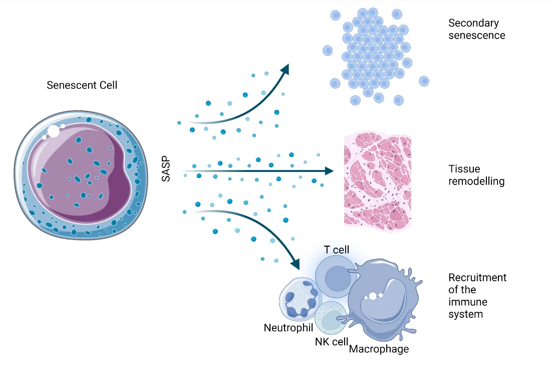
Fig 1. A source diagram reviewed before captioning: SASP factors can propagate secondary senescence, remodel tissue, and recruit neutrophils, natural-killer cells, T cells, and macrophages. These are context-dependent effects, not a quantitative burden assay. Bulbiankova 2023, p. 9. [C][7]
The Fig 1 branching pattern shows why a single visible sign cannot be assigned to a single hallmark: one senescent-cell state can alter neighboring cells, matrix, and immune composition at the same time. [C][7]
> Sources: Fig 1 from the own MEDLIB corpus, Bulbiankova et al. 2023, reviewed from the on-disk image and source article. [C][7]
| Classic trap: | Turning the twelve-hallmark framework into a twelve-item commercial score and assigning each facial sign to exactly one hallmark. |
|---|---|
K3.3 · Cellular senescence, SASP, and skin senotype
| Senotype question | Required evidence domain | False-positive control |
|---|---|---|
| Is arrest stable? | Proliferation plus p16/pRB or p53/p21 context | Exclude reversible quiescence and lineage differentiation. |
| Is persistent damage present? | DDR, telomere-dysfunction foci, or related evidence | Acute injury alone is insufficient. |
| Is cell structure compatible? | Morphology, nuclear architecture, lamin B1, lysosomal state | Large or flat morphology alone is nonspecific. |
| Is metabolism altered? | SA-β-gal, lipofuscin, lysosomal or metabolic readouts | Macrophages and postmitotic cells can confound. |
| Is there a secretory program? | Cell- and inducer-specific cytokines, chemokines, proteases, or growth factors | Plasma mediators do not localize source. |
| Which cell is involved? | Keratinocyte, melanocyte, fibroblast, endothelial, immune, or adnexal identity | A tissue average can hide changing cell mixture. |
| Where is it? | Exact skin site, depth, exposed/protected status, and spatial localization | One biopsy does not quantify whole-face or whole-body burden. |
| Does it matter functionally? | Matrix, pigment, barrier, repair, immune recruitment, or neighboring-cell endpoint | Marker movement alone is not rejuvenation. |
| What state is excluded? | Apoptosis, exhaustion, acute inflammation, wound response, and malignancy as relevant | Senescence is not synonymous with aging or disease. |
Skin-focused evidence and MICSE converge on a contextual multi-marker phenotype; SASP heterogeneity and spatial skin mapping prevent a universal single-marker burden claim. [C][7][A][9][B][10–12]
Senescence identification: use a decision panel, not a marker
| Evidence domain | Compatible findings | What it rules in | What it does not prove |
|---|---|---|---|
| Stable cell-cycle arrest | Reduced proliferation; p16/pRB or p53/p21 pathway evidence; reduced Ki-67/PCNA; low nucleotide incorporation. [A][9][C][7] | Durable arrest within the tested context. | Senescence by itself; quiescence and differentiation require exclusion. |
| Structural/morphological change | Enlarged flattened cells, vacuolation, altered nuclear architecture, lamin B1 loss. [C][7] | A senescence-compatible phenotype. | Specificity across tissues or causes. |
| Lysosomal/metabolic change | SA-β-gal activity, lysosomal expansion, lipofuscin, altered metabolism. [A][9][C][7] | A useful component of a panel. | A universal positive marker; macrophages and other cells can confound. |
| Persistent damage response | γH2AX/53BP1 foci, DNA-SCARS, telomere dysfunction-induced foci. [A][9][C][7] | Persistent damage signaling. | That all damaged cells are senescent. |
| Secretory program | Context-specific cytokines, chemokines, growth factors, proteases, lipids, and extracellular vesicle cargo. [B][10][11] | A SASP-like program. | A fixed universal SASP or the cellular source of plasma cytokines. |
| Anti-apoptotic adaptation | Survival-pathway activity; absence of apoptosis markers in context. [C][7] | Viable arrested-cell adaptation. | Therapeutic target safety or clinical actionability. |
| Tissue localization | Co-localized markers in a defined cell type and anatomical compartment. [A][9][B][12] | Which cells at which site carry the senotype. | Whole-body burden. |
| Functional consequence | Altered matrix production, pigment signaling, repair, immune recruitment, or neighboring-cell behavior. [B][10][12] | Tissue relevance. | Causality without perturbation or longitudinal evidence. |
Ogrodnik 2024 MICSE guidelines emphasize that no marker is specific and broadly applicable enough to identify senescence across tissues and organisms. A credible in-vivo claim specifies context, cell type, marker combination, and exclusions. [A][9]
Senescence is not synonymous with aging
| State | Proliferation | Reversibility | Secretory pattern | Damage | Clinical/research distinction |
|---|---|---|---|---|---|
| Senescence | Stable arrest despite mitogenic context | Usually durable | May develop heterogeneous SASP | Often persistent DDR, but variable | Requires a multi-domain senotype. [A][9][C][7] |
| Quiescence | Arrested | Reversible with stimulus | Not defined by SASP | No required persistent damage | Do not label low proliferation alone as senescence. |
| Terminal differentiation | Lineage-programmed arrest | Usually durable | Lineage-specific | No required DDR | Mature differentiated cells are not automatically senescent. |
| T-cell exhaustion | Functionally altered under chronic stimulation | Variable and context dependent | Immune effector pattern altered | Distinct biology | Do not use p16 or low proliferation without immune context. |
| Apoptosis | Cell-death program | Irreversible | Distinct dying-cell signals | May follow damage | Viable senescent cells resist rather than complete apoptosis. |
| Transient wound senescence | Time-limited arrest and signaling | Resolved by clearance | Can support repair | Injury-linked | Beneficial when appropriately timed. [B][10] |
| Chronic accumulated senescence | Persistent burden and impaired clearance | Sustained | Can promote inflammation and matrix dysfunction | Age/exposure-linked | Potential aging driver, still tissue and cell specific. [B][10,9,11,12] |
Senescence can participate in embryogenesis, wound repair, tumor suppression, fibrosis limitation, and tissue remodeling. Chronic accumulation is the harmful scenario; duration, cell identity, inducer, and clearance determine the net effect. [B][10,9,11][C][7]
Inducers and expected signatures
| Inducer | Common pathway context | Skin relevance | Interpretation trap |
|---|---|---|---|
| Replicative exhaustion | Telomere shortening/dysfunction and DDR | Long-lived proliferative lineages and experimental fibroblast passage | Culture passage is not equivalent to chronological skin aging. |
| UV radiation | DNA damage, ROS, MAPK/AP-1 signaling, mitochondrial injury | Photoexposed keratinocytes, melanocytes, fibroblasts. [C][6,7] | Acute sunburn response labelled permanent senescence. |
| Oncogene activation | Strong arrest with a prominent secretory program | Nevi, premalignant context, experimental models | Tumor biology generalized to normal aged skin. |
| Oxidative stress | Redox signaling, macromolecular damage, mitochondrial feedback | Pollution, UV, inflammation, experimental stress-induced premature senescence | One circulating oxidative marker treated as causal burden. |
| Mitochondrial dysfunction | Altered ATP/redox balance, mitochondrial ROS, metabolic remodeling | Dermal fibroblast and keratinocyte function | Mitochondrial abundance treated as function. |
| Mechanical/ECM stress | Altered cell shape, mechanotransduction, matrix feedback | Fragmented dermal ECM and fibroblast state | Laxity alone used as a molecular marker. |
| Inflammation/infection | Cytokine signaling, immune-cell recruitment, damage | Chronic dermatoses, wounds, local infection | Disease inflammation relabelled as “inflammaging.” |
| Therapy-induced stress | Radiation, cytotoxic or targeted treatment | Cancer history, field injury, wound context | Cosmetic procedure effects assumed identical to oncologic exposure. |
SASP: composition, function, and non-specificity
Coppé 2010 established the SASP as a mechanism by which senescent fibroblasts become pro-inflammatory and alter their microenvironment. [B][11] Later skin-focused evidence shows that composition varies by cell type, inducer, time, tissue, and immune context. [B][10,9][C][7]
| SASP component class | Examples used in research | Potential skin effect | Why plasma measurement is insufficient |
|---|---|---|---|
| Pro-inflammatory cytokines | IL-1 family, IL-6, IL-8, TNF-related signals | Local inflammation, immune recruitment, neighboring-cell effects. [C][7] | Many tissues, infections, adiposity, and acute illness produce the same mediators. |
| Chemokines | Cell-recruitment signals | Neutrophil, macrophage, lymphocyte trafficking | Source and tissue localization are lost in circulation. |
| Matrix proteases | MMP-1, MMP-3, MMP-9 and related enzymes | Collagen/elastin remodeling and basement-membrane effects. [C][6,7] | Proteases reflect multiple inflammatory and repair processes. |
| Growth factors | IGFBP, VEGF, PDGF, HGF families | Repair, vascular response, paracrine state change | A factor may be beneficial or harmful according to phase and tissue. |
| Lipid mediators | Prostaglandin and oxidized-lipid signals | Inflammation and cell-state communication | Preanalytics and systemic metabolism strongly influence levels. |
| Extracellular-vesicle cargo | miRNA and proteins | Fibroblast-keratinocyte-melanocyte cross-talk | Vesicle origin is difficult to assign without tissue methods. |
| DAMP-related signals | Stress-associated molecules | Innate immune activation | Injury, necrosis, infection, and procedure effects overlap. |
Consensus: SASP is heterogeneous; there is no universal SASP panel that quantifies senescent-cell burden in a patient. [A][9][B][11]
Discrepancy: biomarker-discovery view: multi-analyte secretory signatures can support classification and mechanistic studies [B][11] · clinical-caution view: source ambiguity, inflammatory confounding, and changing senotypes prevent a universal circulating diagnostic [A][9] · decide by tissue localization, cell identity, and orthogonal markers.
Skin compartments: the senotype is not uniform
| Cell/compartment | Senescence-compatible changes | Potential phenotype | Required corroboration |
|---|---|---|---|
| Epidermal keratinocyte | Arrest markers, altered differentiation, DDR, barrier-related signaling | Thinning, impaired barrier or repair, field damage | Cell-type-localized panel plus clinical/histological context. |
| Melanocyte | Senescence-linked secretory state and altered pigment signaling | Mottled pigmentation or lentigines | Pigment diagnosis, spatial localization, and melanocyte-specific markers. [B][12] |
| Papillary dermal fibroblast | Altered matrix synthesis, reduced renewal, SASP | Fine texture change, support loss | Dermal layer identification and matrix endpoints. |
| Reticular dermal fibroblast | Decreased collagen/elastin programs and senescence-associated state | Laxity and deeper matrix change | Spatial evidence; Yu 2024 associated reticular fibroblast senescence with reduced matrix synthesis. [B][12] |
| Endothelial/perivascular cells | Arrest, inflammatory and barrier signaling | Vascular fragility or altered repair | Vascular diagnosis and local markers. |
| Macrophage | Age-altered phenotype, impaired clearance, pro-inflammatory signaling | Skin inflammaging and reduced surveillance | Immune phenotyping; macrophage markers cannot be read as senescent-cell markers by default. |
| Hair-follicle niche | Altered stem/progenitor support | Hair aging and reduced regenerative capacity | Follicle-specific evaluation; outside facial-skin inference if not sampled. |
| Adnexal structures | Cell- and gland-specific age changes | Dryness, secretion change | Separate gland and barrier evaluation. |
Yu 2024 used single-cell RNA sequencing and spatial transcriptomics to curate a skin-specific senescence signature. Photoaged skin showed a higher senescent-cell burden than chronological aging; senescent melanocytes aligned with increased melanin synthesis, while senescent reticular fibroblasts aligned with reduced collagen and elastic-fiber synthesis. [B][12] This is tissue-mapping evidence, not a validated office blood test.
Practical senotype panel for research or pathology collaboration
| Panel tier | Minimum elements | Interpretation |
|---|---|---|
| Tier 0: phenotype | Clinical site, exposure, disease, treatment history, repair phenotype | Defines why the tissue is being examined. |
| Tier 1: arrest | At least one arrest-pathway marker plus reduced proliferation | Supports stable cell-cycle exit. |
| Tier 2: damage/structure | DDR or telomere-dysfunction evidence; lamin B1 or morphology where appropriate | Adds persistent stress and structural context. |
| Tier 3: metabolism/lysosome | SA-β-gal, lipofuscin, lysosomal marker, or metabolic change | Supports but does not independently define senescence. |
| Tier 4: secretory state | Cell- and tissue-relevant SASP components | Describes communication phenotype. |
| Tier 5: exclusion | Quiescence, differentiation, apoptosis, immune-cell identity, acute inflammation | Reduces false-positive classification. |
| Tier 6: consequence | Matrix, pigment, repair, barrier, or neighboring-cell endpoint | Connects cell state to tissue function. |
A defensible report states “senescence-compatible multi-marker phenotype in [cell type/site]” rather than “senescence burden” unless burden measurement itself has been validated.
Figure-guided marker reading
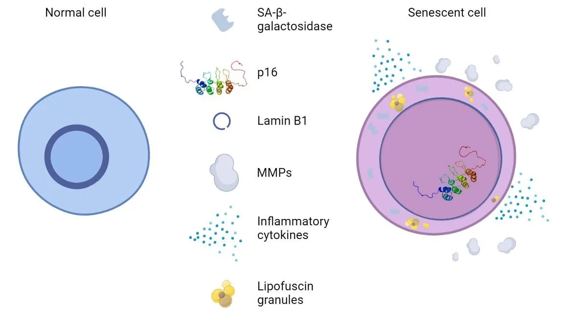
Fig 2. The reviewed diagram contrasts a normal cell with a senescent-cell phenotype: increased SA-β-gal, p16, MMPs, inflammatory cytokines, and lipofuscin with reduced lamin B1. It is a qualitative constellation; none is individually universal. Bulbiankova 2023, p. 15. [C][7]
The Fig 2 constellation should be read vertically: arrest, nuclear structure, lysosomal state, matrix signaling, inflammatory secretion, and accumulated pigment all contribute different information. [A][9][C][7]
> Sources: Fig 2 from the own MEDLIB corpus, Bulbiankova et al. 2023, reviewed from the on-disk image and full source. [C][7]
Chair-side interpretation scenarios
| Scenario | Evidence reading | Action inside K3 | What remains outside K3 |
|---|---|---|---|
| High circulating IL-6 after recent infection | Nonspecific inflammatory signal; not a SASP diagnosis. | Defer novelty testing; use ordinary clinical evaluation. | Treatment of infection/systemic inflammation. |
| p16-positive skin cells in one biopsy | One arrest-pathway marker in one site. | Ask for co-markers, cell identity, morphology, and clinical context. | Any systemic senolytic plan. |
| SA-β-gal positive culture | Compatible lysosomal phenotype under the culture conditions. | Verify arrest, damage, morphology, and controls. | Patient-level burden inference. |
| Elevated lipofuscin fluorescence | Compatible with nondegradable pigment accumulation. | Distinguish cell identity, age pigment, and assay artifacts. | Whole-organ or whole-body aging score. |
| High MMP and low collagen in photoexposed skin | Matrix degradation pattern compatible with photoaging and SASP, but not specific. | Pair with exposure, clinical phenotype, and tissue markers. | Claim of a unique senescence cause. |
| Treatment changes one marker | Target-associated signal only. | Require stable assay, control site, multi-marker concordance, and clinical endpoint. | “Cleared senescent cells” unless directly shown. |
| Marker-positive lesion | Could reflect premalignancy, inflammation, repair, or senescence. | Dermatologic diagnosis takes priority. | Aesthetic procedure until pathology is clarified. |
Longitudinal design controls
- Sample the same site and compartment; exposed and protected skin are not interchangeable. [B][12]
- Record recent UV exposure, inflammation, infection, topical use, and procedures.
- Use identical fixation, storage, staining, imaging, thresholds, and batch strategy.
- Pre-specify the senotype and exclusions; do not select positive markers after viewing results.
- Include cell identity; changing immune-cell infiltration can mimic a changing secretory signature.
- Pair the molecular endpoint with a local functional or structural endpoint.
- Treat small unreplicated changes as indeterminate when assay variation is unknown.
- Preserve raw images and blinded scoring.
(P) If a panel cannot distinguish a changed cell state from a changed cell mixture, it cannot support an individual rejuvenation claim.
Failure signatures
- p16-only diagnosis: p16 is useful but not exclusive to all senescent cells or absent from all non-senescent contexts. [A][9][C][7]
- SA-β-gal-only diagnosis: lysosomal activity can produce false positives in macrophages and postmitotic cells. [C][7]
- SASP-only diagnosis: circulating mediators have many sources.
- Morphology-only diagnosis: large, flat cells are not sufficiently specific.
- Static-autophagy diagnosis: abundance does not equal flux.
- Acute-procedure confusion: transient wound signaling is labelled chronic burden.
- Tissue extrapolation: one punch biopsy becomes whole-face or whole-body burden.
- Therapeutic inversion: a marker panel is used to justify a systemic intervention before clinical utility exists.
| Classic trap: | Calling one p16 or SA-β-gal result “senescent-cell burden,” then treating movement in that marker as proof that the skin or patient was rejuvenated. |
|---|---|
K3.4 · DNA-methylation clocks: target, validation, limits, and aesthetic relevance
| Clock decision | Correct reading | Prohibited conversion |
|---|---|---|
| Horvath | Multi-tissue chronological-age-associated methylation pattern | Facial-skin age or aesthetic outcome. |
| Hannum | Whole-blood chronological methylomic pattern | Universal tissue age. |
| PhenoAge | Methylation surrogate of a phenotypic-age target linked to morbidity and healthspan domains | Literal years added to a tissue. |
| GrimAge | Mortality/healthspan-oriented composite using DNAm surrogates, including smoking exposure | Remaining lifespan or causal damage. |
| DunedinPACE | Rate-like estimate trained from longitudinal multisystem decline | Attained age in years. |
| Reliability | Replicate, batch, specimen, cell-mixture, preprocessing, and version behavior | Decimal precision as proof of analytical precision. |
| Validation | External calibration, construct validity, incremental prediction, longitudinal validity, and utility are separate steps | Population association as individual diagnosis. |
| Aesthetic relevance | Research correlation can pair a locked clock with a validated local skin endpoint | Procedure selection from a clock. |
| Repeat result | Interpret only against assay-specific variation and an independent meaningful endpoint | Lower score as rejuvenation or longevity. |
The foundational clocks operationalize different targets; later method work documents population, tissue, ethical, and technical-reliability limits. They must remain separate axes. [B][13–18][A][19]
Clock comparison at a glance
| Clock | Training target | Original specimen/context | Output meaning | Strong use | Main limitation for aesthetic practice |
|---|---|---|---|---|---|
| Horvath multi-tissue | Chronological age | 8,000 samples, 51 tissues/cell types; 353 CpGs | DNAm age pattern across multiple tissues. [B][13] | Cross-tissue age-associated methylation research | Not an aesthetic outcome or a direct facial-skin measure. |
| Hannum | Chronological age | Whole blood, 656 people aged 19–101; 71 CpGs | Blood methylomic age pattern. [B][14] | Blood-based age association | Cell composition, cohort, and platform constrain transport. |
| DNAm PhenoAge | Phenotypic-age target linked to clinical chemistry and outcomes | Developed from a phenotypic-age composite, then methylation | Morbidity/healthspan-oriented age estimate. [B][15] | Outcome association beyond chronological prediction | “Years” are model units, not observed tissue age or remaining lifespan. |
| DNAm GrimAge | Mortality/healthspan risk target using DNAm surrogates | Blood; DNAm surrogates of plasma proteins and smoking pack-years | Mortality- and morbidity-oriented risk surrogate. [B][16] | Risk prediction in validated populations | Sensitive to smoking and model composition; not proof of causal aging or response. |
| DunedinPACE | Longitudinal pace of multisystem decline | Blood methylation trained on change in 19 organ-system indicators across repeated waves | Pace of aging, not attained age. [B][17] | Longitudinal epidemiology and intervention research | Cannot be read as “biological age in years”; individual action thresholds are absent. |
First-generation versus later-generation targets
| Generation/strategy | Optimization target | What a low error means | What it does not mean |
|---|---|---|---|
| Chronological-age clocks | Predict calendar age | Methylation pattern tracks age in the training/validation data. [B][13,14] | The clock captures morbidity, function, mortality, or treatment response. |
| Phenotypic/mortality clocks | Predict composite phenotype, proteins, smoking exposure, morbidity, or survival | The score carries information about the target in studied populations. [B][15,16] | The score is causal, organ-specific, or directly modifiable. |
| Pace clocks | Reconstruct longitudinal change | One-time methylation estimates a rate-like construct trained from repeated physiological data. [B][17] | The number is attained biological age. |
| Principal-component implementations | Reduce technical noise while preserving clock signal | Replicates and trajectories can become more reliable. [B][18] | The construct becomes clinically validated by improved precision alone. |
| Tissue-specific clocks | Fit a named tissue or cell type | Better alignment within that tissue. | Transfer to blood, saliva, or whole-person outcomes. |
| Commercial proprietary clocks | Varies; may combine multiple models | Only what transparent validation demonstrates. | Equivalence to published clocks, even if names sound similar. |
Clock anatomy: where error enters
collection → cell mixture → DNA extraction → bisulfite conversion → array/sequencing → quality control → normalization → CpG values → algorithm/version → reference cohort → interpretation
| Stage | Failure mode | Direction of distortion | Control |
|---|---|---|---|
| Collection | Acute illness, inflammation, smoking exposure, time or handling differences | Biological state and cell mixture change | Standard operating procedure and contextual record. |
| Specimen | Blood, saliva, buccal, or tissue substituted | Tissue/cell composition changes the methylome | Use the validated specimen. |
| Cell composition | Leukocyte proportions differ | Apparent age acceleration can partly reflect immune composition | Report or adjust composition as validated. |
| DNA quality | Degradation or contamination | Probe failure and noisy methylation values | Laboratory QC thresholds. |
| Bisulfite conversion | Incomplete or variable conversion | Systematic methylation bias | Conversion controls and batch review. |
| Platform | Different array generations or sequencing panels | Missing probes and measurement shifts | Platform-specific validation or justified harmonization. |
| Normalization | Pipeline changed | Reproducible but shifted outputs | Freeze and report preprocessing. |
| Batch | Baseline and follow-up processed separately | Batch appears as biological change | Randomize samples and process paired specimens together where possible. |
| Algorithm | Formula/version changed | Trend discontinuity | Version-lock longitudinal monitoring. |
| Comparator | Different cohort or calibration | Percentile and acceleration change | Use the same reference and disclose its limits. |
| Reporting | Point estimate without interval | False precision | Include repeatability and uncertainty. |
Bell 2019 identifies mechanism, tissue and disease specificity, longitudinal validation, diverse populations, and ethical implications as unresolved clock challenges. [A][19] Higgins-Chen 2022 showed that technical noise could produce deviations up to 9 years between replicates for six clocks; principal-component versions brought most replicate agreement within 1.5 years in their analysis. [B][18] These are method-specific research findings, not universal office tolerances.
The five clock archetypes in clinical language
Horvath
- Measures: a 353-CpG multi-tissue methylation pattern trained to chronological age across 51 tissues and cell types. [B][13]
- Strength: portability across many tissues; foundational cross-tissue research tool.
- Does not measure: aesthetic response, facial-skin age, lifespan, or one organ's function.
- Interpretation: age acceleration is residualized or otherwise defined relative to chronological age; the exact method must be reported.
- Transfer warning: embryonic, stem-cell, cancer, and diseased tissues illustrate that methylation age is biological but context dependent. [B][13]
Hannum
- Measures: a 71-CpG whole-blood model trained in 656 people aged 19–101. [B][14]
- Strength: blood-based age-associated methylation signal.
- Does not measure: a universal tissue age.
- Interpretation: whole-blood cell composition, sex, genetics, cohort, array, and preprocessing matter.
- Transfer warning: claims should remain inside blood and validated populations unless external evidence supports transport.
DNAm PhenoAge
- Measures: a methylation surrogate of a phenotypic-age target designed to capture lifespan and healthspan-related information. [B][15]
- Strength: associations with mortality, cancer, physical function, and other aging outcomes in validation analyses.
- Does not measure: the histological age of facial skin or a procedure's local benefit.
- Interpretation: an “older” value is a model-based risk signal, not years literally added to tissues.
- Transfer warning: a phenotypic target can inherit disease, inflammatory, and population composition effects.
DNAm GrimAge
- Measures: composite of DNAm surrogates for plasma proteins plus a DNAm smoking-pack-years estimator; output is age-like. [B][16]
- Strength: strong mortality and morbidity prediction in large validation datasets.
- Does not measure: causal damage, remaining life, or a direct cellular aging rate.
- Interpretation: AgeAccelGrim is typically adjusted for chronological age; reporting must distinguish raw GrimAge from acceleration.
- Transfer warning: smoking-related signal is integral to the construct, not a nuisance that can be forgotten.
DunedinPACE
- Measures: pace of aging reconstructed from longitudinal change in 19 organ-system indicators across two decades, then distilled into a blood DNA-methylation test. [B][17]
- Strength: test-retest reliability and association with morbidity, disability, mortality, and adversity in validation cohorts.
- Does not measure: attained biological age in years.
- Interpretation: a rate-like score belongs on a pace axis; it must not be subtracted from chronological age.
- Transfer warning: intervention-response use still requires assay stability, pre-specified outcome, and validation in the tested population.
Clock result audit
| Report item | Pass | Fail |
|---|---|---|
| Clock identity | Exact published/proprietary model and version | “Epigenetic age test” |
| Target | Chronological, phenotypic, mortality, pace, tissue, or other | “True age” |
| Specimen | Named and validated | Omitted or substituted |
| Platform | Array/sequencing method and preprocessing | Proprietary without documentation |
| Output | Raw score and defined acceleration/rate calculation | Only persuasive color category |
| Reference | Relevant cohort and calibration | Undefined “optimal” range |
| Precision | Analytical QC and repeatability | One decimal place without error data |
| Longitudinal comparability | Same specimen, platform, version, and processing | Different vendor or model trended as one series |
| Clinical endpoint | Pre-specified and independently measured | Score movement as the only endpoint |
| Actionability | Confirmatory pathway defined | Automatic product or treatment recommendation |
Validation ladder
| Level | Question | Minimum evidence | What remains unproven |
|---|---|---|---|
| 1 | Analytical validity | Accuracy, precision, QC, failed-sample handling | Biological relevance |
| 2 | Test-retest reliability | Same-sample and repeat-sample behavior | Outcome prediction |
| 3 | Internal validity | Cross-validation separated from training | External transport |
| 4 | External validity | Independent cohort, same intended use | Calibration in this patient |
| 5 | Construct validity | Associations behave as predicted against aging domains | Causality |
| 6 | Incremental validity | Adds information beyond age and standard risk factors | Clinical utility |
| 7 | Longitudinal validity | Tracks meaningful within-person change beyond noise | Treatment responsiveness |
| 8 | Surrogacy | Change predicts change in meaningful clinical outcome | Individual treatment benefit unless validated |
| 9 | Clinical utility | Test-guided decisions improve outcomes versus usual care | Broad use outside studied setting |
An association with mortality supports prognostic validity; it does not establish intervention-response validity. Improved repeatability supports precision; it does not create clinical utility. [A][1][19][B][18]
Aesthetic relevance: direct, indirect, and invalid uses
| Use | Status | Required evidence |
|---|---|---|
| Explain that epigenetic alteration is one hallmark | Direct conceptual use | Hallmark framework. [A][4,5] |
| Add a blood clock to a systemic risk discussion | Indirect adjunct | Validated intended use, routine clinical corroboration, and referral pathway. |
| Correlate a clock with standardized skin phenotype in research | Research use | Prospective protocol, skin endpoint, confounder control, and external replication. |
| Monitor a procedure using clock plus local endpoint | Experimental | Stable assay, control site/group, pre-specified clock and skin outcome, change beyond error. |
| Diagnose “aged skin” from blood clock | Invalid | Blood is not facial skin. |
| Choose filler, toxin, device, peel, or surgery from a clock | Invalid | No validated treatment-selection pathway. |
| Prove rejuvenation from a lower clock score | Invalid alone | Requires meaningful concordant endpoint and demonstrated surrogacy. |
| Market systemic longevity from local appearance improvement | Invalid | Local appearance is not lifespan or healthspan. |
Confounder and transport matrix
| Axis | Why it matters | Documentation |
|---|---|---|
| Age range | Models can regress toward the training mean and behave differently at extremes. | Patient age and validation range. |
| Sex | Training composition and methylation patterns may differ. | Sex distribution and sex-specific validation. |
| Ancestry | CpG architecture, exposures, social determinants, and cohort calibration may alter performance. [A][19] | Ancestry representation and external validation. |
| Disease | Disease and treatment can change methylation and immune composition. | Active diagnoses, cancer history, inflammatory state. |
| Smoking | Directly relevant to GrimAge construction. [B][16] | Current and cumulative exposure. |
| Medication | Can influence blood composition or methylation. | Medication history and change between samples. |
| Acute illness | Alters inflammatory and immune-cell profiles. | Defer or annotate collection. |
| Cell mixture | Blood and saliva are mixtures, not stable pure tissues. | Cell estimation or validated adjustment. |
| Menopause/hormonal state | May affect biology and cohort comparability. | Relevant clinical context; treatment remains federated. |
| Weight change | Alters metabolic/inflammatory context. | Record longitudinally. |
| Sleep/exercise/fasting | May introduce short-term state differences. | Standardize as assay documentation permits; no prescription from K3. |
| Laboratory batch | Can mimic longitudinal change. | Paired processing and batch identifier. |
Two-sided clock controversy
Outcome-prediction view: clocks trained on phenotypic or mortality targets and pace measures show reproducible population associations and can support stratification or trial endpoints. [B][15,16,17]
Clinical-diagnostic caution: clocks operationalize different constructs; technical noise, tissue, cell composition, ancestry, disease, calibration, and platform differences prevent interchangeable individual diagnosis. [A][19][B][18]
Decision variable: use a clock only if its construct, specimen, intended use, precision, external validation, and action pathway match the question. Never average different clocks.
Repeat testing adjudication
| Repeat pattern | Plausible explanations | Defensible statement |
|---|---|---|
| Small favorable change | Technical noise, biological variation, cell-mixture shift, true signal | “Change is indeterminate unless it exceeds assay-specific expected variation.” |
| Large favorable change, clinical endpoints unchanged | Batch/version effect, state change, model responsiveness without clinical relevance | “The clock changed; rejuvenation is not established.” |
| Clock stable, local skin improves | Different biological domains | “Local aesthetic endpoint improved without detectable change in this clock.” |
| Two clocks move oppositely | Different targets or reliability | “The models disagree and cannot be averaged.” |
| Same clock changes after platform update | Measurement discontinuity | “Longitudinal series is interrupted.” |
| Pace improves while age clock rises | Pace and attained age are distinct constructs | “No contradiction; interpret each on its own axis.” |
Source figure: what classic clocks actually differ on
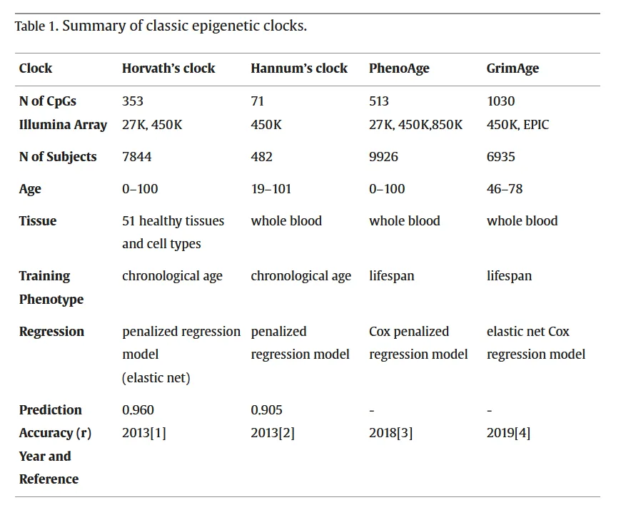
Fig 3. The UPO slide table, reviewed before captioning, contrasts Horvath, Hannum, PhenoAge, and GrimAge across CpG count, array, sample count, age range, tissue, training phenotype, and regression. The slide is a navigation aid; external primary sources govern current interpretation. UPO Sorted, p. 46. [D]
The Fig 3 table makes the non-interchangeability visible: a multi-tissue chronological clock, a blood chronological clock, a phenotypic-age clock, and a mortality-oriented clock were optimized for different targets. [B][13,14,15,16]
> Sources: Fig 3 from UPO Sorted in the own MEDLIB corpus, read on disk; factual interpretation cross-checked against the original clock publications. [B][13,14,15,16][D]
Minimum patient-facing result template
> Assay: [clock/version], [specimen], [platform].
> Construct: [chronological-age pattern / phenotypic risk / mortality surrogate / pace].
> Result: [raw output] with [laboratory-provided precision/repeatability].
> Comparator: [validation cohort and relevant limits].
> Concordance: [routine clinical findings and local skin endpoints].
> Interpretation: [consistent / discordant / indeterminate] for the stated construct.
> Not established: tissue age outside the specimen, lifespan change, procedure selection, or rejuvenation.
> Action: [none / standard confirmation / referral / standardized repeat if decision-relevant].
(P) The clock name should never be allowed to do more interpretive work than its training target.
| Classic trap: | Comparing Horvath, GrimAge, and DunedinPACE as if all reported the same “age,” then treating a lower repeat value as proof of facial or systemic rejuvenation. |
|---|---|
K3.5 · Biomarkers beyond clocks: telomeres, inflammaging, immune age, and multi-omics
| Family | Minimum interpretation | Non-interchangeability rule |
|---|---|---|
| Telomeres | Name tissue/cell mixture, method, unit, and whether mean, distribution, or shortest telomeres are measured. | A leukocyte value is not whole-person age or remaining lifespan. |
| Inflammaging | Interpret a composite inflammatory context after excluding acute and ordinary disease causes. | One cytokine is not an aging diagnosis. |
| Immune age | Combine composition, function, repertoire, latent-infection context, and longitudinal design where validated. | A cell-count shift is not necessarily changed immune aging. |
| Proteomics | Preserve platform, nonlinear life-course pattern, organ function, medication, and inflammatory confounding. | Protein age is not DNAm age. |
| Metabolomics | Standardize diet, fasting state, circadian timing, drugs, processing, and renal/hepatic context. | Dynamic metabolic state is not a stable universal trait. |
| Glycomics | State glycan species, platform, population, and inflammatory/hormonal context. | IgG glycan age is not a universal clock. |
| Composite clinical age | Expose each component and model coefficient. | Disease burden cannot be hidden inside persuasive age units. |
| Multi-omics | Validate feature harmonization, missing-data handling, calibration, and incremental utility. | More features do not automatically create clinical actionability. |
| Longitudinal use | Keep specimen, method, laboratory, model, version, and comparator fixed. | Different assays cannot be averaged into a consensus age. |
Telomere methods, inflammaging, longitudinal immune trajectories, proteomic waves, glycan patterns, omics clocks, and exercise-telomere evidence support several partly shared but distinct domains. [A][20][B][21–25][C][26]
At-a-glance panel
| Biomarker family | Specimen/readout | What it captures | Main strength | Main limitation |
|---|---|---|---|---|
| Telomere length | Leukocyte or tissue mean/distribution; method-specific | Chromosome-end property and replicative/damage history | Established aging and telomere-disorder biology | High interindividual, cell-type, and assay variation; no stand-alone age diagnosis. [A][20] |
| Inflammatory markers | CRP, IL-6 and other mediators | Current and chronic inflammatory burden | Clinically familiar context and outcome associations | Nonspecific; infection, obesity, disease, trauma, and treatment confound. [B][21] |
| Immune composition/function | Cell subsets, signaling, repertoire, response | Immunosenescence and immune-system trajectory | Mechanistic multidimensional view | Platform complexity, state sensitivity, and limited routine standardization. [B][22] |
| Proteomic age | Multi-protein panel | Circulating pathways and nonlinear life-course change | Broad pathway coverage | Platform, cohort, renal/hepatic/inflammatory confounding. [B][23] |
| Metabolomic age | Metabolite panel | Integrated metabolic state and exposures | Dynamic systemic phenotype | Diet, fasting, drugs, time, handling, and platform dominate. [A][3][24] |
| Glycan age | IgG or other glycomic features | Immune-inflammatory glycosylation patterns | Strong age association in studied cohorts | Platform-specific, population-specific, not a universal clock. [B][25] |
| Composite clinical age | Routine measures combined by algorithm | Multisystem physiological state | Feasible and interpretable components | Disease burden may dominate; model-specific output. [A][2,3] |
| Multi-omics age | Combined methylation/protein/metabolite/glycan/etc. | Multiple partially shared aging domains | Potentially broader coverage | Overfitting, missing-data, harmonization, cost, and transport. [B][24] |
Telomere length: what the number contains
| Dimension | Alternatives | Consequence for interpretation |
|---|---|---|
| Specimen | Whole blood, sorted leukocytes, saliva/buccal, skin, muscle, other tissue | Tissues and cell mixtures have different length distributions and turnover. |
| Statistic | Mean length, shortest telomeres, distribution, chromosome-specific length | Mean length can hide a clinically or biologically relevant short tail. [A][20] |
| Method | qPCR, Southern blot/TRF, Q-FISH, Flow-FISH, STELA/TeSLA, sequencing/computational | Outputs, precision, throughput, DNA requirements, and comparability differ. [A][20] |
| Unit | Relative T/S ratio, base-pair estimate, fluorescence or distribution metric | Units are not interchangeable across methods or laboratories. |
| Cell composition | Mixed leukocytes versus sorted cells | Shifts in neutrophils/lymphocytes can look like longitudinal telomere change. |
| Time | Cross-sectional versus repeat | True attrition is small relative to some assay and biological variation. |
| Disease context | Telomere biology disorder versus general aging | Diagnostic utility is established in specific disorders, not universal aging. [A][20] |
Ferrer 2023 reviewed probe hybridization, qPCR, and sequencing/computational methods and concluded that a test combining accuracy, simplicity, and scalability remains lacking. [A][20] Schellnegger 2022 found that most exercise-telomere studies used leukocyte qPCR, while randomized trials were inconsistent and tissue/cell specificity limited causal interpretation. [C][26]
Telomere methods: choose by the question
| Question | Preferred property | Suitable method family | Limitation to state |
|---|---|---|---|
| Population association | High throughput, low DNA requirement | qPCR or scalable sequencing estimate | Relative measure and inter-lab variation. |
| Absolute mean length | Calibrated length estimate | TRF/Southern or validated calibrated method | DNA requirement and subtelomeric contribution. |
| Cell-specific clinical evaluation | Single-cell/cell-subset resolution | Flow-FISH or Q-FISH | Specialized laboratory and reference data. |
| Shortest-telomere burden | Distribution-tail sensitivity | TeSLA/STELA-type approaches | Low throughput and specialized analysis. |
| Chromosome-specific question | Locus resolution | STELA or emerging long-read methods | Coverage and scalability. |
| Longitudinal trend | High repeatability and stable cell composition | Same validated method, laboratory, specimen, and processing | Small change may remain below measurement certainty. |
Telomere result audit
- Report specimen and whether cells were sorted.
- Report method, calibration, unit, QC, and laboratory reference.
- State whether the result is a mean, relative ratio, distribution, or shortest-telomere measure.
- Record acute illness and cell-count context.
- Do not compare values across methods or laboratories as one series.
- Do not translate a percentile into years of life.
- Do not infer skin telomeres from leukocytes.
- Refer suspected telomere biology disorder through the appropriate specialty; that is a disease-diagnostic pathway, not a longevity service.
Consensus: telomere attrition is a hallmark and telomere length is biologically meaningful, but one leukocyte value is not a definitive whole-person biological age. [A][4,5][20]
Discrepancy: epidemiologic view: telomere measures associate with aging and disease in populations [A][3][20] · clinical-caution view: inherited baseline, cell mixture, tissue, method, and within-person variation limit individual age assignment [A][20][C][26] · decide by disease indication, method, and repeatability.
Inflammaging: marker bundle, not a solitary cytokine
Franceschi 2000 described inflammaging as a progressive pro-inflammatory state arising from lifelong antigenic and stress load, with macrophages central to the network. [B][21]
| Layer | Candidate readouts | Interpretation | Confounders/exclusions |
|---|---|---|---|
| Acute-phase | CRP and related proteins | Systemic inflammatory state | Acute infection, trauma, procedure, obesity, liver state. |
| Cytokine | IL-6, IL-1 family, TNF-related signals | Pathway activity; not source specific | Autoimmune disease, malignancy, infection, medication. |
| Myeloid | Monocyte/macrophage phenotypes | Innate immune aging and inflammatory programming | Recent infection, steroids, sample handling. |
| Lymphoid | Naive/memory balance, exhausted/senescent-like phenotypes | Adaptive immune remodeling | CMV/EBV history, vaccination, acute stress, immunosuppression. |
| Coagulation/vascular | Fibrinogen and endothelial-linked markers | Inflammation-risk interface | Cardiovascular and metabolic disease. |
| SASP-related | Multi-protein signatures | Possible senescence-associated communication | Many non-senescent sources; no tissue localization. |
| Skin-local | Tissue cytokines, macrophage state, barrier/inflammatory phenotype | Local inflammaging context | Rosacea, dermatitis, infection, UV injury, recent procedure. |
A high inflammatory marker is first a clinical inflammation problem, not an aging diagnosis. Routine differential diagnosis precedes novelty scoring.
Immunosenescence: composition plus function
| Domain | Readout examples | Why one component is insufficient |
|---|---|---|
| Cell composition | Naive, memory, effector, regulatory, myeloid and lymphoid subsets | Composition changes with infection, stress, medication, and age. |
| Repertoire | T- and B-cell diversity | Diversity does not directly report effector function. |
| Signaling | Response to stimulation, cytokine production | Ex-vivo conditions and timing alter results. |
| Latent infection | CMV/EBV serostatus or related context | Chronic antigen exposure can dominate age associations. |
| Functional response | Vaccine or challenge response in research | Requires prospective standardized testing. |
| Multi-omics trajectory | Integrated longitudinal immune state | High-dimensional models need external calibration. |
Alpert 2019 followed 135 healthy adults longitudinally for nine years and derived IMM-AGE from high-dimensional immune trajectories; the score predicted all-cause mortality beyond established risk factors in a separate cohort. [B][22] This supports prognostic research utility, not a universal office reference range or direct treatment algorithm.
Glycan, proteomic, and metabolomic panels
IgG glycan age
Krištić 2014 analyzed 5,117 individuals from four European populations. A combination of three IgG glycans explained up to 58% of chronological-age variance, and residual variation correlated with physiological parameters. [B][25] The biological link to inflammation is plausible because IgG glycosylation alters immune function.
Limits: European-cohort transport, laboratory platform, batch, sex and hormonal context, inflammation, and lack of universal individual thresholds. A glycan age is not interchangeable with DNAm age.
Proteomic age
Lehallier 2019 measured 2,925 plasma proteins in 4,263 people aged 18–95 and identified nonlinear waves of proteomic change in the fourth, seventh, and eighth decades. [B][23]
Limits: the proteome reflects kidney and liver function, inflammation, medications, disease, hydration, and sample handling. Nonlinearity makes a single universal linear “protein age” particularly fragile.
Metabolomic age
Metabolomic panels integrate endogenous metabolism, microbiome products, diet, fasting, circadian state, medications, renal/hepatic function, and preanalytics. Jylhävä 2017 and Macdonald-Dunlop 2022 place metabolomic clocks among several partially overlapping predictor families. [A][3][B][24]
Limits: a dynamic metabolome can be valuable for state monitoring but may be less stable as a trait marker unless collection and processing are tightly standardized.
Omics clocks are partly shared and partly distinct
Macdonald-Dunlop 2022 compared fifteen omics aging clocks in about 1,000 participants; correlations with chronological age ranged from 0.21 to 0.97, and age-acceleration measures often associated with health. Epigenetic and IgG glycomic clocks appeared to track more generalized aging, while other clocks captured specific risks. [B][24]
| Observation | Correct interpretation | Incorrect interpretation |
|---|---|---|
| Two clocks correlate strongly | They share age-related signal in the studied cohort. | They measure the same biology. |
| One clock predicts one disease better | Its features/target may capture that risk domain. | It is the best universal age test. |
| Multi-omics model has higher accuracy | More features improve model fit or target prediction. | Individual clinical utility is proven. |
| Clock acceleration associates with outcome | The residual carries prognostic information. | Changing the residual will change the outcome. |
| Different clocks disagree | Distinct constructs, platforms, or calibration. | One must be wrong, so average them. |
Composite panel design
| Design choice | Preferred approach | Failure mode |
|---|---|---|
| Construct | Define function, morbidity risk, mortality risk, pace, or organ state | Mixing endpoints into “biological age” without weights. |
| Marker selection | Biological plausibility plus reproducible outcome association | Selecting only strongest results in the development cohort. |
| Preanalytics | Standardized collection, processing, storage, and timing | State noise becomes age signal. |
| Missing data | Pre-specified handling validated externally | Vendor silently imputes values. |
| Scaling | Platform- and cohort-specific normalization | Cross-lab comparison without harmonization. |
| Model | Lock coefficients and version | Continuous proprietary updates break trends. |
| Validation | Independent cohort relevant to intended use | Cross-validation alone. |
| Calibration | Predicted versus observed risk across range | Good discrimination but misleading absolute risk. |
| Incremental value | Compare against age and standard clinical predictors | Novelty replaces rather than improves standard care. |
| Utility | Test-guided pathway improves a meaningful outcome | Attractive dashboard without decision benefit. |
Practical ordering hierarchy
| Tier | Measure | Rationale | K3 action |
|---|---|---|---|
| 1 | History, examination, standard risk and disease assessment | Established clinical utility | Complete first. |
| 2 | Validated skin phenotype/function | Directly answers aesthetic question | Standardize and trend. |
| 3 | Targeted standard laboratory test for a clinical indication | Actionable reference intervals and pathways | Interpret or refer conventionally. |
| 4 | Qualified aging biomarker with a defined construct | Adjunctive or research information | Use only with uncertainty and no automatic treatment. |
| 5 | Multi-omics commercial panel | Highest complexity and transport risk | Research-grade caution; require full documentation. |
Discordance matrix
| Pattern | Likely explanation classes | Action |
|---|---|---|
| Short telomeres, favorable DNAm clock | Inherited baseline, method/cell mixture, distinct constructs | Do not average; assess clinical indication and assay validity. |
| High inflammation, favorable omics age | Acute state or model weighting | Evaluate inflammation clinically; novelty score does not overrule. |
| Older proteomic age, normal routine labs | Panel captures subtle risk or platform noise | Check validation, renal/hepatic context, repeatability, and actionability. |
| Younger glycan age, poor healing | Different biological domains | Investigate standard causes of poor healing. |
| Immune-age acceleration after infection | Transient immune remodeling | Defer age interpretation until clinically stable if repeat is decision-relevant. |
| Multi-omics improvement without functional change | Surrogate movement or technical variation | Do not claim healthspan improvement. |
Corpus controversy: exercise and telomeres
The own-corpus systematic review included 43 studies: 8 randomized trials, 27 observational studies, and 8 interventional studies. Randomized trials were inconsistent; 33 studies reported a positive association under at least one analysis, while methods and populations were heterogeneous. Most used leukocyte qPCR. [C][26]
Discrepancy: observational/athlete studies often report longer telomeres with activity [C][26] · randomized and longitudinal evidence is inconsistent, and intense acute exercise can produce different short-term signals [C][26] · decide: telomere testing does not prescribe exercise here; systemic lifestyle intervention is federated.
Reporting template for a non-DNAm biomarker
> Family: [telomere / inflammatory / immune / proteomic / metabolomic / glycan / composite].
> Construct: [defined target].
> Specimen and preanalytics: [details].
> Platform/version: [details].
> Output and unit: [raw, percentile, acceleration, distribution].
> Reference: [assay- and population-specific].
> Confounders: [disease, acute state, cell mixture, medication, exposure].
> Concordance: [standard clinical and skin-specific findings].
> Clinical status: [research / adjunct / established disease test].
> Action: [none / standard confirmation / referral].
(P) A panel becomes less interpretable, not more, when heterogeneous outputs are compressed into one persuasive number without a declared target.
| Classic trap: | Treating one leukocyte telomere, CRP, glycan-age, or multi-omics result as the definitive age of the whole person, then comparing it directly with an epigenetic clock. |
|---|---|
K3.6 · Mitochondrial, oxidative, and metabolic readouts
| Measurement tier | Question answered | Boundary |
|---|---|---|
| Cellular respiration | How oxygen consumption behaves under a defined assay sequence | Ex-vivo cellular capacity, not whole-body age. |
| ATP-linked/coupling measures | How respiration relates to ATP synthesis under the protocol | Formula and non-mitochondrial contributions must be stated. |
| Membrane potential | Electrochemical state under a defined probe and cell condition | Viability and dye behavior can dominate. |
| Respiratory complexes | Activity of named enzymes or complexes | One complex does not summarize the organelle. |
| mtDNA | Copy number, damage, deletion, or heteroplasmy | Structural/genetic property is not bioenergetic function. |
| Mass/morphology | Abundance, network, fusion/fission, or ultrastructure | Compensation can increase abundance while function falls. |
| Mitochondrial ROS | Compartment-linked redox probe signal | ROS has signaling and damage roles; probe specificity matters. |
| Circulating oxidation products | Formation/repair/clearance balance for lipid, DNA, or protein products | Source cannot be assigned to skin or mitochondria. |
| Composite index | Formula-specific aggregation of component readouts | No universal mitochondrial age or interval exists. |
Human mitochondrial methods require specimen, processing, normalization, and functional/structural separation; UPO's oxidative-marker discordance reinforces the source and clearance problem. [A][27–28][D][29][B][30]
Directness ladder
| Level | Readout | What it measures | Interpretation |
|---|---|---|---|
| 1 | Oxygen-consumption/respirometry profile | Functional oxidative phosphorylation under defined substrates and perturbations | Most direct functional tier, but ex-vivo and specimen dependent. [A][27][28] |
| 2 | ATP production and coupling | Energetic output and efficiency | Requires normalization and context; ATP has non-mitochondrial contributions. [A][27] |
| 3 | Membrane potential | Electrochemical gradient | Functional state proxy; dye, cell type, and viability affect result. [A][28] |
| 4 | Respiratory-complex activity | Enzyme or complex-specific capacity | Identifies pathway limitation but not whole-cell performance alone. [A][28] |
| 5 | mtDNA quantity, damage, deletion, heteroplasmy | Structural/genetic mitochondrial features | Structure and copy number do not equal function. [A][28] |
| 6 | Mitochondrial mass/morphology/dynamics | Abundance, network, fusion/fission, ultrastructure | Compensatory abundance can coexist with dysfunction. [C][7] |
| 7 | Mitochondrial ROS/redox probes | Compartment-linked redox signal | Probe chemistry and handling matter; ROS is also physiological signaling. [A][28] |
| 8 | Circulating oxidative products | Lipid, protein, or nucleic-acid oxidation products | Indirect systemic burden; source and clearance are ambiguous. [D][29] |
| 9 | Clinical metabolic proxies | Glucose/lipid/fitness and organ-function measures | Clinically actionable domains, but not a mitochondrial biological age. |
Acin-Perez 2021 reviews skeletal muscle as a reference tissue for many bioenergetic questions while highlighting circulating white cells and platelets as more accessible alternatives. Sample size, immediate processing, multi-day collection, and site differences constrain human bioenergetic studies. [A][27]
What a functional assay actually needs
| Element | Required specification | Why |
|---|---|---|
| Specimen | Muscle, PBMC, isolated cell subset, platelet, fibroblast, skin biopsy, or other | Mitochondrial phenotype is tissue and cell specific. |
| Cell state | Viability, activation, confluence, passage, differentiation | Cell state can dominate respiration. |
| Processing interval | Time from collection to assay and temperature | Respiration changes rapidly ex vivo. |
| Substrate/medium | Defined assay environment | Determines which pathway is tested. |
| Perturbation sequence | Basal, ATP-linked, maximal, reserve, non-mitochondrial components as appropriate | Decomposes the oxygen-consumption signal. |
| Normalization | Cell number, protein, DNA, mitochondrial mass, or another justified denominator | Different denominators answer different questions. |
| Replicates/QC | Technical replicates, plate controls, exclusion thresholds | Prevents plate or well effects from becoming biology. |
| Batch strategy | Baseline/follow-up placement and calibration | Longitudinal drift can mimic response. |
| Reference | Matched laboratory and cell-type comparator | No universal reference across tissues/platforms. |
| Clinical endpoint | Functional, disease, or skin endpoint | Assay change alone is not healthspan or aesthetic benefit. |
Respirometry terms without over-claiming
| Term | Operational meaning | Common error |
|---|---|---|
| Basal respiration | Oxygen consumption under baseline assay conditions | Assuming all basal oxygen use is ATP production. |
| ATP-linked respiration | Component attributed to ATP synthesis under the assay model | Calling it whole-body energy production. |
| Proton leak | Non-ATP-linked mitochondrial respiration after defined perturbation | Automatically labelling all leak pathological. |
| Maximal respiration | Capacity under uncoupled or maximally stimulated conditions | Treating ex-vivo maximum as exercise capacity. |
| Spare/reserve capacity | Difference or ratio between maximal and basal, method dependent | Comparing across protocols without harmonization. |
| Non-mitochondrial respiration | Residual oxygen consumption after respiratory-chain inhibition | Ignoring it during calculation. |
| Coupling efficiency | Fraction or relation assigned to ATP-linked activity | Using it without reporting formula and conditions. |
Structural versus functional disagreement
| Finding | Possible reading | What is not established |
|---|---|---|
| High mtDNA copy number + low respiration | Compensation, altered cell mixture, impaired quality, assay issue | “More mitochondria” as better function. |
| Low mtDNA copy number + preserved respiration | Efficient network, cell-type effect, normalization issue | Mitochondrial aging from copy number alone. |
| High mitochondrial mass + low membrane potential | Accumulated dysfunctional organelles or altered dynamics | Functional reserve. |
| High ROS + normal respiration | Signaling, inflammatory state, probe artifact, non-mitochondrial source | Mitochondrial failure. |
| Low ROS + low respiration | Reduced electron flow, excessive scavenging, assay artifact | Healthy redox state. |
| Abnormal morphology + normal function | Compensated structure or sampling limitation | Clinical dysfunction. |
| Normal blood-cell function + skin phenotype | Tissue discordance | Normal dermal fibroblast mitochondria. |
Koklesova 2022 discusses composite Mitochondrial Health Index and Bioenergetic Health Index approaches. [B][30] These indices can organize several readouts but remain specimen-, formula-, and context-specific; they are not a standardized mitochondrial age.
Oxidative stress: flux and balance, not “free-radical level”
| Domain | Candidate measures | What changes the result | Clinical caveat |
|---|---|---|---|
| Lipid oxidation | F2-isoprostanes, lipid hydroperoxides, aldehydes | Diet, collection, storage, renal clearance, acute stress | Different products need different methods. |
| DNA oxidation | 8-OHdG and related lesions | Damage formation plus repair and excretion | A high circulating/urinary value may reflect formation, repair, or clearance. [D] |
| Protein oxidation | Carbonyls and modified amino acids | Circulating versus tissue source, turnover | Serum may not represent solid-tissue oxidation. [D][29] |
| Antioxidant enzymes | SOD, catalase, glutathione-related activities | Compartment, induction, nutritional and inflammatory state | Higher activity can mean adaptation to higher stress. |
| Redox couples | GSH/GSSG and related systems | Rapid handling artifacts and compartment differences | Blood ratio is not skin mitochondrial redox state. |
| Direct probes | Superoxide or hydrogen-peroxide-sensitive systems | Specificity, photochemistry, loading, cell viability | Probe signal is method-defined, not a universal ROS unit. |
| Damage panel | Multi-marker oxidative signature | Each marker has separate kinetics and clearance | Do not average unvalidated markers. |
The UPO marker lecture explicitly shows poor agreement across serum and urine oxidative markers and asks whether serum measures represent tissue oxidation. It notes that serum protein carbonyls may reflect the circulation rather than brain, liver, kidney, or muscle, and that 8-OHdG is influenced by oxidation, repair, and excretion. [D] These teaching cautions remain useful; UPO numeric wellness protocols are not adopted without external validation.
Redox is not linearly bad
- Low-level ROS participates in physiological signaling.
- Excess, sustained, or poorly compartmentalized ROS can damage DNA, proteins, lipids, and mitochondria.
- Antioxidant responses can rise with stress; a high enzyme value is not automatically favorable.
- A low damage product can reflect low generation, low repair/excretion, or analytical failure.
- A one-time blood or urine marker cannot localize source to skin or mitochondria.
- Recent exercise, infection, UV exposure, procedures, smoking, and diet can shift results.
- Oxidative damage is downstream of several processes and does not identify one hallmark cause.
Bulbiankova 2023 describes a nonlinear skin-senescence model: low ROS supports signaling and homeostasis, intermediate stress activates adaptive and inflammatory pathways, and severe stress can produce widespread damage and death. [C][7] The model is mechanistic, not an office reference curve.
Mitochondrial readouts in facial skin
| Question | Direct measurement | Indirect measurement | Not acceptable |
|---|---|---|---|
| Are dermal fibroblast mitochondria functionally impaired? | Fresh or appropriately handled skin/fibroblast functional assay | Local mtDNA, membrane potential, redox, morphology panel | PBMC result alone. |
| Did photoexposure alter mitochondrial biology? | Paired exposed/protected skin with local readouts | UV history plus skin mtDNA/damage markers | One systemic oxidative marker. |
| Did a procedure change mitochondrial function? | Controlled local before/after functional assay with timing and tissue controls | Local molecular proxy plus clinical endpoint | Serum “mitochondrial age.” |
| Does the patient have mitochondrial disease? | Specialist diagnostic pathway | Standard clinical work-up | Aesthetic wellness panel. |
| Is visible aging mitochondrial? | No single definitive test | Multi-hallmark local assessment | Causal claim from ROS. |
Skin-specific confounding
| Confounder | Effect |
|---|---|
| UV exposure | Acute and chronic DNA, redox, inflammatory, and mtDNA effects. [C][6,7] |
| Biopsy site/depth | Changes cell populations and metabolic demand. |
| Local anesthetic/vasoconstrictor | May affect tissue physiology and downstream assays. |
| Recent device/peel/injection | Wound response, inflammation, altered cell mixture. |
| Cell culture | Passage, oxygen, confluence, serum, substrate, and freezing alter phenotype. |
| Age and phototype | Change baseline biology and comparator relevance. |
| Smoking/pollution | Systemic and local redox effects. |
| Active dermatosis | Inflammation can dominate the signal. |
| Sample delay | Rapid loss of bioenergetic validity. [A][27] |
Figure: respiratory-chain context
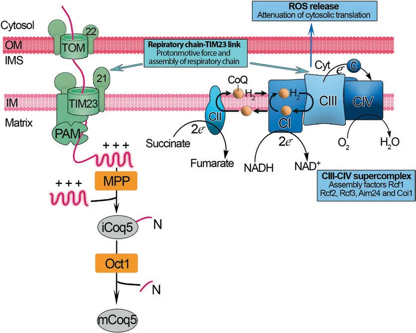
Fig 4. The reviewed source diagram shows respiratory-chain complexes, coenzyme Q, cytochrome c, oxygen reduction, protein-import machinery, and a ROS-release branch. It is used as an orientation map, not as evidence that one circulating oxidative marker measures respiratory-chain function. Tesfaye 2023, p. 307. [C]
The Fig 4 architecture explains why direct respiration, complex activity, membrane potential, mtDNA, and ROS are complementary rather than substitutable measurements. [A][27][28]
> Sources: Fig 4 from the own MEDLIB corpus, Tesfaye 2023, reviewed from the on-disk image; interpretation anchored to human mitochondrial-method reviews. [A][27][28]
Practical interpretation grid
| Result | First question | Confirmatory route | Claim ceiling |
|---|---|---|---|
| Low PBMC reserve capacity | Was collection/processing valid, and is cell composition known? | Repeat under protocol or specialist/research laboratory | PBMC bioenergetic finding only. |
| High urinary oxidative marker | Is this formation, repair, excretion, diet, or renal context? | Standard clinical context and validated assay repeat if decision-relevant | Systemic marker elevation, not skin aging. |
| High mtDNA copy number | Which cells, denominator, and function? | Pair with respiration or other functional measure | Structural/genetic proxy. |
| Low membrane potential | Is viability and dye protocol valid? | Orthogonal functional readout | Assay-specific cellular finding. |
| High MHI/BHI | Which formula, specimen, and reference? | Inspect component values and external validation | Index-specific result. |
| Improved ROS panel after procedure | Were timing, UV, inflammation, and batch controlled? | Local clinical endpoint and replicated assay | Association, not rejuvenation. |
No universal mitochondrial reference interval
Mitochondrial assays use different cells, substrates, perturbations, normalization methods, instruments, formulas, and reference cohorts. Sharma 2025 reviews cellular bioenergetics, mtDNA, membrane potential, mitochondrial ROS, enzyme assays, and emerging PET approaches, underscoring methodological heterogeneity. [A][28] Therefore:
- use the laboratory's validated, specimen-specific interval or comparator;
- do not import cut-offs from another instrument, cell type, or protocol;
- do not convert oxygen-consumption values into “mitochondrial age” without a validated model;
- do not trend values across laboratories as one series;
- report raw components before a composite index;
- route disease-suggestive findings to the relevant medical specialty.
Consensus: mitochondrial function requires functional and structural context; no single proxy can substitute for the panel. [A][27,30,28]
Discrepancy: translational view: blood-cell bioenergetics and composite indices offer accessible systemic phenotyping [A][27][B][30] · tissue-specific caution: cell state, processing, and organ heterogeneity limit inference to facial skin or whole-body aging [A][27][28] · decide by specimen, directness, and intended use.
(P) If the assay can be normalized three plausible ways and each changes the ranking, the result is a method-development finding rather than a patient diagnosis.
| Classic trap: | Calling one ROS, 8-OHdG, mtDNA-copy-number, or blood-cell respiration result “mitochondrial biological age,” then using it to explain facial aging. |
|---|---|
K3.7 · Longevity diagnostics and screening in practice
| Practice gate | Pass condition | Stop condition |
|---|---|---|
| Clinical problem | A named symptom, phenotype, risk, healing concern, or research question | Generic desire for a “full panel.” |
| Standard care | History, examination, and indicated ordinary diagnostics completed first | Novelty testing would delay disease evaluation. |
| Construct | Test target is explicit and matches the question | Vendor says only “true biological age.” |
| Analytical validity | Specimen, platform, version, QC, and repeatability documented | Black-box result or failed QC. |
| Comparator | Assay- and population-specific calibration is relevant | Universal or cross-platform range. |
| Clinical validity | External association and calibration match intended use | Evidence comes from another tissue or outcome. |
| Utility | A result can lead to no action, confirmation, standardized repeat, or referral | Automatic product or treatment bundle. |
| Longitudinal use | Change exceeds expected variation and aligns with an independent endpoint | Score movement alone. |
| Communication | Report distinguishes measurement, association, diagnosis, response, and unproven claims | Years gained, lifespan, or all-hallmark reversal. |
| Hand-off | Systemic treatment requests go to Medicina Integrativa or the relevant specialty | Aesthetic clinic prescribes from the score. |
Current biomarker, clock-method, and biological-age reviews support a decision-first workflow; UPO contributes curriculum context, while current claim frameworks do not create product-level proof. [A][1–3][B][18][A][19][31][D][32][B][33]
Practical diagnostic sequence
| Step | Required output | Proceed only if |
|---|---|---|
| 1. Define the concern | Visible aging, poor healing, systemic symptom, family history, commercial-score question, or research monitoring | The concern is clinically clear. |
| 2. Exclude urgent/standard disease | History, examination, and indicated standard diagnostics | Standard care is not delayed. |
| 3. Define the biomarker construct | Chronological pattern, risk, pace, organ proxy, senotype, telomere, mitochondrial, or multi-omics | The assay documentation names it. |
| 4. Check intended use | Screening, prognosis, stratification, monitoring, research | Use matches validation. |
| 5. Verify specimen/platform | Specimen, preanalytics, laboratory, version, QC | All are documented. |
| 6. Identify comparator | Population and assay-specific interval/percentile | Comparator resembles the patient enough to interpret. |
| 7. Estimate uncertainty | Technical and biological variation, confounders, test-retest data | Change can be classified as signal, noise, or indeterminate. |
| 8. Seek corroboration | Routine clinical and skin-specific measures | At least one independent meaningful domain is available. |
| 9. Decide action | No action, standard confirmation, standardized repeat, or referral | Action does not depend on an unsupported surrogate. |
| 10. Communicate limits | Explicit “not established” statements | No longevity or rejuvenation over-claim remains. |
What to measure in an aesthetic-longevity intake
| Domain | First-line clinical measurement | Optional research/adjunct layer | Do not substitute |
|---|---|---|---|
| Skin phenotype | Standardized photographs, validated severity scales, examination | 3D topography, elasticity, barrier or color measurements | Blood biological-age score. |
| Healing/safety | Wound history, infection history, scarring, medication and disease review | Targeted standard labs if clinically indicated | Senescence or cytokine panel as clearance. |
| Cardiometabolic risk | Standard medical history, blood pressure, anthropometrics, clinically indicated laboratory assessment | Qualified composite aging biomarker | Proprietary age score for established risk thresholds. |
| Inflammatory context | Symptoms, examination, ordinary clinical evaluation | Targeted validated inflammatory markers | “Inflammaging age” as a diagnosis. |
| Functional reserve | Clinically appropriate function/frailty/performance assessment | Research composite age | Methylation clock as functional test. |
| Cognitive or neurologic concern | Standard screening/referral pathway | Research biomarker only inside appropriate setting | Commercial age dashboard. |
| Endocrine concern | Symptom-driven standard diagnostic pathway | None from K3 | Hormone optimization based on age score. |
| Molecular aging | None is universally required | One qualified assay with explicit intended use | Multi-test bundle without a decision pathway. |
| Skin molecular state | Clinical indication or research protocol | Site-specific tissue panel | Saliva or blood as facial-skin tissue. |
Reference-range rules
Biological-age outputs do not share universal clinical reference intervals. Mathur 2024 reviews multiple methods and notes that different approaches can yield different results; no consensus identifies one best measurement. [A][31]
| Output type | Correct reference | Incorrect reference |
|---|---|---|
| Routine clinical analyte | Current accredited laboratory interval plus disease-specific clinical thresholds | A longevity vendor's “optimal” band without validated outcome utility. |
| Epigenetic age | Exact clock's validation/calibration population and defined acceleration method | Another clock's age or percentile. |
| Pace measure | Exact model distribution and units | Years of biological age. |
| Telomere result | Laboratory, method, specimen, age/cell-specific reference | Cross-lab internet percentile. |
| Senescence panel | Study protocol and tissue/cell controls | Universal positive/negative cut-off. |
| Mitochondrial function | Specimen, instrument, protocol, and normalization-specific controls | Whole-body mitochondrial-age range. |
| Proteomic/metabolomic/glycan age | Vendor/platform/version and external validation cohort | Cross-platform age comparison. |
| Multi-omics composite | Locked model and comparator; calibrated risk if validated | Average of several biological ages. |
Result classification
| Class | Definition | Report language | Action |
|---|---|---|---|
| Analytically invalid | QC, specimen, or processing failed | “No interpretable result.” | Recollect only if decision-relevant. |
| Valid but uncalibrated | Assay ran; comparator is inappropriate/unknown | “Result cannot be interpreted for individual status.” | Do not act. |
| Calibrated association | Valid output within a relevant cohort model | “Associated with [target] in [population].” | Corroborate; no automatic treatment. |
| Concordant clinical signal | Novel result aligns with established clinical domain | “Adjunctive concordance.” | Follow standard clinical pathway. |
| Discordant signal | Novel result conflicts with standard assessment | “Discordant; cause and utility uncertain.” | Prioritize standard care; inspect assay. |
| Longitudinal indeterminate | Change does not clearly exceed expected variation | “No established change.” | Do not claim response. |
| Longitudinal supported | Stable method, change beyond expected variation, concordant endpoint | “Biomarker and endpoint changed concordantly.” | Still avoid lifespan/rejuvenation claim unless validated. |
Preanalytics checklist
- Same specimen type, collection conditions, and laboratory for trends.
- Record time, fasting state if relevant to the assay, recent exercise, sleep disruption, infection, vaccination, inflammation, smoking, alcohol, and medication change.
- Record recent UV exposure and aesthetic procedure when skin biology is discussed.
- Avoid comparing acute post-procedure inflammation with a stable baseline as aging change.
- Process paired longitudinal samples in the same batch when feasible and validated.
- Preserve raw assay report, platform, normalization, and version.
- Document failed probes/features and imputation.
- Record whether reference cohorts include the patient's age, sex, ancestry, and disease context.
Reference-interval versus decision-threshold distinction
| Term | Meaning | Aging-biomarker caution |
|---|---|---|
| Reference interval | Distribution in a defined reference population | “Common” is not “healthy,” and vendor cohorts may be selected. |
| Percentile | Rank within comparator | Does not define disease or action. |
| Risk threshold | Cut-off linked to an outcome and decision | Requires calibration, benefit-harm analysis, and utility. |
| Minimal detectable change | Analytical change beyond measurement noise | Does not ensure biological or clinical meaning. |
| Minimal clinically important change | Change meaningful to patients or outcomes | Rarely established for aging clocks. |
| Acceleration residual | Model-specific deviation from expected value | Depends on regression, covariates, and cohort. |
| Z-score | Standardized distance from comparator mean | Assumes an appropriate distribution and comparator. |
Longitudinal monitoring protocol
| Phase | Specification |
|---|---|
| Before baseline | State decision, endpoint, assay, version, specimen, confounders, and stop rule. |
| Baseline | Prefer replicate or documented reliability when precision is central; collect clinical and skin endpoints. |
| Interval | Chosen by expected biology and assay validation, not marketing cadence. |
| Follow-up | Match preanalytics and batch strategy; record intervening disease, procedures, and exposures. |
| Analysis | Raw change, expected technical/biological variation, predefined covariates, and concordance. |
| Interpretation | Supported, discordant, or indeterminate; no averaging across assays. |
| Communication | State what changed and what remains unproven. |
| Archive | Raw report, version, images, clinical endpoint, consent, and referral outcome. |
Higgins-Chen 2022 demonstrates why replicate reliability matters for longitudinal clock use. [B][18] Bell 2019 emphasizes longitudinal, tissue-specific, and diverse-population work. [A][19]
Over-claiming audit
| Claim | Evidence actually required | Default K3 verdict |
|---|---|---|
| “Biological age diagnosed” | Universal or clearly qualified construct, individual calibration, clinical utility | Not established for a generic score. |
| “Aging reversed” | Durable validated biomarker change plus meaningful clinical outcome and demonstrated relation to aging process | Not established by one repeat test. |
| “Lifespan extended” | Appropriate survival evidence | Never infer from surrogate movement. |
| “Healthspan improved” | Validated functional/clinical endpoints over relevant time | Not inferable from appearance or clock alone. |
| “Skin rejuvenated at cellular level” | Site-specific tissue endpoint, control, validated assay, meaningful phenotype | Not inferable from blood/saliva. |
| “All hallmarks improved” | Valid measures across hallmarks and tissues with prespecified criteria | No accepted clinical panel. |
| “Treatment selected by diagnostics” | Prospective evidence that test-guided care improves outcomes | Not established for aesthetic selection. |
| “Personalized protocol” | Validated treatment-response marker and benefit-harm pathway | A score-matched product bundle is not validation. |
| “You gained years” | Validated translation from score change to years of survival/health | Prohibited. |
Referral triggers
| Trigger | Route | Why |
|---|---|---|
| Symptoms/signs of endocrine, cardiometabolic, inflammatory, malignant, neurologic, renal, hepatic, hematologic, or mitochondrial disease | Relevant medical specialty/primary care | Disease diagnostics take priority. |
| Unexpected severe routine laboratory abnormality | Standard urgent or non-urgent pathway according to finding | Novel aging interpretation must not delay care. |
| Suspected telomere biology disorder | Hematology/genetics or appropriate specialist center | Specific disorder pathway differs from general aging testing. |
| Active inflammatory or infectious skin disease | Dermatology/appropriate care | Confounds biomarkers and procedure safety. |
| Changing pigmented or suspicious lesion | Dermatology | Aesthetic treatment must not obscure diagnosis. |
| Marked poor-healing history or recurrent infection | Standard medical assessment | Procedure risk and disease evaluation. |
| Distress, compulsive testing, or score-driven body-image concern | Appropriate psychological/medical support | Prevents diagnostic harm and repeated low-value testing. |
| Request for systemic longevity drugs, hormones, nutraceuticals, or fasting/exercise protocol | Medicina Integrativa | Explicit federation boundary. |
Explicit hand-off boundary
| IN K3 | FEDERATED OUT |
|---|---|
| Explain hallmarks and biomarker constructs | Select or prescribe systemic geroprotective drugs |
| Assess analytical and clinical validity | Hormone replacement or optimization |
| Standardize sampling and trend interpretation | Nutraceutical selection or dosing |
| Corroborate with routine clinical and skin endpoints | Fasting or diet protocol |
| Identify uncertainty and over-claiming | Exercise prescription |
| Trigger standard diagnostics/referral | Management of cardiometabolic/endocrine disease |
| Design research-grade local skin measurement | Experimental systemic senolytics or gene/cell therapies |
The UPO Class 1 lecture teaches initial valuation with questionnaire, laboratory work, and measurements and presents epigenetic clocks, oxidative stress, and senescence. [D][32] The UPO Class 2 lecture explicitly states that no general consensus aging-biomarker panel exists and shows interpretive discordance among oxidative markers. [D][29] It also includes rapid wellness tests and numeric interpretations that are not adopted here because current external validation and clinical utility were not established.
Commercial report teardown
| Page element | Question | Fail signature |
|---|---|---|
| Headline age | Which construct and model? | “True age” without definition. |
| Color band | Which population and decision threshold? | Green/red category with no calibration. |
| Hallmark wheel | Which measured features map to each hallmark? | Scores inferred from a small unrelated panel. |
| Recommendation list | Was treatment response validated? | Product bundle generated from association only. |
| Trend graph | Same platform/version/batch? | Mixed assays connected by one line. |
| Percentile | Comparator relevant? | Undefined “healthy users.” |
| Evidence page | Original validation or marketing summary? | Citations do not validate intended use. |
| Disclaimer | Does the main copy contradict it? | “Not diagnostic” buried under diagnostic language. |
| Skin claim | Was skin sampled? | Blood/saliva result represented as skin age. |
| Longevity claim | Is survival or clinical utility shown? | Surrogate movement represented as lifespan. |
Klinngam 2025 proposes stringent substantiation for “longevity cosmeceuticals”: direct modulation of established skin-aging hallmarks, extension of skin viability/structure/function over time, clinical trials preferably including post-trial skin biopsy biomarkers, and safety assessment. [B][33] This is a proposed scientific framework, not regulatory approval or proof for products in general.
Screening utility grid
| Criterion | Question | Minimum pass |
|---|---|---|
| Burden | Is the target condition important and sufficiently prevalent? | Defined population and outcome. |
| Detectable preclinical state | Does the test identify a meaningful state before symptoms? | Prospective evidence. |
| Accuracy | Does it discriminate and calibrate? | Independent validation. |
| Actionability | Is there an effective, acceptable next step? | Standard pathway, not speculative product. |
| Benefit-harm | Does test-guided care improve net outcomes? | Comparative utility evidence. |
| Equity | Does performance hold across groups? | Diverse validation and access analysis. |
| Repeatability | Is the result stable enough for the decision? | Assay-specific reliability. |
| Transparency | Can clinician/patient inspect method and limits? | Version, cohort, units, uncertainty. |
Population association alone does not satisfy screening utility. Moqri 2023 explicitly separates biomarker characterization from clinical use cases and validation steps. [A][1]
Consent points before optional testing
- The assay measures a defined proxy, not a universal true age.
- Different tests can disagree because they measure different targets.
- No universal reference interval spans platforms.
- An extreme result may not change clinical care.
- Repeat change may reflect technical or biological variation.
- The test may reveal anxiety-provoking information without proven actionability.
- Standard diagnosis and referral override novelty biomarkers.
- Skin and systemic measurements are not interchangeable.
- Commercial recommendations can exceed the evidence supporting the test.
- Systemic longevity intervention is handled outside K3.
Adjudicating four common requests
“Give me the full longevity panel”
- Define concern and decision.
- Complete ordinary clinical assessment first.
- Refuse an undifferentiated bundle when no action pathway exists.
- If one adjunct assay remains justified, choose one construct and lock the method.
“Repeat everything monthly”
- Check expected biology and test-retest reliability.
- Avoid cadence driven by subscription design.
- Repeat only when the interval and precision can resolve a decision.
- Preserve method and comparator.
“Treat the worst hallmark”
- Hallmarks are interconnected and not a validated additive severity scale. [A][5]
- Confirm a real clinical domain.
- Hand systemic treatment to Medicina Integrativa or the relevant specialty.
“Use my score to choose an aesthetic procedure”
- No clock or multi-omics score selects filler, toxin, laser, energy device, peel, or surgery.
- Choose procedure by anatomy, diagnosis, phenotype, goals, contraindications, and procedure-specific evidence.
- Use the score only as contextual uncertainty if it adds anything.
Quality tiers for clinic policy
| Tier | Example | Policy |
|---|---|---|
| 0 | No documentation, persuasive age only | Do not order or interpret. |
| 1 | Analytical description, no relevant external validation | Research only. |
| 2 | External association validation | Optional adjunct with explicit non-actionability. |
| 3 | Reliable longitudinal performance | Research/monitoring with predefined endpoint. |
| 4 | Incremental clinical prediction | Consider in specialist context; still assess utility. |
| 5 | Test-guided care improves outcomes | Eligible for clinical pathway within validated population. |
No commercial branding changes the tier.
Final discrepancy statement
Translational view: qualified biomarkers can stratify risk, enrich trials, and provide earlier evidence that an aging pathway is engaged. [A][1,2]
Clinical-caution view: population association, attractive age units, and longitudinal movement do not establish universal individual reference intervals, treatment-response validity, or clinical utility. [A][1][19,18,31]
Decision: order only when the construct, specimen, platform, comparator, repeatability, corroboration, and action pathway are all explicit. Otherwise document the gap and do not convert uncertainty into treatment.
(P) The clinic protects patients by keeping three records separate: what was measured, what it was associated with, and what action is clinically justified.
| Classic trap: | Letting a commercial biological-age report replace standard diagnosis, then prescribing a systemic longevity protocol or selecting an aesthetic procedure from the score. |
|---|---|
Coverage vs UPO
| Topic taught by UPO | Status in this chapter | What Atlas adds |
|---|---|---|
| Aging as biological rather than merely chronological change | Covered | Defines construct, intended use, and limits rather than a generic “biological age.” |
| Epigenetics and methylation clocks | Covered | Separates Horvath, Hannum, PhenoAge, GrimAge, and DunedinPACE by training target, specimen, validation, and reporting. [B][13,14,15,16,17,19,18] |
| Telomeres | Covered | Method-specific mean/distribution/shortest-telomere interpretation and no stand-alone age diagnosis. [A][20][C][26] |
| Oxidative stress | Covered | Directness ladder, source/clearance ambiguity, functional-versus-structural mitochondrial distinction. [A][27,30,28] |
| Chronic inflammation | Covered | Inflammaging versus acute/systemic disease and tissue-localization limits. [B][21,22] |
| Cellular senescence and SASP | Covered | MICSE-compatible multi-marker senotype, compartment map, and exclusions. [A][9][B][11,12] |
| Initial questionnaire, measurements, and laboratory review | Covered | Decision-first order form, preanalytics, reference-range rules, consent, and referral triggers. |
| “No consensus panel” warning | Covered and retained | Adds platform non-interchangeability and clinical-utility ladder. [D][29] |
| Rapid urine vitamin-C test and “adrenal stress” chloride titration | Not adopted | UPO includes numeric interpretations without sufficient current external diagnostic validation; standard clinical diagnostics supersede them. [D][29] |
| Numeric oxidative-marker wellness interpretation | Not adopted as a diagnostic age | UPO itself demonstrates poor marker concordance; Atlas requires assay-specific validation and tissue/source limits. [D][29] |
| Systemic metformin, senolytics, supplements, hormones, diet, exercise | Federated | K3 stops at measurement and referral; Medicina Integrativa owns systemic intervention. |
| Twelve-hallmark 2023 update | Added by Atlas | UPO emphasizes older theories and selected pathways; Atlas maps all twelve to skin without one-to-one claims. [A][5] |
| PhenoAge, GrimAge, DunedinPACE target differences | Added by Atlas | Prevents incompatible clocks from being averaged or trended as one age. |
| Technical clock reliability and principal-component clocks | Added by Atlas | Makes longitudinal measurement error explicit. [B][18] |
| Single-cell/spatial skin senescence mapping | Added by Atlas | Localizes melanocyte and reticular-fibroblast senotypes. [B][12] |
| Proteomic, glycan, immune, and multi-omics ages | Added by Atlas | Distinguishes partly shared signals and platform-specific validation. [B][22,25,23,24] |
| Commercial-claim and screening-utility audit | Added by Atlas | Separates association, surrogacy, utility, and marketing. [A][1] |
Self-assessment
- Why can DunedinPACE not be subtracted from chronological age?
Answer
It estimates a pace-of-aging construct derived from longitudinal multisystem change, not an attained biological age in years. [B][17]- What is the minimum conceptual tuple for any biological-age result?
Answer
Construct, specimen, preanalytics, platform, algorithm/version, comparator, uncertainty, and intended use.- Why is p16 alone insufficient for a senescence diagnosis?
Answer
No single marker is universally specific across tissues and contexts; arrest, damage/structure, metabolic/lysosomal, secretory, cell-identity, and exclusion evidence should be combined. [A][9]- What did the 2023 hallmarks update add to the original nine?
Answer
Disabled macroautophagy, chronic inflammation, and dysbiosis, producing twelve interconnected hallmarks. [A][5]- Why should Horvath, GrimAge, and DunedinPACE not be averaged?
Answer
They were optimized for different targets: chronological-age-associated methylation, mortality/healthspan-oriented risk surrogates, and pace of aging. Their units and constructs are not interchangeable. [B][13][16,17]- What is the first response to a high inflammatory aging marker?
Answer
Evaluate ordinary clinical inflammation, infection, disease, medication, and acute-state causes before interpreting it as aging.- Why does mtDNA copy number not establish mitochondrial function?
Answer
Copy number is structural/genetic; respiration, ATP-linked activity, membrane potential, complex activity, and other functional readouts can be discordant. [A][27,30,28]- What result can safely be claimed after a clock improves but no meaningful clinical endpoint changes?
Answer
The clock output changed if the change exceeds assay-specific variation; rejuvenation, healthspan improvement, and lifespan change are not established.- Which K3 findings trigger a systemic longevity prescription?
Answer
None automatically. K3 handles measurement, uncertainty, standard confirmation, and referral; systemic drugs, hormones, nutraceuticals, fasting, diet, and exercise protocols are federated.- What does a reference percentile establish?
Answer
Only rank within the named comparator population and assay. It does not by itself establish disease, actionability, or a treatment threshold.What's new and trends
Window: 2023–2025.
| Development | What changed | What did not change | Clinical status |
|---|---|---|---|
| Biomarker terminology/validation framework | Moqri 2023 clarified categories, characterization, validation, and potential use cases. [A][1] | No universal clinically actionable biological-age test emerged. | Use framework to audit claims. |
| Twelve-hallmark model | The 2023 expansion formalized disabled macroautophagy, chronic inflammation, and dysbiosis. [A][5] | Hallmarks remain interconnected mechanisms, not a patient scoring system. | Conceptual map. |
| Senescence experimental standards | MICSE 2024 specified minimum information and multi-marker context for in-vivo senescence work. [A][9] | No universal single senescence marker exists. | Research/pathology quality standard. |
| Human skin mapping | Single-cell and spatial work localized photoaging-associated senotypes in melanocytes and reticular fibroblasts. [B][12] | A routine office blood test for facial senescence burden was not created. | Emerging tissue research. |
| Biological-age method reviews | 2024 review literature compared dynamic, multidimensional approaches and reiterated lack of consensus. [A][31] | Cross-platform scores remain non-interchangeable. | Adjudication aid. |
| Mitochondrial methods | 2025 review integrates cellular bioenergetics, mtDNA, membrane potential, mito-ROS, enzymes, and emerging PET. [A][28] | No universal mitochondrial biological-age reference interval. | Specialist/research methods. |
| Longevity-cosmeceutical claims | 2025 framework proposes hallmark target engagement, durable skin function, clinical trials, biopsy biomarkers, and safety for substantiation. [B][33] | The category itself does not prove any product's efficacy or regulatory status. | Emerging/unsettled claim framework. |
| Maturity class | K3 placement | Operational rule |
|---|---|---|
| clinically actionable now | Standard history, examination, validated skin endpoints, ordinary disease diagnostics, and indicated referral | Act through established clinical pathways; optional aging biomarkers never overrule them. |
| promising but not validated | Qualified clocks, longitudinal multi-omics, skin senotype panels, and blood-cell bioenergetics for matched research questions | Use only with explicit construct, protocol, uncertainty, and no automatic treatment. |
| preclinical/speculative | Cross-tissue causal mapping, minimal skin senotypes, and composite mitochondrial-aging constructs | Research only; no patient-facing diagnostic or treatment claim. |
| unsupported commercial claim | Universal “true age,” years gained, all-hallmark reversal, lifespan extension from a surrogate, or score-selected aesthetic treatment | Do not use or advertise. |
What did not change: chronological-age clocks, phenotypic/mortality clocks, pace measures, telomeres, senescence panels, inflammatory markers, and omics panels still operationalize different constructs. Older foundational references remain state of the art because the key definitions, original model targets, and causal framework originated there; recent work refines reliability, mapping, and validation rather than making the tests interchangeable. [A][4][B][13,14,15,16,17]
Unexplored directions (AI speculation)
> Disclaimer: The following items are AI-generated research hypotheses, not evidence, diagnosis, treatment, product advice, or clinical protocols. Each item requires prospective testing before use.
- [IA-ESPEC] Anchor: blood clocks and facial-skin endpoints measure different tissues. Proposal: build paired blood, photo-protected skin, and photoexposed skin trajectories with locked assays. Expected effect: separate shared systemic signal from local exposure-linked signal. Key confounder: changing cell composition and cumulative UV exposure. What would settle it: prospective replication showing whether within-person changes converge or remain tissue specific.
- [IA-ESPEC] Anchor: photoaging carries higher mapped senescent-cell burden than chronological aging in sampled human skin. Proposal: test whether standardized exposome reduction changes local senotype independently of systemic clocks. Expected effect: identify a locally modifiable senotype trajectory. Key confounder: baseline photodamage and recent procedures. What would settle it: controlled longitudinal tissue mapping with blinded local endpoints.
- [IA-ESPEC] Anchor: MICSE requires multi-marker context. Proposal: derive a minimal skin-specific senotype that preserves cell identity and excludes quiescence, differentiation, and acute wound response. Expected effect: improve classification specificity with fewer markers. Key confounder: marker selection overfitting. What would settle it: external validation against spatial, functional, and longitudinal outcomes.
- [IA-ESPEC] Anchor: principal-component clocks reduce technical noise. Proposal: compare improved clock reliability with the reliability of standardized facial imaging in the same longitudinal cohort. Expected effect: reveal which endpoint detects stable within-person change first. Key confounder: unequal sampling intervals and batch structure. What would settle it: pre-registered variance decomposition and endpoint concordance.
- [IA-ESPEC] Anchor: omics clocks share age signal but capture distinct risk domains. Proposal: replace one global dashboard with a transparent vector of domain scores. Expected effect: preserve clinically relevant disagreement instead of hiding it. Key confounder: correlated features and missing-data imputation. What would settle it: better calibrated prediction and decision utility than a single composite in external clinics.
- [IA-ESPEC] Anchor: mitochondrial structural and functional readouts can disagree. Proposal: model disagreement itself as a compensatory-state phenotype rather than forcing one age score. Expected effect: distinguish compensation from decompensation. Key confounder: specimen processing and normalization choice. What would settle it: prospective links between discordance patterns and tissue function.
- [IA-ESPEC] Anchor: skin macrophages may connect chronic inflammation with impaired senescent-cell clearance. Proposal: map macrophage state, local senotype, and repair quality together. Expected effect: identify directional immune-senescence relationships. Key confounder: occult dermatitis or infection. What would settle it: spatial and perturbational evidence establishing directionality.
- [IA-ESPEC] Anchor: reference intervals are assay and population specific. Proposal: create local calibration audits that expose when commercial comparator cohorts are non-transportable. Expected effect: reduce false extreme classifications. Key confounder: local cohort selection bias. What would settle it: prospective recalibration with improved outcome prediction and fairness.
- [IA-ESPEC] Anchor: a score can change without a meaningful endpoint. Proposal: require dual-key trial success: pre-specified biomarker change plus patient-relevant skin function or standard clinical outcome. Expected effect: reduce surrogate-only success claims. Key confounder: unequal sensitivity and timing of the paired endpoints. What would settle it: randomized evidence that dual-key success predicts durable benefit.
- [IA-ESPEC] Anchor: commercial reports compress uncertainty. Proposal: test an uncertainty-first report against age-only dashboards for comprehension and anxiety. Expected effect: improve calibration of patient understanding and reduce inappropriate action. Key confounder: numeracy and pre-existing longevity beliefs. What would settle it: randomized communication outcomes with retained understanding and less inappropriate action.
Safety
Safety follows the validation and interpretation limits defined by current biomarker frameworks and clock-method recommendations. [A][1][19]
| Hazard | Mechanism of harm | Prevention |
|---|---|---|
| False reassurance | Favorable novelty score overrides disease risk or suspicious lesion | Standard clinical evaluation always prevails. |
| Overdiagnosis | Normal variation labelled accelerated aging | Require construct, calibration, repeatability, and actionability. |
| Diagnostic delay | Testing bundle displaces indicated specialist care | Use referral triggers and stop aesthetic treatment when diagnosis is unresolved. |
| Anxiety/nocebo | Age-like number is interpreted as fate or lifespan | Consent, uncertainty, and non-causal language. |
| Overtreatment | Score directly generates systemic product or treatment | Explicit federation boundary; no biomarker-to-treatment shortcut. |
| Financial harm | Repeated low-value panels and subscriptions | Decision-first ordering and validated repeat interval. |
| Procedure harm | Novel biomarkers are treated as procedure clearance | Standard contraindication and medical assessment. |
| Privacy/discrimination | Genomic/epigenomic data have durable sensitivity | Minimize data, obtain consent, secure storage, clarify secondary use. |
| Equity error | Underrepresented populations receive miscalibrated scores | Demand population reporting and relevant validation. |
| Marketing harm | Association becomes longevity or rejuvenation claim | Claim-level audit and endpoint matching. |
| Tissue overreach | Blood/saliva represented as skin | Report specimen in every interpretation. |
| Longitudinal illusion | Batch, version, or cell mixture appears as biological change | Version-lock, standardize, and report uncertainty. |
Never:
- use a biological-age score to clear or contraindicate an aesthetic procedure without established clinical utility;
- promise reversed aging, gained years, extended lifespan, or improved healthspan from a surrogate;
- diagnose facial-skin aging from blood or saliva;
- diagnose senescent-cell burden from one marker;
- prescribe systemic drugs, hormones, nutraceuticals, fasting, diet, or exercise from K3;
- ignore abnormal standard findings because a novelty score is favorable;
- average incompatible clocks or compare cross-platform values as one unit;
- expose genomic or epigenomic data beyond the minimum clinical purpose.
References
- Moqri M, Herzog C, Poganik JR, et al. Biomarkers of aging for the identification and evaluation of longevity interventions. Cell. 2023. [A] DOI 10.1016/j.cell.2023.08.003 · PMID 37657418
- Justice JN, Ferrucci L, Newman AB, et al. A framework for selection of blood-based biomarkers for geroscience-guided clinical trials: report from the TAME Biomarkers Workgroup. Geroscience. 2018;40:419-436. [A] DOI 10.1007/s11357-018-0042-y · PMID 30151729
- Jylhävä J, Pedersen NL, Hägg S. Biological Age Predictors. EBioMedicine. 2017;21:29-36. [A] DOI 10.1016/j.ebiom.2017.03.046 · PMID 28396265
- López-Otín C, Blasco MA, Partridge L, Serrano M, Kroemer G. The hallmarks of aging. Cell. 2013;153:1194-1217. [A] DOI 10.1016/j.cell.2013.05.039 · PMID 23746838
- López-Otín C, Blasco MA, Partridge L, Serrano M, Kroemer G. Hallmarks of aging: An expanding universe. Cell. 2023;186:243-278. [A] DOI 10.1016/j.cell.2022.11.001 · PMID 36599349
- Shin SH, Lee YH, Rho NK, Park KY. Skin aging from mechanisms to interventions: focusing on dermal aging. Front Physiol. 2023;14:1195272. [C] DOI 10.3389/fphys.2023.1195272
- Bulbiankova D, Díaz-Puertas R, Álvarez-Martínez FJ, et al. Hallmarks and Biomarkers of Skin Senescence: An Updated Review of Skin Senotherapeutics. Antioxidants. 2023;12:444. [C] DOI 10.3390/antiox12020444 · PMID 36830002
- Guimarães GR, Almeida PP, de Oliveira Santos L, et al. Hallmarks of Aging in Macrophages: Consequences to Skin Inflammaging. Cells. 2021;10:1323. [B] DOI 10.3390/cells10061323 · PMID 34073434
- Ogrodnik M, Carlos Acosta J, Adams PD, et al. Guidelines for minimal information on cellular senescence experimentation in vivo. Cell. 2024. [A] DOI 10.1016/j.cell.2024.05.059 · PMID 39121846
- Chin T, Dreesen O. The role of cellular senescence in skin aging and age-related skin pathologies. Front Physiol. 2023;14:1297637. [B] DOI 10.3389/fphys.2023.1297637 · PMID 38074322
- Coppé JP, Desprez PY, Krtolica A, Campisi J. The senescence-associated secretory phenotype: the dark side of tumor suppression. Annu Rev Pathol. 2010;5:99-118. [B] DOI 10.1146/annurev-pathol-121808-102144 · PMID 20078217
- Yu GT, Roustaei M, Sinha S, et al. Mapping epidermal and dermal cellular senescence in human skin aging. Aging Cell. 2025. [B] DOI 10.1111/acel.14358 · PMID 39370688
- Horvath S. DNA methylation age of human tissues and cell types. Genome Biol. 2013;14:R115. [B] DOI 10.1186/gb-2013-14-10-r115 · PMID 24138928
- Hannum G, Guinney J, Zhao L, et al. Genome-wide methylation profiles reveal quantitative views of human aging rates. Mol Cell. 2013;49:359-367. [B] DOI 10.1016/j.molcel.2012.10.016 · PMID 23177740
- Levine ME, Lu AT, Quach A, et al. An epigenetic biomarker of aging for lifespan and healthspan. Aging. 2018;10:573-591. [B] DOI 10.18632/aging.101414 · PMID 29676998
- Lu AT, Quach A, Wilson JG, et al. DNA methylation GrimAge strongly predicts lifespan and healthspan. Aging. 2019;11:303-327. [B] DOI 10.18632/aging.101684 · PMID 30669119
- Belsky DW, Caspi A, Corcoran DL, et al. DunedinPACE, a DNA methylation biomarker of the pace of aging. eLife. 2022;11:e73420. [B] DOI 10.7554/eLife.73420 · PMID 35029144
- Higgins-Chen AT, Thrush KL, Wang Y, et al. A computational solution for bolstering reliability of epigenetic clocks: Implications for clinical trials and longitudinal tracking. Nat Aging. 2022;2:644-661. [B] DOI 10.1038/s43587-022-00248-2 · PMID 36277076
- Bell CG, Lowe R, Adams PD, et al. DNA methylation aging clocks: challenges and recommendations. Genome Biol. 2019;20:249. [A] DOI 10.1186/s13059-019-1824-y · PMID 31767039
- Ferrer A, Stephens ZD, Kocher JA. Experimental and Computational Approaches to Measure Telomere Length: Recent Advances and Future Directions. Curr Hematol Malig Rep. 2023. [A] DOI 10.1007/s11899-023-00717-4 · PMID 37947937
- Franceschi C, Bonafè M, Valensin S, et al. Inflamm-aging. An evolutionary perspective on immunosenescence. Ann N Y Acad Sci. 2000;908:244-254. [B] DOI 10.1111/j.1749-6632.2000.tb06651.x · PMID 10911963
- Alpert A, Pickman Y, Leipold M, et al. A clinically meaningful metric of immune age derived from high-dimensional longitudinal monitoring. Nat Med. 2019;25:487-495. [B] DOI 10.1038/s41591-019-0381-y · PMID 30842675
- Lehallier B, Gate D, Schaum N, et al. Undulating changes in human plasma proteome profiles across the lifespan. Nat Med. 2019;25:1843-1850. [B] DOI 10.1038/s41591-019-0673-2 · PMID 31806903
- Macdonald-Dunlop E, Taba N, Klarić L, et al. A catalogue of omics biological ageing clocks reveals substantial commonality and associations with disease risk. Aging. 2022;14. [B] DOI 10.18632/aging.203847 · PMID 35073279
- Krištić J, Vučković F, Menni C, et al. Glycans are a novel biomarker of chronological and biological ages. J Gerontol A Biol Sci Med Sci. 2014;69:779-789. [B] DOI 10.1093/gerona/glt190 · PMID 24325898
- Schellnegger M, Lin AC, Hammer N, Kamolz LP. Physical Activity on Telomere Length as a Biomarker for Aging: A Systematic Review. Sports Med Open. 2022;8:111. [C] DOI 10.1186/s40798-022-00503-1
- Acin-Perez R, Benador IY, Petcherski A, et al. Utilization of Human Samples for Assessment of Mitochondrial Bioenergetics: Gold Standards, Limitations, and Future Perspectives. Life. 2021;11:949. [A] DOI 10.3390/life11090949 · PMID 34575097
- Sharma E, Kelesidis T. Overview of methods that determine mitochondrial function in human disease. Metabolism. 2025:156300. [A] DOI 10.1016/j.metabol.2025.156300 · PMID 40389059
- Ayala A. Manejo de marcadores. UPO Sorted, M6/T17, class 2. [D] MEDLIB corpus file:
Aesthetic_Medicine/UPO Sorted/M6_Antienvejecimiento/T17_Medicina_Antienvejecimiento/03_PRESENTACION_Clase2_Manejo_de_Marcadores-Prof_Ayala.md. - Koklesova L, Samec M, Liskova A, et al. Mitochondrial health quality control: measurements and interpretation in the framework of predictive, preventive, and personalized medicine. EPMA J. 2022. [B] DOI 10.1007/s13167-022-00281-6 · PMID 35578648
- Mathur A, Taurin S, Alshammary S. New insights into methods to measure biological age: a literature review. Front Aging. 2024;5:1395649. [A] DOI 10.3389/fragi.2024.1395649 · PMID 39743988
- Ayala A. La medicina antienvejecimiento como complemento a los tratamientos en medicina estética, cosmética y regenerativa: bases del envejecimiento. UPO Sorted, M6/T17, class 1. [D] MEDLIB corpus file:
Aesthetic_Medicine/UPO Sorted/M6_Antienvejecimiento/T17_Medicina_Antienvejecimiento/02_PRESENTACION_Clase1_Bases_del_Envejecimiento-Prof_Ayala.md. - Klinngam W, Iempridee T, Panich U. Longevity cosmeceuticals as the next frontier in cosmetic innovation: a scientific framework for substantiating product claims. Front Aging. 2025. [B] DOI 10.3389/fragi.2025.1586999 · PMID 40475790
Verification: K3.1–K3.7 authored in English from the 20-concept scope map; seven temporary-curriculum MEDLIB runs completed because K3 was absent from the global curriculum; external PubMed currency searched with BioMCP; all 15 figure candidates opened before selecting and captioning 4; systemic interventions kept federated; no extra subchapter added because all required measurement and diagnostic content has a defined home.